Home » MRI » Possible contraindications for the MRI procedure
September 15, 2020 MRI
Magnetic resonance imaging has become widespread due to its safety for health and the absence of pain. This is a non-invasive way of scanning the body, that is, medical instruments do not penetrate the patient's body. Despite the positive reviews about the procedure, there are contraindications to MRI of the brain - categorical and relative. Let's consider why in some cases diagnostics cannot be carried out.
MRI for hypertension
The main cause of hypertension is vascular atherosclerosis. Scientists have identified the dependence of hypertension on hereditary factors, age characteristics and the presence of chronic diseases in humans.
Among the risk factors that provoke high blood pressure are:
- stressful situations and emotional turmoil;
- diabetes;
- disruption of the adrenal glands;
- malfunction of the thyroid gland;
- passive lifestyle and bad habits;
- violation of fat metabolism in the body.
Hypertension is more often diagnosed in older women. People who consume excessive amounts of salt are considered at risk.
There are several types of hypertension:
- hyperkinetic with increased hemodynamic shock;
- hyperkinetic with a relative increase in peripheral blood vessels;
- normal kinetic with increased peripheral resistance.
Types of hypertension
Depending on the cause, hypertension is divided into primary and secondary. It is known that the trigger for the development of hypertension is unfavorable heredity.
Secondary hypertension manifests itself as a result of damage to organs and systems involved in the regulation of blood supply. Hypertension occurs as a complication of another disease.
To determine the cause of the pathological condition, patients are prescribed a comprehensive examination of the body, including:
- MRI of the brain, its arteries and vessels;
- MRI of the thyroid gland;
- MRI of the kidneys and adrenal glands
Using the latter type of scan, a specialist can detect the presence of benign and cancerous tumors in the kidney, which lead to increased pressure in the organ, as well as damage to its walls.
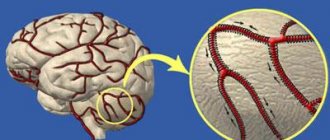
When examining cerebral vessels, the specialist pays attention to the presence of anomalies in their development (tortuosity, loops, additional vessels), and the degree of malformation. These factors lead to arterial hypertension and, if medical care is untimely, are complicated by hemorrhagic or ischemic stroke.
A list of tests for each patient is compiled by a doctor. If the disease first appears in a young person, then to identify its cause, the kidneys and pituitary gland are examined using MRI. For long-term symptoms of hypertension, MRI of the brain and blood vessels is necessary to determine the extent and nature of changes in these structures. Next, the main methods for diagnosing various forms of the disease will be discussed.
Bottom line
Indications and contraindications for magnetic resonance imaging are determined by the attending physician and radiologist. The patient must warn doctors in advance about the presence of implants and electronic devices in the body - the former will interfere with the formation of a magnetic field, and the latter will simply fail after diagnosis.
MRI is completely safe for patients of any age, so there are very few prohibitions on the examination. If there are contraindications, resonance tomography is replaced by another type of diagnosis.
Tags: MRI of the head and brain
Classification of hypertension
Primary and secondary hypertension are classified into several forms of the disease. In the first case, there are:
- Hyperadrenergic form, which is more often observed in young patients. Its symptoms: rapid change in facial skin color, throbbing headaches, anxiety for no reason.
- Normo- and hyporenin form. It is detected mainly in elderly people. The cause of the pathology is the retention of sodium salts in the body, which changes the volume of circulating blood. Signs of the disease are swelling of the face, swelling of the limbs.
- The hyperrenin form develops in individuals with rapidly progressive hypertension and is diagnosed in young men. The pathology is characterized by an increase in blood pressure up to 230 mmHg, accompanied by dizziness, nausea, and kidney pain.
Secondary hypertension is also classified into several forms:
- Renal, developing due to diseases of the renal system (pyelonephritis, polycystic disease, nephropathy, tumors of various etiologies).
- Endocrine, associated with increased or weakened thyroid function, Cushing's syndrome, tumors, hypothalamic syndrome.
- Cardiovascular, caused by heart defects.
- Neurogenic, occurring against the background of encephalitis, atherosclerosis of the arteries and brain tumors
- Drug-induced, which develops with long-term use of drugs that increase blood pressure.
Main types of headaches
Neurologists determine the types of cranialgia by clinical symptoms:
- The most common type is tension pain. Characterized by a feeling of circular soreness along the skull, inside the eye sockets;
- Cluster pain syndrome is accompanied by pulsation of a separate area of the head;
- Tumor pain occurs in the morning, is accompanied by nausea and vomiting, and can gradually progress;
- Intracranial hemorrhage leads to pain in a certain part of the skull, characterized by secondary symptoms (vision loss, unsteadiness of gait, hearing loss, hallucinations);
- Migraine is long-term pain on one side, intensifying against the background of photophobia and loud noises. Often before an attack there are precursors - visual photophobia (rings), auditory hallucinations;
- Temporal arteritis - characterized by local pain in the area of the temporal bone (lateral parts of the skull).
Treatment depends on the type of disease. The combination of several forms at the same time complicates therapeutic tactics. Magnetic resonance scanning makes it possible to study the morphology of the cerebral parenchyma and verify changes in the arteries.
4. Diagnosis of the primary form of the disease
The procedure includes 4 main stages:
- daily blood pressure control;
- collecting information about the disease and related disorders;
- physical examination using a phonendoscope;
- laboratory and instrumental studies.
Patients suffering from the primary form of the disease undergo: a general blood and urine test; blood for biochemistry to determine the level of glucose, potassium, uric acid and creatine.
Among the instrumental diagnostic methods for the primary form of the disease, the following should be noted:
- Echocardiological examination to determine the contractility of the heart and the structure of its ventricles.
- Examination of target organs using MRI, ultrasound or x-ray. They help identify the risk of possible complications from the heart, kidneys and brain.
- Dopplerography, which determines the speed of blood flow in the carotid and cerebral arteries.
X-ray
Using an x-ray, you can determine whether there is hydrocephalus, as well as any other injuries. This simple process is carried out due to the fact that so-called X-rays pass through the human body and all its tissues.
You need to understand that not all tissues can receive such rays, since they are different in density. A similar feature can be recorded on a certain film in the form of a less bright color.
As for soft tissues, they appear as a darker color.
Due to such features, the doctor can identify damaged tissue, find out the cause of this damage and prescribe direct treatment. This examination method is simple and does not require the use of serious equipment. In addition, it has a much lower price compared to the previous ones. It is for this reason that it is used as one of the first in all types of diagnostics. But it is worth knowing that x-rays in the head area are carried out to determine the integrity of the human brain bones.
Diagnosis of the secondary form
A common cause of secondary hypotension is kidney disease. Diagnosis of pathology in this case will be aimed at studying the condition of the target organs. For this purpose, patients are prescribed an ultrasound of the genitourinary system and heart. The advantage of the method is that there is no need to administer contrast agents, which have a toxic effect on the body.
It is not always possible to examine diseases of the kidneys and adrenal glands using ultrasound. This is especially true for benign and malignant tumors in the early stages, narrowing or damage to small arteries. In this case, preference is given to MRI examination with contrast.
5.1. Kidney MRI
Modern tomographs with a power of 3 Tesla are able to detect neoplasms with a diameter of up to 1 mm, which do not give characteristic clinical signs. On MRI images, the lesion appears as a heterogeneous structure. Despite the reliability of the study, MRI cannot accurately determine the malignancy of the tumor. Diagnosis should be complemented by a biopsy of suspicious tissue.
If the neoplasm is located in the pelvicalyceal region, then MRI images will show signs of organ obstruction - hydronephrosis, deformation. The tumor itself is visualized as an area with altered tissue density. Abundant blood supply to the kidneys according to MRI images indicates multiple metastases of the neoplasm. To clarify the localization of the pathological focus and determine its boundaries, contrast MRI is used, the cost of which differs significantly from the standard scanning procedure.
MRI of the kidneys is also prescribed to patients with suspected polycystic disease, which, like tumors, leads to the development of a secondary form of hypertension. In the pictures, the doctor can see: multiple cysts in both kidneys, their deformation and enlargement; brain matter disorder; signs of chronic renal failure.
5.2. MRI of the thyroid gland
The slightest disturbances in the functioning of the endocrine system lead to the development of hypertension. MRI diagnostics is indispensable for identifying thyroid tumors and other pathologies. Scanning allows doctors to clarify not only the clarity of the boundaries of the tumor, its size and location, but also the presence of metastases.
Another disease characterized by increased blood pressure and detected using MRI is Graves' disease. The pathology is associated with increased production of thyroid hormones. An increase in size of the thyroid gland and structural changes in its tissues are clearly visible on MRI images.
5.3. MRI of the brain
The study is often prescribed to determine the causes and consequences of hypertension. MRI can reveal:
- A stroke in which oxygen starvation of certain parts of the brain occurs. The lesions in the images are visualized as white areas with uneven edges and an asymmetrical shape. When contrast is administered, a deterioration in blood supply at the site of the lesion is revealed.
- Tumors of various etiologies. Rapidly growing tumors compress small and large vessels of the brain, leading to pressure surges. If a tumor is suspected of being malignant, an MRI with contrast is performed.
- Viral encephalitis, determined by MRI on days 3-5 of development. The images visualize symmetrical zones of inflammation, localized in the temporal lobes and spreading to the brain stem. In advanced cases, areas of atrophy of the soft tissues of the organ, foci of necrosis and microhemorrhages are observed.
- Vascular atherosclerosis. In this case, doctors prescribe MR angiography to patients.
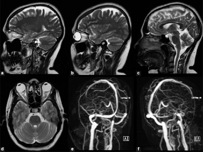
MRI image of the brain. Sagittal (a, b, c) and axial (d) T2 images show vertical flexion of the optic nerves, protuberance of the perioptic nerve sheaths, flattening of the posterior sclera, and a partially empty pituitary fossa. Contrast MR angiography of cerebral vessels (e, f) show narrowing of the distal right transverse sinus and the entire left transverse sinus.