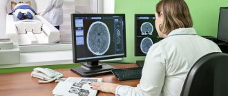
A study of the functionality and structure of a child’s brain may be required from birth, and for this purpose the safest methods are selected. Electroencephalography (EEG) is a painless and harmless way to diagnose abnormalities in the brain, which can be prescribed to a small child.
The study is indicated to assess the functionality of the cerebral cortex by recording the activity of electrical impulses. The method is recognized as informative for obtaining results at any age .
An EEG shows a graphical representation of the activity of neurons, brain cells that form its bioelectrical activity. The result obtained looks like a set of lines , which the specialist deciphers, seeing the course of physiological processes and pathological changes.
The essence of the study and what it shows
Diagnostics is based on the registration of special signals from cerebral structures, which precisely indicate certain pathological processes and changes. To better understand what's what, let's turn to the anatomical certificate.
Normally, the human brain works due to spontaneous excitation, the generation of an electrical impulse and its transmission through neurons, the construction of specialized circuits and networks.
This activity causes cerebral structures to vibrate at specific frequencies. Based on this characteristic, experts identify rhythms of different intensities.
For example:
- Alpha waves, which correspond to a state of complete relaxation, half-asleep.
- Beta - during moderate concentration.
- Gamma impulses are associated with increased stress on the psyche and intellectual pursuits.
- And so on, the list goes on.
Special equipment can detect and record such electrical signals and vibrations. In a convenient form, the specialist receives information about the peculiarities of the brain.
Based on the correlation of certain cerebral impulses, the doctor can draw conclusions about the nature of the nervous system and pathological processes. Although the exact data still depends on the results of several procedures.
What can be tracked based on the results of such instrumental diagnostics:
- Brain rhythms, features of electrical activity. In general, regardless of possible pathological processes.
- The emergence of foci of increased excitation. Similar anomalies occur against the background of epilepsy, after injuries and injuries.
- Decrease or increase in the activity of certain areas of cerebral structures.
Thus, the EEG shows inflammatory, destructive, tumor, neurological disorders or indirectly indicates them.
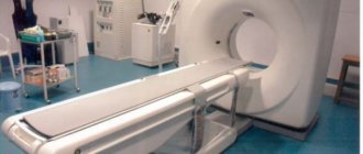
The technique is universal in its way, but does not provide 100% data. Additional measures are needed: MRI, computed tomography, skull x-ray, ultrasound of cerebral vessels.
The encephalography technique itself is heterogeneous. There are several modifications of the diagnostic method. Including EEG, MEG, PEG and others. We will talk about them further.
The essence of the diagnostic method
Back in the 19th century, it was established that the human brain is capable of generating electrical impulses, but more than eighty years passed before doctors were able to use this ability in medical practice. This happened after the creation of the first electroencephalograph apparatus, and the graphic image that was obtained by recording the biocurrents of the human brain was called an electroencephalogram.
Signals emanating from brain neurons change under the influence of various factors, while different parts of the brain give coordinated impulses, mutually weakening or enhancing each other’s activity. An encephalogram records various changes in impulses and helps to identify abnormalities in the functioning of the brain or its individual parts. Electroencephalography allows you to track the activity of each part of the brain separately, study neural impulses and the coordination of the actions of brain parts (rhythm).
What diseases can be detected
The instrumental method is nonspecific. This means that it is impossible to make a diagnosis based on the results alone.
There will remain room for doubts and questions until additional clarifying procedures are carried out.
Still, the method may prompt some thoughts. What diagnoses can be suspected based on the results of encephalography:
Epilepsy
The disease is complex and multifaceted. Contrary to popular belief, it does not always manifest itself “in all its glory”: foam at the mouth, spasms and convulsions, loss of consciousness and falling.
Insidious, silent variants of the pathological process are also possible. When the disease is discovered by chance and as a result of diagnosis for other disorders.
The diagnosis of epilepsy can be made based on the results of encephalography. As a rule, the technique is used several times. To finally verify the essence of the deviation.
Organic and other psychoses
That is, those disorders that directly disrupt the normal biochemistry of cerebral structures. For example, schizophrenia, the consequences of consuming drugs, alcohol, and certain drugs.
An imbalance in brain rhythms is detected as a result of insufficient or excessive signal movement along neurons.
Of course, it is impossible to establish the origin of the violation. To do this, you need to collect anamnesis and monitor the patient for at least several weeks.
Insomnia
As an independent disease or syndrome against the background of one or another pathological process.
The EEG technique is used for routine assessment of the situation, and to obtain more data, doctors resort to studying the circadian rhythm of the brain. This is much more difficult in terms of conducting and deciphering the results.
Psychosomatic disorders
For example, the notorious vegetative-vascular dystonia, which, in fact, is not an independent diagnosis at all. Here again problems of interpretation arise. To understand the essence of the violation, you need to know the context of the situation - it will help you draw conclusions and make an accurate diagnosis.
An EEG is an examination that allows you to identify inflammatory, degenerative, intoxicating, and tumor processes, but only halfway, since a lot of questions remain.
Based on the diagnostic results, the doctor either puts forward a hypothesis regarding the situation or verifies a previously defined pathological process. Depends on the order of examinations.
What is the purpose of an EEG?
If symptoms indicating dysfunction of neurons are identified, the cause for diagnosis should be determined. When performing an EEG on patients such as children, you need to know exactly what the child’s brain encephalogram shows . It is carried out for the following purposes:
- To assess the degree of pathology;
- Determining the ability to work of a patient who is unconscious: under anesthesia or in a coma;
- Analysis of all processes occurring in nerve cells in order to prevent attacks of epilepsy and seizures;
- Make sure of the positive result of the prescribed treatment and, if no effect is detected, change the concept of treatment methods;
The grounds preceding diagnosis using an encephalogram are the following indicators:
- Suffered head injuries;
- Developmental inhibition, applicable to children;
- Increased frequency of fainting, decreased sensitivity of body parts;
- Determining the presence of cancer in the body;
- Epilepsy attacks and convulsions;
- Hypertension;
- Sleep disturbance;
Using this diagnostic technique, such as an electroencephalogram of the brain, what it is and how it is possible to identify complications in the form of a mental disorder or the development of schizophrenia, you can be confident in the accuracy of the result obtained. The versatility of the method is used for examination before passing exams for the right to drive vehicles or handle and own weapons. The issuance of a permit depends on .
Indications
The reasons for a brain encephalogram are inflammatory, tumor, neurological diseases and individual symptoms that, theoretically, may indicate a problem.
Specific indications include:
- Insomnia. Various types. Some patients cannot enter a state of rest at all. Others quickly “pass out”, but just as quickly jump up and remain awake all night. And so on.
Insomnia is often associated with neurological problems. Therefore, encephalography is one of the first to be performed to exclude functional disorders.
- Pathologies of the endocrine system. Hormonal imbalances have the potential to alter normal brain function. An encephalogram is used to study the extent to which certain abnormalities affect the activity of cerebral structures.
First, the level of hormones as a whole is measured, and expanded data on the diseased organ is obtained (thyroid, pituitary gland, adrenal glands, pancreas, reproductive structures).
- Dizziness. Disturbances in normal coordination of movements. Movement disorders. What they are connected with, what caused the symptoms, is another question. We'll have to look into it separately and factually.
- Fainting. Impaired consciousness. Syncope is usually caused by disorders of cerebral blood flow and ischemia of brain tissue. There are options. Encephalography will just help to understand the situation.
- Suffered a stroke. Acute disturbance of blood flow in the brain. Affects the nature of the work of cerebral structures, the speed and quality of transmission of nerve impulses. A break in some of the established neural circuits cannot be avoided.
An encephalogram of the head provides an opportunity to assess the nature of the problems, their degree, and the exact location of the disorder.
In addition, tomography is performed to study anatomical and structural disorders in addition to functional ones.
- TBI, neck injuries. Because they rarely go away without consequences.
- Headache. Any location and intensity. The origin again remains a mystery for the time being. Until a full and comprehensive diagnosis is carried out.
- Nausea, vomiting for no apparent reason. Especially if they are repeated on a regular basis.
- Seizures similar to epileptic.
- Convulsions of unknown origin. May be associated with disorders of the central nervous system.
- Previous or suspected inflammation of the brain. Encephalitis, meningitis. The point is to study how much cerebral structures are affected. Also, in this way it is possible to study the effectiveness of the treatment.
- Coma. As part of monitoring the electrical activity of nerve tissue. As soon as the rhythms begin to fade, this is an unfavorable sign. It is corrected in the format of maintaining basic body functions.
- Suspected brain tumors, neoplastic processes.
- Mental development disorders. Usually we are talking about younger patients. Special stimulant drugs are prescribed as needed.
- Mental disorders. Oddly enough, in this way it is possible to study the effectiveness of medications, antipsychotics and others. Based on the results, one can judge the restoration or, on the contrary, regression of brain functions and the quality of therapy.
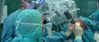
- Preoperative preparation. Encephalography is mandatory.
- Study of the patient's condition after surgery.
These are the key indications.
Electroencephalography is well tolerated, can be repeated several times, does not create a harmful load on the body and is recommended for patients of all ages; contraindications include acute conditions and pathologies that preclude adequacy.
Contraindications
Here are some reasons for refusing the examination:
- Extensive wounds after surgery. Because the affected area simply cannot be disturbed. Infectious problems, suture dehiscence and other pathological consequences are possible.
- Acute psychoses. Because the patient simply cannot sit through the entire procedure calmly and without moving. This eliminates the physical possibility of conducting research.
- Untreated injuries, wounds in the head area. Which is due to the same reasons: you can get an infection or harm a person even more.
- Septic diseases, processes. Up to the banal ARVI, which is associated with obvious distortions of brain rhythms. We need to wait for the right moment.
The list of contraindications is minimal. These are relative options. Once the base is gone, exploration can be done.
What is electroencephalography of the brain
Carrying out such a study of the body as an encephalogram of the brain is a complex instrumental process, the implementation of which is prescribed to study the degree of activity of tissue cells and identify pathologies in them. Based on the results of the study, which is revealed by the encephalogram, it is possible to find out the causes of tissue dysfunction, which becomes a disruption in the functioning of certain areas of the hemispheres of the head. A procedure such as finding out what an encephalogram of the brain shows is the best option for identifying illnesses such as dementia, mental disorders, and epilepsy.
Types of EEG and their differences
There are 5 modifications of the method.
- The classic method is electroencephalography - a non-invasive diagnosis that is based on measuring the electrical potentials of the brain and the total activity of cerebral structures. This is the most accurate way to study the condition of nerve tissue.
On the other hand, he is very sensitive. An awkward micro-movement is enough to blur the results.
At the same time, the results are affected by stress factors and the patient’s tension. And decoding is very difficult when the doctor is not experienced enough. There is a high risk of error.
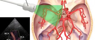
- If classical EEG examines the electrical activity of the brain, rheoencephalography (abbreviated REG) focuses on the state of blood vessels. The essence of the technique is the effect on cerebral structures of a low-power current.
Based on the response, the doctor draws conclusions about the condition of the vascular walls, their conductivity and other characteristics. This is a very specific examination method.
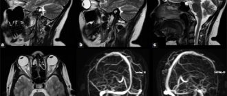
- Echoencephalography (EchoEG) is used to assess the anatomical state of the brain. The technique is based on the reflective ability of the tissues of cerebral structures and the density of the substance.
Using echoEG, you can diagnose tumors and other space-occupying formations, and displacement of brain tissue. Which is very important in the early stages, because later treatment becomes unlikely.
- Pneumoencephalography (PEG) is extremely specific. The technique is based on filling the drainage system of the brain with air. It is injected in small quantities through a puncture in the lumbar region.
The technique allows you to measure pressure in the conduction system, assess the likelihood of ventricular dislocation and intracranial hypertension. These are indirect signs of several possible pathologies.
- Finally, magnetoencephalography (MEG), a visual method for assessing the electrical activity of the brain. Using special sensors, the desired indicator is recorded, after which, in visual form, the doctor receives a full-format picture of the distribution of impulses.
This is very convenient, since both foci of increased activity and shifts relative to the norm are visible. The method is used both in medicine and in theoretical scientific research.
As can be judged from the descriptions, none of the methods practically overlaps with the other. They face completely different goals and objectives.
Therefore, these methods can be prescribed in a system determined by a doctor.
Preparation
No special events needed. All you have to do is come to the clinic. The only exception is pneumoencephalography, but this is a very specific technique that is rarely used due to its invasiveness and a lot of discomfort for the patient.
During the examination, the person must sit or lie still. The fewer voluntary movements, the higher the information content of the technique.
You also need to maintain maximum peace. If there is such a need, it is permissible to use sedatives several hours before the test.
Standard herbal remedies such as chamomile, motherwort or valerian will do. The main thing is not with alcohol. Decoctions and infusions are allowed.
It is highly advisable to follow these basic rules the day before the examination:
- No smoking.
- Do not drink alcohol, coffee, tea.
- You need to remove metal jewelry right in the office.
Preparation for an EEG of the brain means abstaining from psychotropic substances, stimulants, including those of natural origin, and also following all instructions from the doctor’s assistant or the specialist himself.
Progress of the procedure
It all depends on the specific technique and its modifications. In general, the algorithm will be like this:
- The patient lies down on a couch or sits on a chair. It is important to relax as much as possible and suppress the feeling of internal tension.
- The doctor or his assistant lubricates the areas where the sensors are applied with a special gel. This is necessary in order to facilitate the conduction of the signal and its registration.
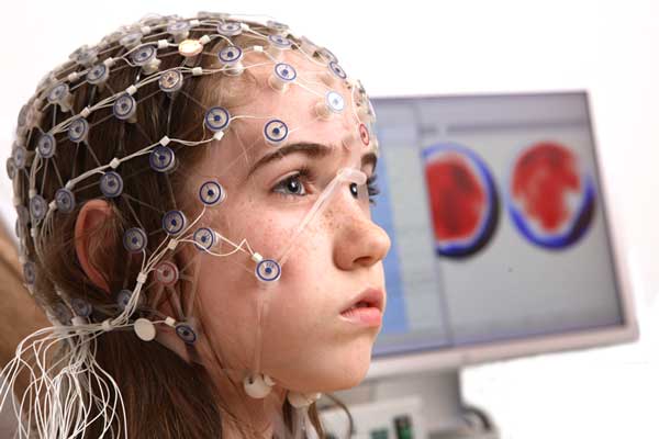
- A special network of sensors (cap) is placed on the head, which will record brain activity.
- The next step is to check the operation of the device.
- The encephalograph records the diagnostic results, builds a special graph and displays the information on the monitor or in printed form (depending on how old the device is in a particular institution).
If necessary, special functional tests are carried out. They do not pose any particular difficulty for the patient. The doctor gives instructions.
Among them:
- Close/open your eyes.
- Make a fist, relax your hand.
- Breathe hard, quickly and often.
- Noise and light stimuli are also used. This is somewhat uncomfortable, but quite tolerable.
The readings are then recorded again. The sensors are removed from the patient. A person receives the result in their hands after 10-20 minutes or at any other time by agreement.
An EEG with stress tests shows the brain’s reaction to a stress factor, a voluntary motor act, and describes the adaptive abilities of the nervous system. This is significant.
There are other options. For example, when conducting 24-hour encephalography, rhythms are recorded longer, and so on.
Carrying out an EEG
Before the procedure, it is necessary to exclude coffee and avoid the presence of hairsprays, lotions, and gels. It is necessary to get rid of metal objects, earrings, hairpins, piercings. You should not eat heavily. EEG is not performed during acute colds. The process is absolutely harmless and not painful. The patient sits down, closes his eyelids and relaxes. Special sensors are coated with gel, attached to the skin, a cap is put on, and a process begins that records the biological tone of the brain. The electrodes are connected to an amplifier because the activity signals are weak. The readings are recorded by a device called an electroencephalograph. The indicators according to which the deviation from the norm is determined are displayed. They can be recorded either on tape or in a file on a computer. The procedure takes no more than 30 minutes.
During EEG, you can use provoking moments to close and open your eyes, breathe deeply and often through your mouth. At certain points, the doctor tells when the patient needs to do this. The encephalogram is carried out in a quiet position, but can also be done in sleeping mode.
to contents ^
Decoding the results
Interpretations should be carried out by specialists. Because it’s not easy to figure it out on your own.
Here is a specific example of a diagnostician’s conclusion based on a patient’s EEG:
If translated from medical into Russian, these are minor disorders of the brain. Let's figure it out in order.
- Alpha rhythms are predominantly recorded during diagnosis, since they are responsible for the patient’s state during relaxation. The normal frequency is 8-13 Hz (we have 6-7).
- As for microvolts, the indicator is no more than 100. There are mild deviations from the adequate limit.
- Disorganization can be either an acceptable option (with fatigue, stress), or the result of pathology against the background of blood flow disorders.
- RFS is rhythmic photostimulation. Special diodes are placed in front of the patient's eyes, irritating cerebral structures. In our case, the indicators increased slightly, within normal limits. This rules out epilepsy (which is the first thing they look for).
The reaction in the occipital leads is within the desirable range, since this is where the visual cortex, the final segment of the visual analyzer, is located.
- HV is hyperventilation. That is another functional test. The replacement of the alpha rhythm is due to the gradual relaxation of blood vessels, which is also explainable from an anatomical point of view by the saturation of the body with oxygen and changes in blood acidity.
In any case, deciphering EEG indicators falls on the shoulders of the doctor and is based on the study of rhythms of different types, their frequency and intensity, and the results of functional tests.