Types of cysts
With lacunar cysts of the brain, the size, norm and location determine their type. But first of all, it is taken into account how they began to develop - in utero, which means they are congenital, or in the process of ordinary life, here the reasons for their appearance are much more diverse.
Moreover, there is a difference between one type of cyst and another based on the location of their appearance in the brain. A retrocerebellar cyst is a neoplasm that arises under the arachnoid membrane of the brain.
If the tumor appears on the outer arachnoid membrane, it is called an arachnoid cyst. Lacunar cerebrospinal fluid cysts occur between the membranes of the brain. A vascular cyst is a tumor that arises in the plexus of blood vessels in the brain. Lacunar cyst of the basal ganglia occurs in the cerebellum, pons, or subcortical ganglion.
You have received only a basic understanding of what it is - a lacunar cyst of the brain. This topic is being studied in medical institutes to this day, which is why quite a lot of types and types of this neoplasm have been discovered, and new tumors and cysts are added to the resulting list every year.
Causes of lacunar cysts
You need to understand that if a patient was diagnosed with a cyst that developed during the period of intrauterine growth, then it is most likely a hereditary disease. It is impossible to insure yourself against this type of tumor. There are cases where a person did not know that he had a congenital cyst until it was accidentally discovered during a routine medical examination. The tumor was 10 cm in diameter and did not interfere with the person’s life at all. What is a cerebral lacunar cyst? This is a formation that creates problems, but not in every case of its occurrence.
If the cyst is diagnosed as an acquired pathology, this means that it was the result of some kind of somatic disease. For example, it appeared as a complication after meningitis or traumatic brain injury. Diabetes mellitus, thrombosis, and hypertension can lead to the appearance of a cyst.
Often, frequent concussions can cause the appearance of a tumor in the brain. This phenomenon occurs in professional sports, where an athlete often receives traumatic brain injuries, for example, boxing or other martial arts.
Post-ischemic lacunar cyst has become widely known in medical circles. From the name it is clear that it was a consequence of ischemic brain disease, which, in fact, leads to chronic hypertension. Postischemic lacunar cysts of the brain are studied and treated simultaneously with stroke. Often they become the cause of craniotomy and brain surgery. But lacunar cerebrospinal fluid cyst is amenable to radiation therapy, and surgery in this case is not always required.
What accompanies a choroid plexus cyst in a newborn?
Let's consider the most commonly diagnosed genetic defect, accompanied by the presence of a cyst. We are talking about Edwards syndrome or trisomy 18. With this anomaly, the 18 pair of chromosomes does not diverge, another chromosome 18 is added to it. Thus, normally there are two of them, but with this disease there are three. The resulting genotype in the embryo consists of 47 chromosomes.
A copy of chromosome 18 can cause the death of the embryo, or the baby will have multiple defects and anomalies at birth. This leads to:
- neural tube defect;
- hammer feet;
- curled fingers;
- hygroma cysts;
- hydrocephalus;
- micrognathia;
- rocker foot;
- limited growth.
There is also trisomy 21 or Down's disease, but for some reason, a cyst of the choroid plexus of the brain forms less often with this disease.
The significance of the cyst is zero even in the presence of Edwards syndrome, since it is the deviations that accompany this developmental anomaly that become important.
Signs of a cyst in the brain

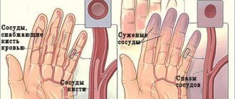
Lacunar cysts of the brain may not show symptoms for many years. At the same time, it grows and develops. The first signs that a person has it appear only when the enlarged tumor begins to put pressure on nearby blood vessels, thereby disrupting blood flow or creating pressure on certain parts of the brain. And depending on what lobes these are, characteristic signs appear:
- The patient experiences hallucinations.
- Severe headache, and the location of the pain is located opposite the cyst.
- A person begins to feel sick or vomit for no apparent reason.
- In severe cases, seizures with convulsions and loss of consciousness are possible.
- Coordination of movements is impaired.
- Speech and a person’s ability to write texts are impaired.
- If a cyst appears in the frontal lobe, then a person’s vision may be impaired or completely lost.
- If the tumor is in the temporal lobes, the sense of smell is impaired and the ability to distinguish tastes is lost.
But the neoplasm does not always have at least some manifestation. Often, a lacunar cyst of the brain does not receive any treatment at all, because it has not manifested itself in any way throughout a person’s life. There was no pain or any disturbances in brain function.
Symptoms
The distinctive signs of the disease largely depend on the location of the pathological focus and may differ in each specific case. When a spinal cord cyst is localized in the cervical region, the cavity usually does not grow to a large size and often does not have pronounced symptoms.
However, if it increases, a person may experience a number of negative sensations:
- pain in the neck area with varying intensity;
- sudden changes in blood pressure, dizziness, headaches;
- restriction in movement and numbness of the fingers.
In the case of the development of a spinal cord cyst in the thoracic region, the signs may be different due to the high probability of negative interaction with internal organs. The most common of them are:
- a feeling of squeezing in the thoracic region during movement;
- violation of the swallowing process, nausea, heartburn;
- feeling of heart rhythm disturbances;
- muscle tension ;
- pinched intercostal nerves.
General symptoms of this disease include muscle atrophy, changes in gait, decreased quality of vision, convulsions, and short-term loss of consciousness.
Diagnosis of the presence of a cyst in the brain
Treatment of lacunar post-ischemic cyst of the brain is prescribed only after a complete comprehensive diagnosis. The same applies to any type of cyst - it is important to know its size and location.
After all, if the cyst does not pose a danger to the functioning of the brain, does not grow, then they simply observe it, without trying to cure it in any way - there is no need. But if it begins to grow and put pressure on the surrounding vessels and brain tissue, then urgent therapy begins.
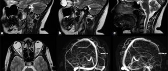
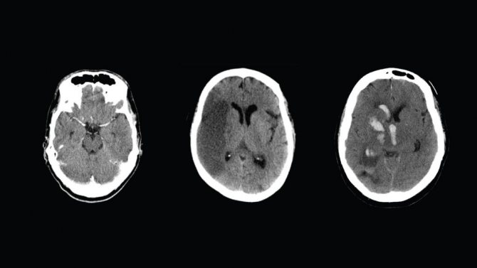
The main method for diagnosing brain tumors is computed tomography. It most clearly and accurately determines the presence, size and location of the cyst.
In order to understand whether its contents are dangerous, that is, whether it can turn into a malignant tumor, a histological analysis of the cells obtained from the cyst as a result of a biopsy is done.
The diagnosis can be clarified by examining the vessels of the head and neck using Doppler sonography. At the same time, the condition of the patient’s blood is studied, the presence of elevated cholesterol levels in it, in order to eliminate the likelihood of cholesterol plaques.
The blood is also examined for the presence of pathogenic microorganisms that can cause inflammation.
Diagnosis may include daily monitoring of blood pressure fluctuations. For this purpose, special sensors are attached to the patient, which accompany all his activities throughout the day.
Diagnostics
Unlike neoplasms in other organs, a brain cyst is not palpable. And a person cannot detect it in himself. Only unpleasant symptoms force the patient to see a doctor. But even in this case, special studies are not immediately prescribed. Sometimes a specialist can associate headaches or blurred vision with another cause and prescribe treatment. And only after there is no positive dynamics for a certain period, the patient is sent for additional examination:
- MRI;
- Ultrasound;
- radiography;
- dopplerography;
- ECG;
- EEG;
- blood test for infections;
- blood pressure monitoring.

Usually a comprehensive examination is prescribed, which includes several of the listed methods. Based on their results, doctors find out the size of the cyst, its location and preliminarily determine the type. If it was not possible to identify the tumor the first time, after a while a series of studies are carried out to observe changes in the cyst over time.
By the way! Often, a cystic formation of the brain is discovered accidentally, during an MRI or X-ray, the purpose of which was to diagnose a completely different disease.
Drug treatment of lacunar cyst
If the cyst does not bother the patient, does not increase in size and does not pose a danger, then it is not subject to special treatment. In this case, therapy is aimed at eliminating the cause of its occurrence. For example, if a patient has suffered a severe viral disease such as meningitis or encephalitis, then he is treated with antibiotics and immune-strengthening drugs. The specific type of drug, its dosage and regimen of use are prescribed individually depending on the general condition of the patient.
A neurologist may prescribe a drug that breaks up adhesions in connective tissue, blood thinners, and antioxidants. All this allows you to restore impaired blood flow in the vessels of the brain and stabilize pressure.
Preventive measures
To avoid the formation of a hollow formation in the temple area, it is necessary to follow the rules for the prevention of primary and secondary tumors.
Prevention of congenital tumors is carried out during pregnancy and indicates a healthy lifestyle, a balanced diet and timely registration of the expectant mother.
Preventive measures for acquired hollow formations: complex treatment of viral diseases of the brain, consequences of head trauma.
An arachnoid cyst is a dangerous pathology with serious consequences if treatment methods are ignored. If the cyst is diagnosed at an early stage and removed on time, the patient follows medical recommendations, and undergoes regular medical examinations, then there is no risk of complications. Otherwise, the cerebrospinal fluid neoplasm causes discomfort, complications and death of a person.
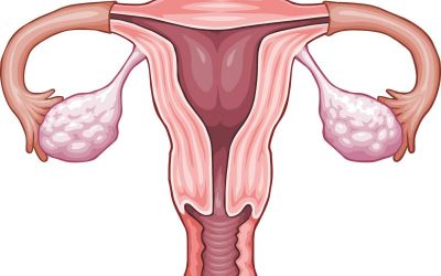
The appearance of a large cyst on the right ovary in a woman
Previous article

What is a vaginal cyst?
Next article
Surgery

If the cyst poses a danger to the patient’s condition, then surgery is indicated. It can be performed using various methods - bypass surgery, endoscopy and craniotomy.
Each method has both positive and negative sides. Endoscopy can only reach those cysts that are located directly under the skull bone. The instrument, which is a thin tube, does not reach the neoplasms lying deep in the brain matter.
Bypassing a cyst involves suctioning out the fluid filling the cyst using a thin needle. As a result of this procedure, the cyst is reduced until only one shell remains. The negative side of this procedure is the risk of introducing an infection into the brain, which will cause complications.
Craniotomy involves opening the skull to provide access to the cyst at any depth. This type of operation provides a 100% opportunity to completely remove the tumor, but has a long postoperative period.
The type of operation is chosen by the attending physician, based on the patient’s condition, location and size of the cyst, and many other medical indicators. The wishes of the patient in this situation are not taken into account.
Treatment of a tumor of the left temporal lobe
If it is small in size and there is no pressure on the brain structures and ventricles, the pathology does not need to be treated; only medical supervision over the dynamics of growth and character and regular MRIs and CT scans are necessary. But in this case, it is important to determine the root causes so that new benign tumors do not develop.
With progressive tumors, conservative treatment is required. Medicines are prescribed to relieve inflammation, normalize cerebral circulation, and restore damaged neurons. The duration of the course is selected individually. Prescribed:
- anti-inflammatory, decongestant medications;
- drugs that improve blood circulation;
- nootropics to start metabolic processes;
- means for maintaining immunity;
- hepatoprotectors.
There are several indications for surgery: rapid growth of the tumor, rupture, or if medical assistance is ineffective. Apply:
- Radical surgery - craniotomy. The method contributes to traumatization and leads to dangerous consequences.
- Shunting - ensuring the outflow of fluid into the abdominal or subdural cavity using drainage tubes. They make a hole in the skull and install a drainage system equipped with special valves. The catheter is placed under the skin. The use of the latest equipment helps to perform the operation accurately.
- Endoscopy - cystic contents are removed by puncture.
- Drainage is carried out mainly for newborns. This needle aspiration method effectively removes fluid from the cerebrospinal fluid cyst. The liquid mass moves into the natural circulation zone, which promotes the resorption of the bubble. The method does not guarantee that the anomaly will reoccur.
- Fenestration - the formation is completely excised. The method is performed using an endoscope or laser.
Only the neurosurgeon decides which method to use during surgery, because each case is individual and has its own characteristics.
Postoperative rehabilitation
The time allocated for postoperative rehabilitation depends on the complexity of the surgical intervention and the severity of the disease that caused the cyst.
For example, after removal of a congenital cyst, which did not complicate the patient’s condition, using bypass surgery, 10-15 days are allotted for rehabilitation.
And if the tumor was caused by an infectious disease such as meningitis, the cyst disrupted some functions in the body, vision, hearing or musculoskeletal function, trepanation was required to remove it, then a complete cure may take 5-6 months.
Special diet

During treatment and in the postoperative period, the patient adheres to a special diet. To reduce cholesterol in the blood, which can cause a blood clot, dishes with fatty, fried meat are removed from his diet. Meat products suitable for consumption in this situation are boiled fish, chicken and veal.
To normalize blood pressure and strengthen the immune system, fresh fruits and vegetables are included in the diet. The patient's diet is adjusted so that he eats 6-7 times a day, but in small portions. This reduces the load on the stomach, but allows all the beneficial substances from food to be fully absorbed into the intestines.
Drinking coffee and alcoholic beverages is strictly prohibited.
Possible complications
If any type of lacunar cyst is left untreated, the patient may develop various complications.
Thus, a pineal cyst leads to encephalitis or hydrocephalus - the accumulation of fluid in the brain. And an arachnoid cyst can lead to epilepsy. In colloidal cysts, complications are even more dangerous - cerebral hernia, hydrocephalus and death.
If an untreated cyst remains in a child's brain, it can delay his intellectual and even physical development. The most dangerous complication is cyst rupture. In this case, a quick and painful death awaits the person.
Traditional medicine treatment

Despite the seriousness of the disease, lacunar cysts have several methods of treatment with folk remedies. In general, it, like conservative therapy, is aimed at eliminating the causes of the tumor. Therefore, traditional methods must be used in combination with drug therapy and only with the permission of the attending physician.
Medicinal plants that can positively affect a person’s condition in this situation are hemlock, elecampane, wormwood, chamomile, calendula, yarrow, raspberry, corn silk, and Dioscorea Caucasica.
From these plants you can make decoctions or alcohol tinctures. The decoction is simple to make - 1 tbsp. a spoonful of the plant is poured into a glass of boiling water and simmered over low heat for no more than 15 minutes. After the broth has cooled, it should be strained and taken in a glass half an hour before meals.
The alcohol infusion takes longer to make - the dry, crushed plant is filled with alcohol in a ratio of 1 to 3, that is, 300 ml of alcohol per 100 g of plant, and infused for 2 months in a dark place. The infusion should be shaken once a week. After straining, the product is taken 1 teaspoon per day, half an hour before meals.
Prevention

To prevent the occurrence of a cyst in the brain, it is necessary to take a number of measures aimed at protecting a person from situations in which a cyst begins to develop:
- It is necessary to avoid stress and nervous tension.
- Treat viral diseases in a timely manner, preventing them from becoming chronic.
- Protect your head from injuries at work or in sports. That is, wear a protective helmet or hard hat.
- It is necessary to monitor blood pressure and take measures to reduce it during periods of increase.
- You need to give up bad habits - smoking and drinking alcohol, because they cause many diseases. A person who smokes is several times more likely to get cancer.
Description of education
A cyst in the vascular (choroid, choroid, villous) plexus does not form so often. In general, this is only 1-3% of all pregnancies that are monitored. This formation should disappear by 27-28 weeks of pregnancy. Half of the cysts are bilateral. But there are cases when the cyst is visualized before birth. There's nothing wrong with that either.
The fetus is not in danger. In addition, if it is later detected in a newborn or in an adult (in very rare cases, a person has it throughout his life), it does not matter. There may be several choroid plexus cysts; this does not affect the prognosis in any way.
What is this choroid plexus cyst? Inside the plexus, cerebrospinal fluid or cerebrospinal fluid accumulates, which is produced in it. It nourishes the fetal brain and spinal cord. The choroid plexus is a sign of the early formation of the central nervous system in the fetus, and there are two of them, just like the cerebral hemispheres (right and left).
Science does not know why the accumulation of liquid is localized in a certain place. There is no point in understanding this. After all, this choroid plexus cyst in the fetus is not of particular significance. It is called this because on ultrasound the accumulation is visualized in exactly this form.