The human brain is completely unique. It performs a huge number of functions, controlling absolutely all activities of the human body. The complex structure of the brain is more or less known only to specialists. Ordinary people have no idea how many different components form their “biological computer.” The result of dysfunction of even one part can be serious problems with health, behavioral reactions and the psycho-emotional state of a person. One of these parts is the 4th ventricle of the brain.
Types of ventricles of the brain
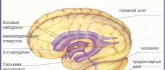
The lateral ventricle(s) are small cavities in the cerebrum that produce a specific cerebrospinal fluid. They are considered the largest of the ventricular system. This is a paired formation, and there is a specific topography for it.
The left lateral ventricle, according to tradition, is called the first. And the right one is second. They are symmetrical with each other and adjacent anatomical formations, and are located below the epiphysis on the sides of the midline. Each ventricle has a body and horns: anterior, posterior and inferior. The lateral ventricles are connected to the third ventricle through the foramina of Monroe.
The third ventricle is located between the areas responsible for vision. It has the shape of a ring and in its wall is the gray matter of the brain containing the autonomic ganglia. In addition to the lateral ventricles, this cavity is connected to the cerebral aqueduct.
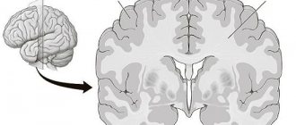
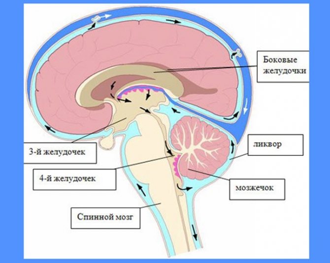
The fourth ventricle is located between below the cerebellum. It is shaped like a pyramid and is more correctly called a diamond-shaped fossa. In addition to the cerebrospinal fluid, most of the nuclei of the spinal nerves are located at the bottom of this fossa.
The norm for the lateral ventricles is to produce half a liter of cerebrospinal fluid per day, but only one hundred and forty milliliters of this amount constantly circulates in the subarachnoid space. Despite the fact that the basis for cerebrospinal fluid is blood plasma, they have significant differences in the amount of electrolytes and protein.
— detoxification (transportation of metabolic products);
— depreciation (when walking, falling, sharp turns);
— formation of a hydrostatic membrane around the elements of the nervous system;
— maintaining a constant composition of fluids in the central nervous system;
— transport (transfer of hormones and some drugs).
Anatomy
The vascular base of the fourth ventricle (tela choroidea ventriculi quarti) is a fold of the pia mater, protruding along with the ependyma into the fourth ventricle, and has the appearance of a triangular plate adjacent to the inferior medullary velum. Its base is directed forward and upward, its apex is directed towards the lower corner of the rhomboid fossa, and its edges are directed towards the lateral edges of the inferior medullary velum. It forms, together with the inferior medullary velum, the posterior part of the roof of the fourth ventricle. In the vascular base, blood vessels branch, forming S. s. fourth ventricle (plexus choroideus ventriculi quarti). In this plexus there is a middle, oblique-longitudinal part, located in the fourth ventricle, and a longitudinal part, extending into its lateral recesses. S. s. the fourth ventricle is formed by the anterior and posterior villous branches of the fourth ventricle (rr. choroidei ventriculi quarti ant. et post.). The anterior villous branch of the fourth ventricle departs from the anterior inferior cerebellar artery (a. cerebelli inferior anterior) near the flocculus and, branching in the vascular base, forms the S. s. lateral recess of the fourth ventricle. The posterior villous branch of the fourth ventricle branches from the posterior inferior cerebellar artery (a. cerebelli inferior posterior) and branches in the middle part of the cerebellar artery. Outflow of blood from S. s. The fourth ventricle is carried out through several veins that flow into the basal or great cerebral vein. From the S. s., located in the area of the lateral recess, blood flows through the veins of the lateral recess of the fourth ventricle (w. recessus lateralis ventriculi quarti) into the middle cerebral veins (vv. mesencephalicae).
The vascular basis of the third ventricle is a thin plate located under the cerebral vault between the right and left thalamus (see), which can be seen after removal of the corpus callosum and cerebral vault. Its shape depends on the shape and size of the third ventricle. In the vascular basis of this ventricle, 3 sections are distinguished: the middle one, enclosed between the medullary stripes of the thalamus, and two lateral ones, covering the upper surfaces of the thalamus; in addition, the right and left edges, the upper and lower leaves are distinguished. The upper one closes the triangular gap between the crura of the cerebral vault, the lower one is adjacent to the ependyma of the third ventricle. Together with the ependyma, the vascular base forms the roof of the third ventricle. At the back, the layers of the vascular base diverge. The upper one extends to the corpus callosum, fornix and further to the cerebral hemispheres, where it is the pia mater of the brain; the lower one covers the upper surfaces of the thalamus. From the lower leaf on the sides of the midline, villi, lobules, and nodes of the S. s. are introduced into the cavity of the third ventricle. third ventricle. In front, the plexus approaches the interventricular foramina, through which it connects with the S. of the village. lateral ventricles.
In S. s. of the third ventricle, the medial and lateral posterior villous branches (rr. choroidei posteriores med. et lat.) of the posterior cerebral artery (a. cerebri post.) and villous branches (rr. choroidei ventriculi tertii) of the anterior villous artery (a. choroidea ant.) branch out. . The medial posterior villous branches (1-3) usually arise from the post-communication part of the posterior cerebral artery. More often there is one branch with a diameter of 0.4-0.8 mm. It follows medial to the posterior cerebral artery, surrounds the cerebral peduncle, fits under the splenium of the corpus callosum and branches in the vascular base of the third ventricle, taking part in the formation of S. of the s. Through the interventricular foramina, this branch anastomoses with the lateral posterior villous branch. The lateral posterior villous branch (1-3) usually branches from the posterior cerebral and less often from the superior cerebellar artery (a. cerebelli sup.) and, located along the thalamic cushion, spreads into the vascular base of the lateral ventricles. More often there is one trunk of a branch, which in the area of the interventricular foramina sends branches to the vascular base of the third ventricle. The villous branches of the third ventricle, originating from the anterior villous artery, anastomose with the posterior villous branches of the posterior cerebral artery. Outflow of blood from the veins of S. s. The third ventricle is supplied by several (3-5) thin veins belonging to the posterior group of tributaries of the internal cerebral veins (vv. cerebri int.).
S. s. lateral ventricles (plexus choroidei ventriculorum lateralium) is a continuation of S. s. of the third ventricle, which protrudes into the lateral ventricles from the medial sides, through the gaps between the thalami and the fornix. From the side of the cavity of each ventricle S. s. covered with a layer of epithelium (lamina choroidea epithelialis), which is attached on one side to the fornix, and on the other to the attached plate of the thalamus (lamina affixa). After separation of S. s. on the edge of the fornix there remains the tape of the fornix (tenia fornicis) and the fimbria of the hippocampus (fimbria hippocampi), and on the attached plate there is a vascular tape (tenia choroidea), the edges are located above the thalamus and stretch from the interventricular foramen to the end of the inferior horn. S. s. each lateral ventricle is located in its central part and passes into the lower (temporal) horn. S. s. formed by the anterior villous artery, partly by branches of the medial posterior villous branch. The anterior villous artery is usually a branch of the internal carotid artery (see), but can arise from the middle cerebral or posterior communicating arteries. On the way to the lateral ventricle, it gives off branches to the basal ganglia. Vienna S. s. The lateral ventricle is formed by numerous convoluted ducts. Between the villi of the plexus tissue there are a large number of veins connected to each other by anastomoses. Many veins, especially those facing the ventricular cavity, have sinusoidal expansions and form loops and semirings. Arteries S. s. entwined with venous vessels. Outflow of blood from S. s. the lateral ventricle occurs in the superior and inferior villous veins (vv. choroideae sup. et inf.). The superior villous vein is formed from the veins of the S. s. in the lower (temporal) horn and the central part of the lateral ventricle. It often flows into the thalamostriatal vein, less often into the internal cerebral vein; forms anastomoses with the inferior villous vein. Sometimes, instead of the trunk of the superior villous vein, there are numerous small veins that flow directly into the internal cerebral vein. The inferior villous vein is formed in the central part of the lateral ventricle and passes, receiving tributaries, through the S. s. in the inferior horn and flows into the basal vein.
Vascular basis and S. s. innervate the periarterial nerve plexuses c. n. pp., spreading to the villous arteries and branches from the internal carotid and main (basilar, T.) arteries. In this case, the sources of sympathetic fibers are the superior cervical and stellate ganglia of the sympathetic trunk, and the sources of parasympathetic fibers (see Autonomic nervous system) are the vagus nerve (see). Sensitive innervation is carried out by the branches of the trigeminal nerve (see), forming sensitive nerve endings in the vascular base and in the vessels of the plexuses.
Choroid plexuses
The lateral ventricle(s) are only partially involved in such a concept as the choroid plexus. The bulk of these structures are located in the roofs of the third and fourth ventricles. They are responsible for most of the production of cerebrospinal fluid. In addition to them, this function is performed directly by the nervous tissue, as well as by the ependyma, which covers the ventricles of the brain from the inside.
Morphologically, the choroid plexuses are outgrowths of the pia mater, immersed in the ventricles. On the outside, these protrusions are covered with cuboidal specific choroid epithelium.
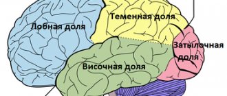
Lateral divisions
Conventionally, they are called the first and second. Each lateral ventricle of the brain includes three horns and a central section. The latter is located in the parietal lobe. The anterior horn is located in the frontal, the lower - in the temporal, and the posterior - in the occipital zone. In their perimeter there is a choroid plexus, which is distributed quite unevenly.
So, for example, it is absent in the posterior and anterior horns. The choroid plexus begins directly in the central zone, gradually descending into the lower horn. It is in this area that the size of the plexus reaches its maximum value. For this reason, this area is called a tangle. Asymmetry of the lateral ventricles of the brain is caused by a disturbance in the stroma of the tangles.
Origin and role
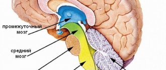
In ancient animals, the primary nervous system was formed - the central vesicle and neural tube. During the process of evolution, the central bubble was divided into three. In humans, the anterior one transformed into the hemisphere, the second into the midbrain, and the posterior into the medulla oblongata and cerebellum. In addition to them, on the basis of the third bubble, internal cavities of the brain, the so-called ventricles, were formed: two lateral ones, a third and a fourth.
The lateral (left is called the first, right - the second) ventricles are the largest cavities of the brain and contain cerebrospinal fluid. Their walls are formed by adjacent brain structures, such as the frontal lobes, corpus callosum, and visual thalamus. Their posterior parts continue into the occipital lobe.
The third ventricle is formed by the fornix of the brain, the optic chiasm, and the “plumbing” to the fourth ventricle.
The 4th ventricle was formed from the posterior wall of the third bladder. It has the shape of a doubly curved parallelepiped. The lower surface is formed from special fibers of nervous tissue connecting the cerebellum and the brain, and there are also pathways from the vestibular apparatus (inner ear) to the base and cortex of the brain.
The lateral walls contain the nuclei of the cranial nerves from the fifth to the twelfth pairs, which, in turn, are responsible for:
- facial sensitivity and chewing (fifth pair);
- peripheral vision (sixth pair);
- movement of facial muscles, facial expressions, tears, salivation (seventh pair);
- taste sensations (seventh, ninth and tenth pairs);
- hearing, sense of balance, coordination of movements of the whole body (eighth pair);
- voice, its timbre, pronunciation of sounds (ninth, tenth, eleventh pairs);
- heart rate, regulation, composition and quantity of digestive juices, lung capacity (tenth pair);
- movements of the head, neck, upper shoulder girdle, chest muscle tone (eleventh pair);
- work of the tongue (twelfth pair).
The upper wall of the fourth ventricle is formed in the shape of a tent. In fact, the lateral and superior fornix are elements of the cerebellum, its membranes and pathways, including blood vessels.
All four ventricles regulate intracranial pressure and are interconnected by a vascular network and connecting channels.
Ependymocytes
The lateral ventricles of the brain are lined from the inside with a special tissue that can both produce cerebrospinal fluid and absorb it. This helps maintain the optimal amount of fluid in the cavity and prevent increased intracranial pressure.
The cells of this epithelium have many organelles and a large nucleus. Their outer surface is covered with a large number of microvilli; they help promote the cerebrospinal fluid, as well as its absorption. Outside the ependyma are Kolmer cells, which are considered a special type of macrophages capable of moving throughout the body.
Through multiple small gaps in the basement membrane of epindemocytes, blood plasma sweats into the ventricular cavity. Proteins produced directly by the cells of the internal epithelium of the brain cavities are added to it, and this is how cerebrospinal fluid is obtained.
Structure
Inside the 4th ventricle is lined with special tissue similar to epithelial tissue. Its composition is regulated and controlled by special receptors with very fine chemical sensitivity. Its cells carry out the interpenetration of blood elements, hormones and other biologically active substances between the circulatory system and the cerebrospinal fluid (cerebrospinal fluid). It should be noted that the 4th ventricle is the area of responsibility of the immune system for protection against the penetration of infectious and parasitic agents. Since the 4th ventricle is directly connected to the arachnoid membrane, which covers the entire brain and is in contact with the choroid, any infection that enters the 4th ventricle can spread to any part of the cerebral cortex or to its base through the third and lateral ventricles.
General system structure and some important terms
Cavities containing cerebrospinal fluid communicate with a number of organs. In particular, with the spinal cord canal, subarachnoid space. The structure of the system is as follows:
- 2 lateral ventricles;
- third and fourth ventricles;
- choroid plexuses;
- choroid ependymocytes;
- tanycytes;
- blood-cerebrospinal fluid barrier;
- liquor fluid.
Contrary to their name, the ventricles are not bags filled with cerebrospinal fluid, but hollow spaces, or cavities, located in the brain. The produced liquor performs a huge number of functions. The common cavity, formed from the ventricles of the brain with canals, overlaps with the subarachnoid space and the median canal of the spinal part of the central nervous system.
Most of the total cerebrospinal fluid is produced in the area of the choroid plexuses located above the 3rd and 4th ventricular cavities. A little substance is located in the wall areas. Soft membranes emerge into the lumen of the cavities, from which plexuses of blood vessels are created. Ependymal cells (choroid ependymocytes) play a huge role and are quite functional in stimulating nerve impulses.
An important criterion is the promotion of cerebrospinal fluid with the help of special cilia. Tanycytes provide connections between blood cells and spinal cord fluid in the ventricular lumens; they have become a specialized variety of ependymal cells. The blood-cerebrospinal fluid barrier is a highly selective filter. It performs the function of selectivity in the flow of nutrients into the brain. It also displays metabolic products. Its main purpose is to maintain the homeostasis of the human brain and the multifunctionality of its activities.
This ventricle is located in the diencephalon. It connects the lateral sections with the fourth. As in other ventricles, the third contains choroid plexuses. They are distributed along its roof. The ventricle is filled with cerebrospinal fluid. In this department, the hypothalamic groove is of particular importance.
Lateral ventricles of the brain
The brain is a complex closed system protected by many structures and barriers.
These protective supports carefully filter all material approaching the sinuous organ. However, such an energy-intensive system still needs to interact and maintain communication with the body, and the ventricles of the brain are one of the tools for ensuring such communication: these cavities contain cerebrospinal fluid, which supports the processes of metabolism, transport of hormones and removal of metabolic products. Anatomically, the ventricles of the brain are a derivative of the expansion of the central canal.
So, the answer to the question of what the cerebral ventricle is responsible for
This cerebrospinal fluid serves as a shock absorber, that is, it provides mechanical protection for parts of the brain (protects against various types of injuries). Liquor, as a liquid, is in many ways similar to the structure of lymph.
Like the latter, cerebrospinal fluid contains a huge amount of vitamins, hormones, minerals and brain nutrients (proteins, glucose, chlorine, sodium, potassium).
Different ventricles of the brain in an infant have different sizes.
Types of ventricles
Each part of the brain's central nervous system requires its own self-care, and therefore has its own storage facilities for spinal cerebrospinal fluid. Thus, the lateral stomachs (which include the first and second), third and fourth are distinguished. The entire ventricular organization has its own message system. Some (the fifth) are pathological formations.
Lateral ventricles - 1 and 2
The anatomy of the ventricle of the brain involves the structure of the anterior, inferior, posterior horn and the central part (body). These are the largest in the human brain and contain cerebrospinal fluid. The lateral ventricles are divided into the left - the first, and the right - the second. Thanks to the foramina of Monroe , the lateral cavities connect to the third ventricle of the brain.
The lateral ventricle of the brain and the nasal bulb as functional elements are closely interconnected, despite their relative anatomical distance.
Their connection lies in the fact that between them there is, according to scientists, a short path along which pools of stem cells pass.
Speaking about this type of ventricles, it can be argued that the normal size of the ventricles of the brain in adults depends on their age, skull shape and somatotype.
In medicine, every cavity has its normal values. Lateral cavities are no exception. In newborns, the lateral ventricles of the brain normally have their own dimensions: the anterior horn is up to 2 mm, the central cavity is 4 mm.
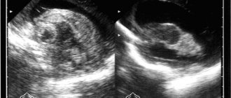
These dimensions are of great diagnostic importance when studying pathologies of the infant’s brain (hydrocephalus, a disease discussed below). One of the most effective methods for studying any cavity, including the cavities of the brain, is ultrasound.
It can be used to determine both the pathological and normal size of the ventricles of the brain in children under one year of age.
3rd ventricle of the brain
The third cavity is located below the first two, and is located at the level of the intermediate section of the central nervous system between the visual tuberosities. The 3rd ventricle communicates with the first and second through the foramina of Monroe, and with the cavity below (4th ventricle) through the aqueduct.
Normally, the size of the third ventricle of the brain changes with the growth of the fetus: in a newborn – up to 3 mm; 3 months – 3.3mm; in a one-year-old child – up to 6 mm.
In addition, an indicator of the normal development of cavities is their symmetry. This stomach is also filled with cerebrospinal fluid, but its structure differs from the lateral ones: the cavity has 6 walls.
The third ventricle is in close contact with the thalamus.
4th ventricle of the brain
This structure, like the previous two, contains cerebrospinal fluid. It is located between the Sylvian water supply and the valve. The fluid in this cavity enters the subarachnoid space through several channels - two foramina of Luschko and one foramen of Magendie.
The rhomboid fossa forms the bottom and is represented by the surfaces of the brain stem structures: the medulla oblongata and the pons. Also, the fourth ventricle of the brain provides the foundation for the 12th, 11th, 10th, 9th, 8th, 7th, and 5th pairs of cranial nerves.
These branches innervate the tongue, some internal organs, the pharynx, facial muscles and facial skin.
5th ventricle of the brain
In medical practice, the name “fifth ventricle of the brain” is used, but this term is not correct. By definition, the stomachs of the brain are a set of cavities interconnected by a system of messages (channels) filled with spinal cerebrospinal fluid.
In this case: the structure called the 5th ventricle does not communicate with the ventricular system, and the correct name would be “cavity of the septum pellucida.”
From this follows the answer to the question of how many ventricles there are in the brain: four (2 lateral, third and fourth).
In most cases, the size of this structure does not correlate with the frequency of pathology, however, there is evidence that in patients with schizophrenia, stress disorders and people who have suffered a traumatic brain injury, this part of the nervous system is enlarged.
Choroid plexuses of the ventricles of the brain
As noted, the function of the abdominal system is the production of cerebrospinal fluid. But how is this liquid formed? The only brain structure that provides the synthesis of cerebrospinal fluid is the choroid plexus. These are small villous formations belonging to vertebrates.
The choroid plexus is a derivative of the pia mater. They contain a huge number of vessels and carry a large number of nerve endings.
Ventricular diseases
In case of suspicion, an important method for determining the organic state of the cavities is puncture of the ventricles of the brain in newborns.
Diseases of the ventricles of the brain include:
Ventriculomegaly is a pathological expansion of cavities. Most often, such expansions occur in premature babies. The symptoms of this disease are varied and manifest themselves in the form of neurological and somatic symptoms.
Ventricular asymmetry (individual parts of the ventricles change in size). This pathology occurs due to an excessive amount of cerebral fluid. You should know that violation of the symmetry of cavities is not an independent disease - it is a consequence of another, more serious pathology, such as neuroinfections, massive contusion of the skull or tumor.
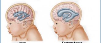
Hydrocephalus (fluid in the ventricles of the brain in newborns). This is a serious condition characterized by the excessive presence of cerebrospinal fluid in the gastric system of the brain. Such people are called hydrocephalus.
The clinical manifestation of the disease is excessive volume of the child's head. The head becomes so large that it is impossible not to notice. In addition, the defining sign of pathology is the “sunset” symptom, when the eyes shift to the bottom.
Instrumental diagnostic methods will show that the index of the lateral ventricles of the brain is higher than normal.
Pathological conditions of the choroid plexus occur against the background of both infectious diseases (tuberculosis, meningitis) and tumors of various locations. A common condition is cerebral vascular cyst. This disease can occur in both adults and children. The cause of cysts is often autoimmune disorders in the body.
Thus, the norm of the ventricles of the brain in newborns is an important component in the knowledge of a pediatrician or neonatologist, since knowledge of the norm allows one to determine pathology and find deviations in the early stages.
You can read more about the causes and symptoms of diseases of the cerebral cavity system in the article enlarged ventricles.
Didn't find a suitable answer? Find a doctor and ask him a question!
Source: https://sortmozg.com/structure/zheludochki-golovnogo-mozga
Fourth cavity
This section is located between the pons, cerebellum and medulla oblongata. The shape of the cavity is similar to a pyramid. The floor of the ventricle is called the rhomboid fossa. This is due to the fact that anatomically it is a depression that looks like a diamond. It is lined with gray matter with a large number of tubercles and depressions.
The roof of the cavity is formed by the lower and upper brain sails. It seems to hang over the hole. The choroid plexus is relatively autonomous. It includes two lateral and medial sections. The choroid plexus attaches to the lower lateral surfaces of the cavity, extending to its lateral inversions.
Changes in structure
The expansion of the ventricles of the brain negatively affects the activity of the nervous system. Their condition can be assessed using diagnostic methods. For example, a computed tomography scan reveals whether the ventricles of the brain are enlarged or not. MRI is also used for diagnostic purposes.
Asymmetry of the lateral ventricles of the brain or other disorders can be caused by various reasons. Among the most popular provoking factors, experts call increased formation of cerebrospinal fluid. This phenomenon accompanies inflammation in the choroid plexus or papilloma.
Asymmetry of the ventricles of the brain or changes in the size of the cavities may be a consequence of impaired outflow of cerebrospinal fluid. This happens when the holes of Luschka and Magendie become impassable due to inflammation in the membranes - meningitis. The cause of obstruction may also be metabolic reactions due to venous thrombosis or subarachnoid hemorrhage.
Methods for examining the 4th ventricle
The method of examining the 4th ventricle of the brain that has the highest reliability is magnetic resonance imaging (MRI). In most cases, it must be carried out using a contrast agent in order to obtain a clearer picture of the state of the vessels, the speed of blood flow and, indirectly, the dynamics of the cerebrospinal fluid.
Positron emission tomography, which is a more high-tech version of x-ray diagnostics, is becoming widespread. Unlike MRI, PET takes less time and is more convenient for the patient.
It is also possible to take cerebrospinal fluid for analysis through a spinal cord puncture. In the cerebrospinal fluid one can detect various changes in its composition: protein fractions, cell elements, markers of various diseases and even signs of infections.
From an anatomical point of view, the 4th ventricle of the brain cannot be considered a separate organ. But from the point of view of functional significance, the importance of its role in the work of the central nervous system, its activity certainly occupies one of the most important positions.
Ventricular diseases
When one lateral ventricle (or both) produces more fluid than it can absorb, a pathological condition called hydrocephalus develops. The internal volume of the ventricles of the brain gradually increases, compressing the brain tissue. Sometimes this leads to irreversible ischemia and necrosis.
In newborns and young children, the symptoms of this disease are disproportionate sizes of the skull compared to the facial skull, bulging of the fontanelles, and causeless restlessness of the child, turning into apathy. Adults complain of headache, pain in the eye area, nausea and vomiting.
For diagnosis, neuroimaging methods are used: magnetic resonance therapy or computed tomography. Timely detection and treatment of this disease allows you to avoid a significant number of complications and maintain the possibility of normal life.
MRI with asymmetry of the lateral ventricles
The ventricles of the brain are designed to regulate the pressure of cerebrospinal fluid (CSF) inside the brain. Tanks have a certain space that can increase in size.
The largest are the lateral ventricles. Through the interventricular foramina the cavities are connected to the third ventricle. Anatomical connections ensure the flow of cerebrospinal fluid when intracerebral pressure changes.
The fourth ventricle is located between the medulla oblongata and the cerebellum. This anatomical formation has a diamond shape.
In a healthy person, a constant relationship between the described structures is ensured. Blockage of one of the ventricles leads to an increase in the amount of cerebrospinal fluid with a subsequent increase in pressure. MRI detects ventricular asymmetry early, before clinical symptoms occur. Without signs, it is difficult to determine indications for referral to magnetic resonance therapy.
MRI asymmetry of the lateral ventricles
Causes of enlargement of the cerebral ventricles
Throughout life, there is a gradual increase in the spaces containing cerebrospinal fluid. Some pathological conditions lead to compression of the ventricular structures:
- Hydrocephalus - accumulation of fluid inside the brain with the development of dropsy;
- Birth injury;
- Long-term inflammation of the meninges with the formation of adhesions;
- Hemorrhagic hemorrhages;
- Strokes, hemorrhages.
There is a fundamental difference between the concepts of asymmetry and ventricular dilatation. The second nosology is an independent disease. It is formed under the influence of intrauterine infections and acquired pathology.
Asymmetry is caused by additional cerebral formations (tumors, limited accumulation of blood). Immediately after birth, the condition is detected by ultrasound through open fontanelles.
After healing of the cranial sutures, soft tissue components are shown by MRI.
The clinical picture of the asymmetrical arrangement of cerebral structures depends on the location and degree of compression. Small imbalances in the accumulation of cerebrospinal fluid are compensated by the reverse absorption of cerebrospinal fluid.
The cause of dilation may be overproduction of cerebrospinal fluid. In most cases, the etiological factor of the nosology is a violation of oxygen supply (hypoxia). The condition provokes increased formation of cerebrospinal fluid in order to improve the supply of nutrients and vitamins to the brain formations.
Pathogenesis of ventricular asymmetry
Main morphological options:
- Atrophic;
- Hypertensive.
The latter option is characterized by an increase in intracranial pressure. The condition causes compression of the ventricles. Formed in people with hypertension, secondary essential hypertension.
Atrophic forms are provoked by hydrocephalus, hemorrhages, and infections. Nosology in children causes a large number of cerebral palsy.
Symptoms of dilatation of the lateral ventricles
In an adult, clinical symptoms are rarely observed. Symptoms of dilatation in children:
- Dizziness;
- Nausea and vomiting;
- Apathy;
- Feeling of constant anxiety;
- Impaired walking coordination.
The clinical picture is nonspecific, so manifestations vary from person to person. In more rare cases, pathology of sensitivity, muscle paresis, cognitive disorders, and problems with head tilt occur.
Manifestations in newborns
The clinical picture in newborns is determined by the severity of the pathology. Immediately after birth, a number of external signs are noted:
- Frequent regurgitation;
- Tendency to throw the head back;
- Low muscle tone;
- Tremor;
- Paralysis;
- Nervous excitability;
- Decreased peripheral sensitivity;
- Constant crying;
- Refusal to eat.
Identification of any symptom requires an ultrasound scan of the brain. Ultrasound rays penetrate well through unclosed fontanelles.
Hydrocephalus in a baby is determined by the appearance of the skull. The increase in head size is caused by excess accumulation of cerebrospinal fluid. Liquor causes dilatation (expansion) of the ventricles (lateral, third, fourth).
Inflammatory processes in the cerebral parenchyma provoke bacterial and viral infections that newborns contract in utero (syphilis, chlamydia, mycoplasma, cytomegalovirus, herpes).
What is the MRI normal for the lateral ventricles in an adult?
The dimensions depend on the cause of the pathology. An asymmetrical arrangement of cerebral structures is observed in neoplasms, intracranial hypertension, and hydrocephalus. Some differences in the measured values depend on the type of examination - native or contrast magnetic resonance imaging.
Tomographic indicators of the cerebral ventricles
MRI of the head shows the lateral ventricles as anechoic zones (density below surrounding structures). Three-dimensional modeling allows us to distinguish the anatomical structures of the lesion:
- Triangle (atrium);
- Body;
- inferior temporal horn;
- Occipital (back) part;
- Frontal (front) zone
The size of the ventricular spaces increases with age. In the gestational period, the value is several tenths of a millimeter. In a mature child, the ventricular spaces are slit-like. MRI determines the width of the space in the area of the foramen of Monroe, where the norm is 2 mm.
The expansion in adults begins posteriorly, so any examination requires clear visualization of the occipital structures.
Pathological dilatation is diagnosed when the diameter of the anterior horns, determined on coronal sections, increases over five millimeters.
Magnetic resonance scanning can accurately determine when the lateral ventricles are dilated and when disposition occurs.
Consequences of asymmetry of the lateral ventricles and hemispheres
A mild degree of pathology in children leads to minor clinical symptoms. The child develops slowly, but the condition gradually passes. The lag behind peers is observed during the first year of life.
High intracranial pressure and hydrocephalus are dangerous conditions. Excessive accumulation of cerebrospinal fluid and hemorrhages cause damage to the cerebral parenchyma and provoke cerebral palsy.
The expansion of the ventricular spaces provides changes in brain properties. The nosology causes nervous disorders and a drop in intracranial pressure. Disruption of liquorodynamics is first observed at the level of the brain, then from the spinal cord.
Symptoms of intracranial hypertension syndrome
- Rapid decrease in vision;
- Severe headaches;
- Deterioration of health in the evening;
- Nausea;
- Frequent vomiting (regurgitation in small children).
- Congestive changes in the fundus.
Severe forms of nosology lead to death. Dangerous consequences can be prevented if the disease is detected early in its development.
Source: https://mrt-kt-golovnogo-mozga.ru/article/mrt-assimetriya-zheludochkov