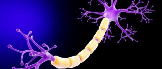
The nervous system helps regulate the functioning of the entire body. It determines its functional unity and guarantees the interaction of the body with external factors. The central nervous system is considered the main component of the entire nervous system. It includes neurons and nerve endings, as well as supporting glia.
Every cell, system and organ is a single whole. They ensure interaction and coordinated functioning of the body. The central nervous system helps him with this. This component of the body is presented as structural units and endings of different lengths and purposes.
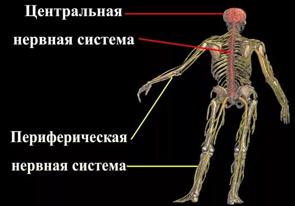
Central nervous system device
The central nervous system is formed from several components - the brain and spinal cord, interacting through the peripheral nervous system. The human central nervous system is responsible for the following feelings and sensations:
- organs of hearing and vision, perception of sounds and light, response to external stimuli;
- smell and touch, with the help of which the external world and the environment are perceived;
- emotionality, sensitivity;
- memory and thought processes of the body, intellectual activity.
The brain structure of the central nervous system consists of gray and white matter. The gray substance is represented by nerve cells with small branching processes. This substance occupies the center of the spinal cord, affecting the spinal canal. In the brain, the gray matter is the main component of the cortex, having scattered formations that are essentially white. The white layer is located under the gray layer and is structurally formed from fibers involved in the formation of nerve bundles. Similar bundles of bundles build the nerve.
Shells of the central nervous system
Surrounding the central NS are shells, each of which is different:
- Solid - external. It is this membrane that is formed inside the cranial cavity, as well as inside the hollow formation of the spinal column.
- Cobweb cover. This membrane is equipped with nerve endings and blood vessels and is located under the outer membrane.
- Vascular. Between the second and third membranes there is another cavity, the space of which is filled with brain matter. The choroid, as the name suggests, is formed from a collection of arteries, capillaries, and veins that perform the functions of blood vessels. This cover is connected directly to the brain, penetrating its folds.
Brain
This organ has a simple structure and is represented by the following elements: an extended formation - the trunk, a small brain called the cerebellum, which is responsible for muscle tone, coordination and balance, as well as the cerebral hemispheres.
The main element, which includes the higher centers representing reason, mental abilities, and speech abilities, is the hemispheres of the brain. Each of them is formed from a core with gray matter, a white shell and a cerebral cortex that protects the remaining layers.
The cerebellum, which provides coordinated actions, is represented by gray matter, a shell of white matter, and a layer of gray located outside.
The trunk is a part that has no division into layers, is formed from one mass that is not divided into colors. This part directly communicates with the rest and corrects the work of breathing, circulatory systems, movement and feelings.
Spinal cord
This cylindrical organ is located in the depths of the spinal column and has protection in the form of bone tissue formation. The spinal cord itself is located under the membranes.
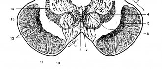
If you look at the organ in section, you can see gray matter in the form of a butterfly or shaped like an H, covered with a white membrane on top. Some of the pathways originate in white matter and end in gray matter and vice versa. Many fibers located in the white mass of the shell organize the interaction of many parts of the gray matter located in the spinal cord.
General information
The central nervous system is the main component of the nervous system.
It includes 2 sections: the brain and the spinal cord. Its purpose is to control all important processes within the body. The brain is responsible for thought processes, speech, and spatial orientation. It promotes the proper functioning of the senses, ranging from normal temperature sensitivity to visual and auditory functions. The spinal cord is involved in regulating the functioning of the body, helps ensure coordination of their activities and is responsible for motor activity. Taking into account the large number of functions of the central nervous system, clinical symptoms that allow one to suspect a tumor in the brain or spinal cord can be varied: from disruptions in behavioral functions to the inability to make movements of body parts.
Functionality of the central nervous system
The structure of any individual is represented by many structures and organs that interact with each other, but all of them are aimed at promoting the normal functioning of the human structure, its protection, support, and nutrition. The interconnection between the systems is ensured by the central nervous system. It is she who regulates the processes that occur in the body; with its help, the direction of work is changed, the pace of functioning is set and all the conditions necessary for this are provided.
The central nervous system performs a number of basic functions without which the body cannot exist:
- Integration. Occurs by combining functions. Integration is divided into 3 forms:
- nervous - a combination of departments of the central nervous system. For example, let's take food that has color and aroma, which is a conditioned reflex stimulus. Various reflexes occur in the body at the sight of food: saliva is secreted, gastric juice is produced. In this particular case, one can observe the integration of behavioral, nutritional and bodily prescriptions;
- humoral. It is a combination of various functions based on body fluids together with hormones. For example, various hormones of internal secretions tend to act synchronously, only increasing the effect of each other, but there is a variant of sequential production, when one hormone increases the effect of another. The process ends with the activation of a number of different functions. So, adrenaline can increase heart rate, increase blood glucose levels, start ventilation, etc.;
- mechanical. This shape is necessary to perform a specific function that ensures the structural integrity of the organ. If any of the organs or parts of the body is injured, structural changes are formed, which subsequently leads to a malfunction of the entire organism.
- Correlation. It is necessary in order to most effectively form the relationship between systems, internal organs and processes, and bring them together.
- Regulation. Ensuring the functioning of the entire central nervous system, it is necessary to regulate and monitor the main indicators of the body. The basis of this regulation is reflexes, the formation and organization of processes, self-regulation, thanks to which the body adapts to the constantly changing internal conditions of the surrounding world. It occurs in forms that are corrective as the action progresses, and are nourishing. The nerve processes related to the body and stimulation have all sorts of effects.
- Coordination. Synchronization and consistency of actions of all parts of one unified system. Change of position or posture, various forms of movement, movement in space, adaptability of reactions to what is happening, work activity, physical activity - all these components must be clearly coordinated and directed by the central nervous system.
- Connection with the environment. The central nervous system is a center that forms the connection and transmission of data from the outside world to the organs and systems of the body for subsequent coordinated actions.
- Cognition and adaptation. In order to adapt to certain circumstances, to select the model of behavior needed at that moment in special situations, to adapt to the activity, this function of the central nervous system is necessary. With the help of this system, comfortable adaptation to the circumstances surrounding a person is ensured.
General overview of the autonomic nervous system (ANS) - structure, divisions and functions
Already from the very beginning of the 19th century, European science clearly realized that in addition to the animal functions of the body, there are also vegetative functions that are carried out without the participation of consciousness.
Of course, research methods were imperfect at that time, knowledge about electricity was in its infancy, so a full-fledged study of this unconscious nervous system by experiment became possible only in the 20th century.
Currently, electrophysiological methods, stress modeling, and the use of imaging diagnostic methods are used.
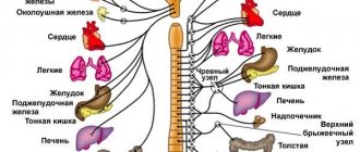
The autonomic nervous system performs the functions of maintaining homeostasis: it regulates the metabolism of various substances, and controls the functioning of the digestive system, excretion, as well as the respiratory system and circulatory system.
In addition, the autonomic nervous system (this is its second name) is involved in the regulation of the body’s growth, its maturation, and also controls the processes of reproduction. It is the vegetative nervous system that is responsible for:
- proper functioning of all internal structures and organs;
- regulates the functioning of smooth muscles of the gastrointestinal tract and blood vessels;
- ensures the secretion of endocrine and exocrine glands of the body;
- finally, it ensures the function of the nervous system itself.
Few people think about the fact that the autonomic department of the peripheral nervous system also takes part in the work of skeletal, striated muscles. Only it does not order the muscles to contract, but provides them with nutrition and removes accumulated harmful substances, for example, lactic acid.
In addition to the listed functions, the autonomic (or autonomic) nervous system has another localization of nuclei (clusters of neurons) in the brain and spinal cord.
https://www.youtube.com/watch?v=ytpressru
Autonomic nerves do not have a segmental structure; they do not form symmetrical roots, like animal ones. Another feature of the structure of autonomic nerves is their small diameter (compared to animal nerves).
Unmyelinated fibers, which conduct nerve impulses at low speed, also belong to autonomous structures.
The structure of the autonomic nervous system is such that the fibers that come from the brain to “control” the internal organs do not reach them in the same way as in the animal system.
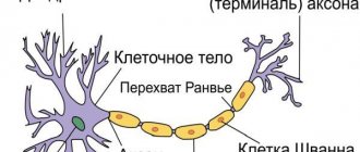
Recall that the body of the neuron, which is located in the anterior (motor) horns of the spinal cord, gives off an axon that enters the sciatic nerve, the longest nerve in the human body. Essentially, this nerve is an extended extension of a group of cells.
Therefore, when answering the question “which cell is the largest in the human body”? - you can answer that these are motor neurons of the central nervous system, lying in the lower lumbar and upper sacral segments, and forming the sciatic nerve.
The difference between the autonomic innervation of internal organs is such that its nerves are necessarily interrupted on the way from the spinal cord to various internal organs in special nodes or ganglia. Therefore, we can say that there is a “ganglionic nervous system”, that is, the first level of control over the work of organs and blood vessels, extended beyond their limits.
In addition, the peripheral ganglia themselves, being part of the autonomic nervous system, are capable, due to their structure, of being even more “autonomous”. The general principles of the structure of the ganglion are such that it can “in itself” close the reflex autonomic arc, “without disturbing” the management over trifles.
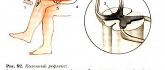
But even here, the reflex circuit involves switching the impulse from sensory to motor neurons within the spinal cord, but not in the periphery. The knee reflex arc is two-neuron and the simplest.
But the arc of autonomic reflexes necessarily consists of at least three neurons: sensitive, motor and intercalary.
How does the ANS work?
Since the departments of the ANS will be discussed in detail in the relevant articles, we will say here the most basic data.
The autonomic system is divided into two sections: sympathetic and parasympathetic. The main differences between them are:
- the sympathetic system controls the stress response, and the parasympathetic system controls the protective response;
- the ganglia of the sympathetic system are located not in the organs, but in a special formation next to the spine - the sympathetic trunk;
- these parts of the ANS have different localization of centers within the boundaries of the brain and spinal cord;
- structures of the sympathetic nervous system are more common.
The brain stem and spinal cord are the “base” for the location of centers, from where the autonomic innervation of internal organs is carried out. They are located:
- In the mesencephalic region (midbrain), for the arc of the pupillary reflex;
- Anatomical interval from 1st thoracic to 2nd-3rd lumbar segment of the spinal cord. Along this length, the thoracolumbar autonomic centers are located in the spinal cord;
- Sacral or sacral centers. They regulate the activity of the pelvic organs, the fibers exit as part of the pelvic nerves.
It would be a mistake to assume that the ANS is responsible for the activity of all systems of the body, being localized only in these departments. As elsewhere in living nature, the main principle of the organization of living matter to ensure the constancy of the internal environment of the organism is the strict subordination of the underlying sections to the overlying ones.
Therefore, there are even higher and “perfect” command bodies.
While the visceral nervous system controls the work of internal organs, the higher autonomic centers - the hypothalamus, striatum and even the cerebral cortex are busy with the most complex work: they perform “fine tuning” and regulation of higher autonomic, including endocrine functions. Let us briefly consider the importance of the hypothalamus for the autonomic nervous system.
About the hypothalamus
The central part of the autonomic nervous system is divided into the hypothalamus, pituitary gland (united in the hypothalamic-pituitary system), striatum, and cerebral cortex.
This division is largely arbitrary, since there are extensive and bilateral connections, for example, with the reticular formation, the limbic system, and this connection is so complex that all its smallest details and details are still unknown.
The hypothalamus is a cluster of paired nuclei, and the number of these pairs is 32. There are anterior, posterior and middle nuclei. The work of these nuclei is closely related to osmoregulation, body temperature and the production of certain pituitary hormones.
https://www.youtube.com/watch?v=https:accounts.google.comServiceLogin
In order to understand how complex the hypothalamus is, here are a few examples:
- When irritated, the posterior nuclei show signs of stress: the pupils and eyes dilate, blood vessels constrict, tachycardia occurs, gastric and intestinal motility is inhibited, adrenaline is released, glycogen breaks down in the liver under the influence of glucagon, blood sugar levels rise, “just in case.” Everyone knows that glucose is a “stress” substance that is released into the blood to provide food to the muscles in case of intense physical activity, for example, chasing prey or escaping.
In humans, stress is always a “headless” activity, and that is why it is so harmful. Any stress should find a way out through physical exercise, then heart attacks, strokes, diabetes and other serious diseases will occur much less frequently.
- When the anterior nuclei are irritated, the opposite effects occur, and urination and defecation are also stimulated;
- The middle nuclei of the hypothalamus control many metabolic processes. Their irritation leads to gluttony and obesity, and their destruction leads to refusal of food, exhaustion, anorexia;
- The paraventricular nuclei control water metabolism. When they are irritated, for example by a tumor, pathological thirst appears.
There are also centers that help increase blood lipid levels; the anterior nucleus regulates body temperature. When it is destroyed, overheating or hyperthermia occurs, in which a person quickly dies, as sweating is impaired.
More “higher spheres”, such as the cortex, with the help of innervation, as well as through the integration of the ANS and the ANS under a “single principle”, allow for the final connection of all nervous systems, above which are emotions and personality.
Structure of the motor sensory system (peripheral, conductive and central sections).
Topic: Physiology of the motor sensory system.
Plan:
Structure of the motor sensory system (peripheral, conductive and central sections).
Muscular - joint feeling.
Improving the motor sensory system in physical education classes and sports training.
Introduction
Complex acts of human behavior in the external environment require constant analysis of the surrounding world, as well as awareness of the nerve centers about the state of internal organs.
Human Analyzers
Internal (chemical, parasthetic sensory systems);
- Receptors of a certain sensitivity are sent ... to their zone of the cortex along specific paths, and to other zones along non-specific paths. As a result, a complex mosaic of excited zones of the cortex (sensitive, associative, motor) and other parts of the brain appears in the cerebral cortex, reflecting analytical and synthetic activity. This activity allows us to more fully perceive the events of the external world, determine our attitude towards it and respond with conscious behavior.
12Next ⇒
Date added: 2014-01-07; ; Copyright infringement?;
Functions and types of human analyzers (Table)
Thin and wedge-shaped beam; anterior and posterior spinocerebellar tract.
3. Postcentral gyrus. Cerebellar cortex.
Proprioception allows a person to navigate the position of his limbs in relation to each other and perceive his own movements.
STRUCTURE AND FUNCTIONS OF SKIN
Skin is the outer covering of the human body. Its area is 1.5-2 m2.
In direct contact with the external environment, the skin performs the following functions:
Protects the body from external influences, including mechanical ones.
Participates in thermoregulation of the body.
Releases sweat and sebum (excretory function).
Contains energy reserves (subcutaneous fatty tissue).
Synthesizes vitamin D.
It is an active component of the immune system.
Participates in water, mineral and other types of metabolism.
Previous28293031323334353637383940414243Next
Date added: 2016-11-02; ;
SEE MORE:
Conducting section of the visual sensory system.
It begins in the bipolar cells of the retina, then passes into ganglion cells, the axons of which, underlying the vitreous chamber, are directed to the area of the blind spot, forming the optic nerve (II pair of CN). The optic nerve leaves the orbit through the optic nerve canal and the optic chiasm is formed in the region of the lower parts of the diencephalon - the hypothalamus.