Description: What is the conducting function of the spinal cord? Although almost every schoolchild knows the answer to the question, an ordinary person is unlikely to be able to answer right away. Its conducting function is simple - it is the transmission of a nerve signal. It is because of this feature of the NS that a person is a single system.
And to ensure control over organ functions, the ability to move, timely transmission or receipt of reflex, sympathetic impulses, pathways are needed. Failures in the transmission of impulses entail serious disruptions in the functioning of the body.
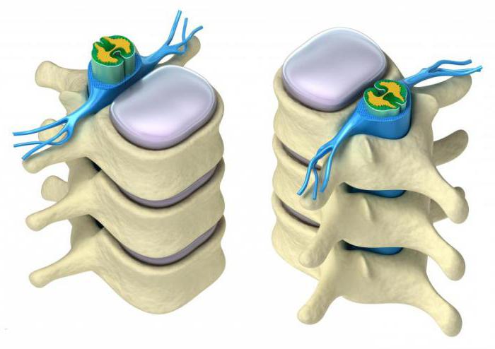
What does the spinal cord look like?
Not everyone knows what the spinal cord looks like. Moreover, not all people have an idea of what its role is in the life of every person. It is therefore worthwhile to fill this knowledge gap. In addition, many people mistakenly believe that the brain and spinal cord are separate parts.
To find out what the reflex function of the spinal cord is for, let's try to determine what it looks like. It is impossible to clearly understand where the spinal cord begins and ends. It starts from the first vertebra just below the skull, smoothly connecting to the brain in this area. The division into the spinal cord and the brain is formal, but in reality the spinal cord smoothly passes into the brain. Thus, we can conclude that these two parts are a single whole.
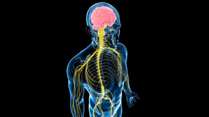
Pathways of the brain and spinal cord
In the nervous system, neurons form synapses with each other, forming chains and networks through which nerve impulses travel only in certain directions. From receptor (sensitive) neurons, impulses travel through intercalary nerve cells to effector neurons. At synapses, impulses are conducted in only one direction - from the presynaptic membrane to the postsynaptic membrane.
Along one chain of neurons, the impulse propagates centripetally
- from the place of its origin in the skin, mucous membranes, organs of movement, blood vessels, tissues and organs to the spinal cord or brain.
Along other neuron circuits, impulses are carried out centrifugally
- from the brain to the periphery, to the working organs: muscles, glands, tissues. Nerve fibers carrying impulses from the spinal cord to the brain or in the opposite direction are folded into bundles that form pathways. Pathways are a set of closely spaced nerve fibers passing through certain areas of the white matter of the brain and spinal cord, connecting various nerve centers and conducting identical nerve impulses.
In the spinal cord and brain there are three groups of nerve fibers (pathways): associative, commissural and projection.
Association nerve fibers
(short and long pathways) connect nerve centers located in one half of the brain.
Short (intralobar) connect nearby areas of gray matter and are located within one lobe (section) of the brain or adjacent segments of the spinal cord. Long (interlobar) association bundles connect areas of gray matter located at a considerable distance from each other, usually in different lobes (sections) of the brain or segments of the spinal cord. The long associative pathways of the cerebral hemispheres include the superior longitudinal fasciculus,
which connects the cortex of the frontal lobe with the parietal and occipital lobes,
the inferior longitudinal fasciculus,
which connects the gray matter of the temporal lobe with the occipital lobe, and
the uncinate fasciculus,
which connects the cortex in the frontal pole with the anterior part of the temporal lobe.
In the spinal cord, association fibers form their own bundles of the spinal cord
(intersegmental bundles), which are located near the gray matter.
Commissural
(commissural)
nerve fibers
(conducting pathways) connect identical nerve centers of the right and left hemispheres of the cerebrum. Commissural pathways pass through the corpus callosum, commissure of the fornix, and anterior commissure. The corpus callosum connects new, younger sections of the cerebral cortex of the right and left hemispheres, in which the fibers diverge fan-shaped, forming the radiance of the corpus callosum. In the anterior commissure there are fibers connecting the areas of the cortex of the temporal lobes of both hemispheres, belonging to the olfactory (more ancient) brain.
Projection nerve fibers
(tracts) connect the spinal cord with the brain, the nuclei of the brain stem with the basal ganglia and cerebral cortex (ascending tracts), and the brain with the spinal cord (descending tracts).
Ascending projection paths
(conducting pathways), afferent, sensitive, conduct nerve impulses to the cerebral cortex that arise as a result of exposure to various environmental factors on the body, including impulses coming from the sensory organs, the musculoskeletal system, internal organs and blood vessels. Depending on this, the ascending projection pathways are divided into three groups: exteroceptive, proprioceptive, interoceptive.
Exteroceptive pathways carry
pain, temperature, tactile impulses from the skin, from the senses (vision, hearing, taste, smell).
Pain pathway
and
temperature sensitivity
(lateral spinothalamic tract) consists of three neurons. The receptors of the first (sensitive) neuron, which perceive these irritations, are located in the skin and mucous membranes, and its body lies in the spinal ganglion. The central process of the sensory neuron as part of the dorsal root is directed to the dorsal horn of the spinal cord and ends with synapses on the cells of the second neuron. The axons of the second neurons, the bodies of which lie in the dorsal horn, pass through the anterior commissure to the opposite side of the spinal cord and enter the lateral cord, forming the lateral spinothalamic tract. This pathway ascends into the medulla oblongata, passes through the pontine tegmentum, the midbrain tegmentum, and ends in the thalamus (ventral posterior nucleus and medial nuclei). The axons of thalamic cells (III neuron) are directed to the internal granular plate of the cortex (IV layer) of the postcentral gyrus, where the cortical end of the general sensitivity analyzer is located.
Pathway of touch
and
pressure
(anterior spinothalamic tract) carries impulses from skin receptors to the cells of the cortex of the postcentral gyrus. The course of the fibers of the first neuron of this path is similar to the previous one. Most of the axons of the second neuron also pass through the anterior commissure on the opposite side of the spinal cord into the anterior funiculus and, as part of it, follow upward to the thalamus and then to the postcentral gyrus. Part of the fibers of the second neuron goes as part of the posterior cord of the spinal cord on its side along with the axons of the pathway of proprioceptive sensitivity in the cortical direction.
Proprioceptive pathways
conduct impulses from the organs of the musculoskeletal system (from muscles, tendons, joint capsules, ligaments).
To the cortex of the postcentral gyrus, this path carries information about the position of body parts, range of motion, muscle tone, and tendon tension. Proprioceptive sensitivity allows a person to assess the position of parts of his body in space, analyze his own complex movements and makes it possible to carry out their targeted correction. The cell bodies of the first neuron of this pathway also lie in the spinal ganglion. Their axons, as part of the dorsal roots of the spinal nerves, without entering the dorsal horn, are directed to the posterior cord, where they form thin
and
wedge-shaped bundles.
Nerve fibers travel upward into the medulla oblongata to
the gracilis
and
sphenoid nuclei.
The axons of the second neurons emerging from these nuclei pass to the opposite side, forming a medial lemniscus, pass through the tegmentum of the pons and the tegmentum of the midbrain and end in the thalamus with synapses on the bodies of the third neurons (the anterior part of the ventral posterior nucleus).
The axons of thalamic neurons are directed to the cortex, located in front of the postcentral gyrus in the depths of the central sulcus, to neurons of layer IV. Some of the fibers of the second neurons, upon exiting the gracilis and cuneate nuclei, are directed through the inferior cerebellar peduncle to the vermis cortex of their side. The other part of the fibers passes to the opposite side and also goes through the inferior cerebellar peduncle to the vermis cortex of the opposite side. These fibers carry proprioceptive impulses to the cerebellum to correct subconscious movements of the musculoskeletal system. There are also proprioceptive anterior
and
posterior spinocerebellar tracts,
which carry information to the cerebellum about the state of the musculoskeletal system and motor centers of the spinal cord.
Interoceptive pathways
conduct impulses from internal organs and blood vessels. The receptors located in them (mechano-, baro-, chemo-) perceive information about the state of homeostasis, the intensity of metabolic processes, the chemical composition of tissue fluid, blood, pressure in blood vessels, etc.
Descending pathways
carry impulses from the cerebral cortex and subcortical centers to the nuclei of the brain stem and to the motor and intermediate nuclei of the anterior horns of the spinal cord. The descending tracts are divided into two groups: pyramidal (main motor tract) and extrapyramidal.
Main motor
or
pyramidal, the path
is a system of nerve fibers along which voluntary motor impulses from giant neurons (Betz pyramidal cells) located in the cortex of the precentral gyrus (layer V) are sent to the motor nuclei of the cranial nerves and the gray matter of the spinal cord.
Here a synaptic switch occurs and the signal is then sent to the skeletal muscles. Depending on the direction and location of the fibers, the pyramidal tract is divided into three parts. This is the corticonuclear tract
going to the nuclei of the cranial nerves,
the lateral
and
anterior corticospinal tracts
going to the intermediate nuclei and the anterior horns of the spinal cord (Fig. 2.10).
| Rice. 2.10. Scheme of pyramidal tracts: 1 - precentral gyrus; 2 - thalamus; 3 - cortical-nuclear path; 4 - cross section of the midbrain; 5 — cross section of the bridge; 6 - cross section of the medulla oblongata; 7 - intersection of pyramids; 8 - lateral corticospinal tract; 9 — cross section of the spinal cord; 10 - anterior corticospinal tract; →—direction of movement of nerve impulses |
Corticonuclear pathway
passes through the knee of the internal capsule and the base of the cerebral peduncle. In the midbrain, pons, and medulla oblongata, the fibers of the corticonuclear tract pass to the opposite side to the motor nuclei of the cranial nerves, where they end at synapses on their neurons. The axons of the neurons of the motor nuclei leave the brain as part of the corresponding cranial nerves and are directed to the skeletal muscles of the head and neck.
Lateral
and
the anterior corticospinal tract
pass through the anterior part of the posterior limb of the internal capsule, then through the base of the cerebral peduncle and pons they pass into the medulla oblongata, where they form the pyramids. At the border of the medulla oblongata with the spinal cord, the main part of the fibers of the corticospinal tract passes to the opposite side, continues into the lateral cord of the spinal cord (lateral corticospinal tract) and gradually ends with synapses on the motor and intermediate cells of the gray matter. Other fibers of the cortical spinal tract that do not pass to the opposite side at the border of the medulla oblongata with the spinal cord descend down as part of the anterior cord of the spinal cord. This bundle of fibers forms the anterior corticospinal tract. Its fibers pass segment by segment through the commissure alba and end at synapses on neurons on the opposite side of the spinal cord. The axons of the motor cells of the anterior horns emerge from the spinal cord as part of the anterior roots and innervate skeletal muscles.
Extrapyramidal pathways are phylogenetically older than pyramidal ones. They have many connections with both the brain stem and the cerebral cortex, which controls and controls the extrapyramidal system. Extrapyramidal pathways originate in different parts of the cerebral cortex and brain stem, and they end on the cells of the motor nuclei of the brain stem and gray matter of the spinal cord. The influence of the cerebral cortex on the extrapyramidal system and extrapyramidal pathways occurs through the cerebellum, red nuclei, reticular formation, and vestibular nuclei. One of the functions of the red nucleus is to maintain muscle tone necessary for involuntary posture maintenance, as well as flexion of the limbs during locomotion. From the red nuclei, nerve impulses are sent to the motor nuclei of the spinal cord along the red nucleus-spinal cord (rubrospinal) pathway.
In the coordination of movements of the human body in case of imbalance, the vestibulospinal cord
(vestibulospinal)
tract
that connects the vestibular nuclei with the anterior horns of the spinal cord.
In addition, the vestibular nuclei are connected through the posterior longitudinal fasciculus
with the motor nuclei of III, IV, VI and other pairs of cranial nerves. This connection provides corrective movements of the eyeballs during movements of the head and neck. The axons of the first neurons of the vestibulospinal tract descend as part of the anterior cord of the spinal cord. The vestibular nuclei and the activity of their associated pathways are controlled by the ancient part of the cerebellum (the tent nucleus).
The cerebral cortex controls the functions of the cerebellum, which is involved in the coordination of movements, through the bridge along the corticopontine-cerebellar pathway,
Signal switching occurs through the bridge's own cores.
Thus, the pathways of the brain and spinal cord establish connections between afferent and efferent (effector) centers and close complex neural arches in the human brain.
Some of them are closed on phylogenetically older nuclei, lying in the brain stem and providing functions that have a certain automaticity, without the participation of consciousness, although under the control of the cerebral hemispheres. Others are closed with the participation of the higher parts of the cerebral cortex and provide voluntary actions of organs and organ systems. The pathways unite the body into a functional integrity and ensure the coordinated activity of all its components.
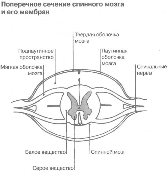
Location of the spinal cord and its membrane
The brain is protected by the cranium, and the spinal cord is hidden in the spine and surrounded by three membranes. The first of them is the most delicate, thin and soft. It contains blood vessels that deliver nutrients to the brain. In other words, the spinal cord is a kind of “courier” for delivering food.
Continuing to talk about how the reflex function of the spinal cord works, we cannot ignore the analysis of the structure of the second arachnoid membrane. There is a special space here called the subarachnoid space. Along the entire length of the spine it is filled with cerebrospinal fluid (CSF). It is this that is taken during puncturing for analysis in order to determine the health status of the spinal cord.
The last shell is located on the outside and has a harder surface, which allows it to provide protective functions against various types of external damage.
Characteristics of the spinal cord
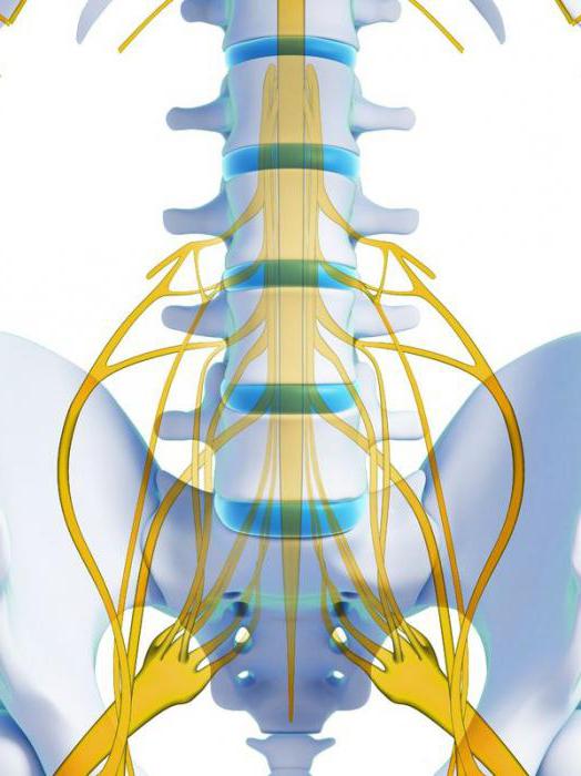
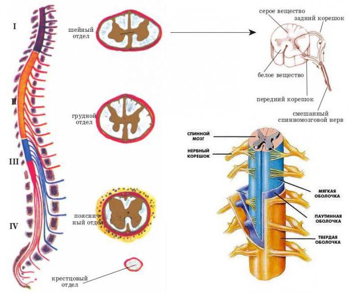
In adults, the spinal cord reaches 45 cm in length and 1.5 cm in thickness. Its weight, by the most conservative standards, is no more than 35 grams. The entire brain is divided into several sections, from which various roots extend:
- cervical;
- chest;
- lumbar;
- cross;
- coccygeal
Since the reflex function of the spinal cord is carried out, the cervical and lumbosacral regions are the most important parts of the spine. In this regard, they are best protected - nature itself took care of this, making them significantly thicker and denser. It is in these places that important nerve endings are located, the defeat of which can lead to serious consequences. In the cervical region there is a cluster of roots responsible for the movement of the arms. The roots of the lower section are responsible for the movement of the lower extremities.
The human spinal cord controls the activity of all internal organs. Each of them is associated with a specific department. In addition, the entire spinal canal is divided into segments and each of the listed sections has its own number. There are 8 of them in the cervical, 12 in the thoracic, 5 in the lumbar and sacral, and one or two in the coccygeal.
Motor cortex
In the projection motor cortex, the functional principle of somatotopic localization is implemented: the representation of muscles that carry out the most complex and significant voluntary movements occupies the maximum area. This applies to facial muscles (facial expressions are a means of biocommunication), muscles of the tongue, pharynx, larynx (articulation is the basis of motor speech), as well as the hands, especially the fingers and the hand itself, represented respectively in the lower and middle parts of the projection motor cortex (Fig. 1.2.2). The latter occupies the posterior part of the outer surface of the frontal lobe (precentral gyrus). Anterior to the projection motor cortex is the premotor cortex, which plays an important role in transforming movements into actions, and anterior to the premotor cortex is the prefrontal cortex, responsible for the implementation of holistic activities. The premotor cortex is also part of the extrapyramidal system. When complex motor skills are mastered, they are performed automatically according to programs read from the premotor cortex.
Lesions of the projection motor cortex cause central paralysis, the premotor cortex causes impairment of action (praxis), and the prefrontal cortex causes impairment of activity. The prefrontal cortex is also important in humans for upright posture, and its damage leads to disorders of standing and walking.
Categories: Nervous system Neurology
On this page there is material on the following topics:
abstract on the topic of the pyramidal system, pyramidal tract and its disorders
facial nerve corticonuclear tract
corticospinal tract briefly
corticospinal tract in humans
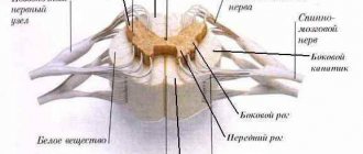
Gray matter
Gray matter or substantia grisea is represented by several columns connected to each other by two plates (anterior and inferior), called commissures. On a cross-section of one of these pillars, you can see that the gray matter in its shape resembles a butterfly with spread wings or the Latin letter H.
In addition, you can also notice that there are projections extending from the substance, which are otherwise called horns. They can be either front, located on the front wall, or rear, running along the back wall. Both the first and second pairs, and have a narrow and wide shape. But in addition to the rear and anterior ones, there are also lateral horns, which contain the centers of the autonomic nervous system.
What is the reflex function of the spinal cord? The fact is that in the anterior horns there is a special type of motor neurons, the processes of which form nerve roots.
In the middle of the gray matter there is a central canal, which is also filled with cerebrospinal fluid. In the upper part, the canal is connected to the ventricles of the brain. In this case, all sections: the ventricles, the central canal and the subarachnoid space take an active part in the circulation of cerebrospinal fluid.
Anatomy of the nervous system pathways
Content
After this, the fibers of the tractus tectospinalis are “directed” through the trunk to the segments of the spinal cord. In the tegmentum of the bridge, this path occupies a dorsomedial position, somewhat ventral to the longitudinal fasciculi. A similar topography is observed in the medulla oblongata, where the tractus tectospinalis is located ventral to the medial longitudinal fasciculus and gradually moves ventrally, approaching the dorsal border of the pyramids. In the spinal cord it is located in the medial part of the anterior funiculus. Gradually, the roof-spinal tract becomes thinner, as some of its fibers end on the motor neurons of the motor nuclei of the cranial nerves in the trunk (roof-nucleus bundle, fasciculus tectonuclearis) and in the overlying segments of the spinal cord. Here, through interneurons, fibers of the tractus tectospinalis influence the alpha small motor neurons of the motor nuclei of the anterior horns.
Motor neurons of the trunk and spinal cord transmit influence through their axons from the integration center of the roof of the midbrain through the cranial and spinal nerves to the innervated skeletal muscles.
Damage to the tractus tectospinalis leads to the loss of starting reflexes to sudden light, sound, olfactory and tactile influences.
Reticulospinal tract
This pathway is considered the most phylogenetically oldest and nonspecific. In this case, the name “tractus reticulospinalis” is understood as a set of efferent fibers starting from various centers of the reticular formation and having functional and topographical features. In a simplified form, the reticulospinal tract can be depicted without decussation, without interneurons, without indicating the specific nucleus from which it originates, and as a single rather than multiple projection (Fig. 18).
Rice. 18. Reticulospinal tracts: 1 - reticular nuclei, 2 - reticulospinal tract, 3 - motor nuclei of the anterior horns of the spinal cord, 4 - spinal nerves
It should be taken into account what the target nucleus is in the spinal cord: in the case of the animal reflex arc, these are the motor nuclei of the anterior horn, and in the case of the sympathetic reflex arc, the intermediate-lateral nucleus of the lateral horn.
In other words, there are several parallel reticulospinal tracts.
The medial reticulospinal tract (tractus reticulospinalis medialis) is the most powerful and longest of the reticulospinal tracts. It begins from the oral and caudal reticular nuclei of the pons and from the reticular nuclei of the medulla oblongata: giant cell and ventral. In the spinal cord, it extends to the sacral segments, gradually thinning and ending segment by segment on the dendrites of the gamma motor neurons of the anterior horns of the spinal cord.
The lateral reticulospinalis tract (tractus reticulospinalis lateralis) begins from the lateral reticular nucleus of the pons, located near the middle cerebellar peduncle (regio parabrachialis). This path, partially crossed, includes the axons of the reticular neurons of the respiratory cord in the spinal cord, where it is located in the lateral funiculus next to the lateral corticospinal tract. Tractus reticulospinalis lateralis has an activating effect on small alpha motor neurons of the anterior horns of the spinal cord. Another part of its fibers ends on the neurons of the intermediate-lateral nucleus of the spinal cord (the center of the sympathetic division of the autonomic nervous system). Therefore, regulation of the organs of “plant life” by the reticular formation becomes possible.
The anterior reticulospinalis tract (tractus reticulospinalis anterior) begins from the tegmental reticular nuclei of the midbrain and pons and, located in the anterior cords of the spinal cord, “reaches” the tenth thoracic segment. This pathway ends at the motor neurons of the anterior horns of the spinal cord.
All reticulospinal tracts are characterized by best expression in the cervical and upper thoracic segments of the spinal cord. More distally, the influence of the reticular formation spreads along the propriospinal tract. In other words, the reticulospinal tract is characterized by the shape of a chain of several sequentially located neurons (polysynaptic organization). Another feature is that the reticulospinal tracts are predominantly uncrossed. All these pathways have an indirect connection with the motor neurons of the anterior horns, since they end on the dendrites of interneurons 7 and 8 of the Rexed plates and through them influence the motor neurons. These influences can be either inhibitory or activating. As a result, the reticular formation, through its reticulospinal tracts and spinal nerves, ensures the tone of skeletal muscles and the performance of complex reflex acts that require the simultaneous participation of many skeletal muscles or even muscle groups (respiratory, grasping movements). Similar relationships exist between the centers of the reticular formation and the nuclei of the cranial nerves.
vestibulospinal tract
This path also refers to very ancient projections in evolutionary terms, closely related to the vestibular analyzer. Tractus vestibulospinalis is involved in the body’s rapid response to such a change in body position in space, which leads to imbalance. In this case, unconditional reflex body movements occur, leading to the fact that a person, having slipped, falls on his outstretched arms and does not hit his head or torso.
This path begins from the lateral vestibular nucleus (Deiters nucleus) (nucl. vestibularis lateralis), located in the tegmentum of the bridge near the border of the latter with the medulla oblongata (Fig. 19).
Rice. 19. Vestibulospinal tract: 1 - vestibular nuclei, 2 - vestibular tract, 3 - motor nuclei of the anterior horns of the spinal cord, 4 - spinal nerves
According to a number of researchers, the tractus vestibulospinalis also includes axons of neurons whose bodies are located in the inferior vestibular nucleus (Roller’s nucleus). The latter is located next to Deiters' nucleus, but somewhat more caudally. The Deiters nucleus has an indirect effect (in particular, through the alpha motor neurons of the motor nuclei of the anterior horns of the spinal cord) on the extensor muscles and is thus a kind of antagonist of the red nucleus. In the medulla oblongata, the vestibulospinal tract is located dorsal and lateral to the pyramids, and in the spinal cord - on the border of the anterior and lateral cords (here it is penetrated by fibers of the anterior roots of the spinal nerves). The path is mostly uncrossed.
Olivespinal tract
Tractus olivospinalis is involved in the unconditioned reflex maintenance of neck muscle tone and in performing movements designed to maintain body balance. This path is relatively young in evolutionary terms, like the olive nucleus (nucleus olivaris) of the medulla oblongata, from which it begins. The olive nucleus has a regulatory influence on the cerebellar hemispheres (cortex and dentate nucleus), the red nucleus and the cortex of the frontal lobe of the cerebral hemisphere.
Axons of neurons nucl. olivaris as part of the tractus olivospinalis reach the sixth cervical segment of the spinal cord, ending segment by segment on the alpha motor neurons of the motor nuclei of the anterior horns on their side of the body (Fig. 20).
Rice. 20. Olivospinal tract: 1 - nuclei of the inferior olive, 2 - olivospinal tract, 3 - motor nuclei of the anterior horns of the spinal cord, 4 - spinal nerves, 5 - neck muscles
The axons of these motor neurons as part of the spinal nerves reach the neck muscles, which innervate them. In the spinal cord, the olivospinal tract is located in the anteromedial part of the lateral funiculus.
3.2. Pyramid paths
These pathways, collectively called the “pyramidal system,” are involved in the conscious control of skeletal muscle function (stimulating or inhibiting contraction). In particular, it is possible to perform voluntary movements characterized by complexity and accuracy. The pyramidal system consists of two pathways: the corticospinal tract (tractus corticospinalis) and the corticonuclear tract (tractus corticonuclearis). The pyramidal system received its name due to the fact that the tractus corticospinalis “passes” through the pyramids of the medulla oblongata. It is clear that the name is not very good, since the main thing here is not topography, but function.
Corticospinal tract
This pathway conducts volitional motor impulses that allow control of skeletal muscles innervated by spinal nerves, i.e. muscles of the limbs, trunk and neck. The corticospinal tract also conducts impulses that can inhibit the activity of motor neurons in the anterior horns of the spinal cord.
VK
Buffer
White matter
White matter - substantia alba, envelops the gray matter, is formed by a collection of nerve fibers, which also come in three types:
- front;
- rear;
- lateral.
Moreover, all the roots have a different direction, and some of them are connected directly to the brain and central nervous system (hereinafter simply the CNS). And if the reflex function of the spinal cord is to transmit signals from the motor neurons of the gray matter, then the task of the white matter neurons is the prompt delivery of impulses from the muscles and joints to the medulla oblongata. Thus, the transmission of all commands along the entire spinal cord is realized.
Here are the paths through which all information regarding sensitivity and pain is transmitted. Only before entering the cerebral cortex, the information first reaches the diencephalon, and only then rushes further to its destination.
Introduction
The pathways of the nervous system and the complex reflex arcs consisting of them are the most important and complex section of neurology. It is important because it affirms the cellular nature of the nervous system (neural doctrine) and shows the ordered nature of the arrangement and connections of neurons (in the form of reflex arcs), which underlies its regulatory function.
There is a significant difference from the method of descriptive anatomy. The latter makes it possible to demonstrate the shape, size and localization of a particular formation of the nervous system, as well as its belonging to gray or white matter, but does not reveal at all the structural organization of the nervous system and the mechanisms of its functioning.
This “separation” of structure from function, which is dangerous for the worldview, eliminates the systematic approach to the nervous system in the form of the study of reflex arcs. Here the emphasis is placed precisely on the presence of connections between neurons, on their interaction, leading to the functioning of both the nervous system itself and the entire organism. However, at the same time, the number of mental operations among students increases (synthesis is added to analysis), which increases the complexity of mastering the material and its subjective complexity. Nevertheless, only the study of the nervous system as a set of reflex arcs allows us to understand its organization and functional significance. Finally, only knowledge of neural connections and interactions allows for topical diagnosis of damage to the nervous system, i.e. take a meaningful approach to the diagnosis and treatment of nervous and many other diseases and injuries.
How do the concepts of “conducting path” and “reflex arc” relate to each other? Here it should be clearly understood that any conductive path is part of one or another reflex arc. Since there are two main links in the reflex arc: afferent and efferent, the pathways are classified into afferent and efferent. Taking into account the hierarchical principle of construction of the central nervous system (the presence of higher and lower nerve centers subordinate to them) and the possibility of closing reflex arcs at the level of higher nerve centers, it is clear that both afferent and efferent pathways should be localized in both the peripheral and central parts nervous system. Since the closure of somatic reflex arcs (the connection of afferent and efferent links through interneurons) always occurs in the central nervous system, the latter also contain an associative link and corresponding associative pathways, localized only within the central nervous system.
Afferent nerve pathways conduct impulses from the receptor to the nerve center and are sensitive. Afferent nerve pathways ending in the projection centers of the cerebral cortex are classified as pathways of conscious sensitivity. The same afferent pathways that end in the subcortical sensory nerve centers are classified as pathways of unconscious sensitivity.
Efferent nerve pathways conduct impulses from nerve centers to the working organ. Since we are talking here only about the somatic nervous system, the working organ is the skeletal muscle, therefore the efferent nerve pathways are called motor. Depending on which nerve centers the efferent pathways are connected to, the latter are responsible for performing both conscious and unconscious movements.
Any conducting pathway (afferent, associative or efferent), depending on the level of closure and complexity of the reflex arc, can be single-neuron or multi-neuron (several neurons connected in series in a chain). If we consider a multineuron pathway as a chain, then within its boundaries we can distinguish links represented by the corresponding neurons. Compactly located neuron bodies form nerve centers (nodal, nuclear or screen type), and axons collected in bundles form nerve tracts. Thus, a multineural pathway consists of nerve centers and tracts. In this case, the nerve centers and tracts of the same pathway are localized in certain but different parts of the nervous system. Each tract within the central nervous system conducts nerve impulses usually in one direction and in most cases - of the same functional content. It should be clearly understood how the tracts within the central nervous system differ from the bundles of fibers that form the cranial or spinal nerves. Nerves contain both afferent and efferent fibers, and different afferent fibers can carry different sensory impulses.
In the future, material will be presented that concerns primarily the somatic part of the nervous system.
How our brain works
Ascending and descending pathways are responsible for the fast and correct functioning of our body. The latter flows are formed using the red nuclear and lateral pathways. It is thanks to these pathways that the reflex and conduction functions of the spinal cord are carried out. Thanks to the red nuclear spinal tract, involuntary motor impulses are produced. While the lateral corticospinal tracts are responsible for voluntary impulses.
All roots are supplied by personal veins and arteries, which as a result form neurovascular bundles. Each such beam is responsible only for its own segment and works autonomously, analyzing incoming information and transmitting the necessary impulses.
Damage to these bundles leads to serious pathological and sometimes irreversible changes in the human body. And so that specialists can determine which particular bundle is damaged and localize the pain, it is necessary to conduct a whole range of studies.
Reflex function
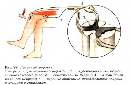
Everything in our body is thought out to the smallest detail, and our body reacts differently to every external stimulus. The defense mechanism is based on reflexes. We sneeze, cough, get burns, flinch from a sharp sound, or react in our own way to gusts of wind. These are all examples of the reflex function of the spinal cord and such actions occur outside of our control.
So that we can respond in a timely manner to any irritant, including critical situations, pain receptors are located throughout the surface of our skin. As a striking example: after touching a hot kettle or any surface, we almost instantly withdraw our hand. The reaction speed is so fast that it is impossible to understand the time frame. In a split second, a reflex ring is formed, which causes the muscles to contract.
Another common case can be cited. If you accidentally swallow a portion of smoke or sniff dust particles with your nose, you will start sneezing or coughing. Thus, it became clear that in such a short time the information was received, processed, and our “defenders” received instructions to free the body from the presence of foreign bodies.
Betz cells
The main efferent structure is the central motor neuron, represented by Betz giant pyramidal cells of layer V of the projection motor cortex (prerolandic gyrus and paracentral lobule, 4th field). The set of processes of Betz cells is part of the pyramidal tract. A significant part of its fibers originates from other parts of the cerebral cortex: the secondary motor cortex of the inner surface of the frontal lobe, the superior frontal gyrus, the premotor cortex (6th field), as well as the postcentral gyrus, and not only from the large pyramidal cells of layer V, but also from small pyramidal cells of layer III and from others. Most of the fibers of the pyramidal tract end in the formations of the extrapyramidal system - the striatum, globus pallidus, substantia nigra, red nucleus, as well as in the reticular formation of the brain stem, interacting between the pyramidal and extrapyramidal systems. Other fibers, especially thickly myelinated ones, originating from Betz giant cells of the projection motor cortex, end on the dendrites of the peripheral motor neuron.
The motor neuron is located in two places - the anterior horns of the spinal cord and in the motor nuclei of the cranial nerves, and therefore the pyramidal tract consists of two pathways - corticospinal and corticonuclear (Fig. 1.2.1).
Conductor function
So, it is now clear what the reflex function of the spinal cord is expressed in, we can move on to another, also significant task - conduction. It involves transmitting signals along ascending pathways to the main brain. From it, depending on the situation, the impulse is directed along descending pathways to some organ.
The conductor function allows us to perform meaningful actions:
- take or throw;
- stand up or sit down;
- walk slowly or run;
- draw;
- cut off.
We perform all these actions in everyday life: at home or at work, and usually we simply do not notice.
This whole connection of the brain, spinal cord, the entire central nervous system, internal organs and all limbs makes the human body unique in nature. Even the most modern robot cannot boast of the number of movements that any biological organism can perform.
What is the conducting function?
Indeed, what is the conductive function of the spinal cord? The very concept of “pathways” implies a common thread of nerves that conduct signals to different areas of the gray matter. The descending and ascending pathways of the spinal cord are subordinated to one function - the transmission of impulses. As a rule, there are 3 types of fibers:
- projection;
- commissural connections;
- associative pathways of nerves.
However, in addition to this classification, there is another one. It distinguishes between motor and sensory pathways. The former provide a reflex reaction and organize the supply of impulses from the brain to the spinal cord, as well as to muscle tissue. They are also responsible for coordinating movements. In addition, these threads stretch to the optic nerve and to the plate of the roof of the midbrain, whose task is to ensure the function of vision and hearing. Thanks to the nerve fibers that make up the sensory pathways, a person is endowed with the ability to recognize the following 4 types of impulses: pain, temperature, tactile feeling, joint-muscular feeling (movement, body position).