Anatomy of the human telencephalon - information:
The telencephalon is represented by two hemispheres, hemispheria cerebri. Each hemisphere includes: the cloak, or mantle, pallium, olfactory brain, rhinencephalon, and basal ganglia. The remnant of the original cavities of both vesicles of the telencephalon are the lateral ventricles, ventriculi laterales.
The forebrain, from which the telencephalon is released, first arises in connection with the olfactory receptor (olfactory brain), and then it becomes an organ for controlling the behavior of the animal, and centers of instinctive behavior based on species reactions (unconditioned reflexes) arise in it - the subcortical nuclei and centers of individual behavior based on individual experience (conditioned reflexes) are the cerebral cortex. Accordingly, in the telencephalon the following groups of centers are distinguished in the order of historical development:
- The olfactory brain, rhinencephalon, is the oldest and at the same time the smallest part, located ventrally.
- The basal, or central, nuclei of the hemispheres, the “subcortex,” are the old part of the telencephalon, paleencephalon, hidden in the depths.
- The gray matter of the cortex, cortex, is the youngest part, neencephalon, and at the same time the largest part, covering the rest as if with a cloak, hence its name “cloak”, or mantle, pallium.
In addition to the two forms of behavior noted for animals, a third form arises in humans - collective behavior, based on the experience of the human collective, created in the process of human labor activity and communication between people through speech. This form of behavior is associated with the development of the youngest superficial layers of the cerebral cortex, which constitute the material substrate of the so-called second signal (verbal) system of reality (I. P. Pavlov). Since in the process of evolution, the telencephalon grows the fastest and strongest of all parts of the central nervous system, it becomes the largest part of the brain in humans and takes on the appearance of two voluminous hemispheres - the right and left, hemispheria dextrum et sinistrum.
In the depths of the longitudinal fissure of the brain, both hemispheres are connected to each other by a thick horizontal plate - the corpus callosum, corpus callosum, which consists of nerve fibers running transversely from one hemisphere to the other. In the corpus callosum, there is an anterior end bent downward, or knee, genu corporis callosi, a middle part, body, truncus corporis callosi, and then a posterior end, thickened in the shape of a roller, splenium corporis callosi. All these parts are clearly visible on a sagittal section of the brain between both hemispheres. The genu of the corpus callosum, curving downwards, sharpens and forms a beak, rostrum corporis callosi, which passes into a thin plate, lamina rostralis, which in turn continues into lamina terminalis.
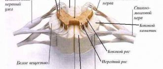
Under the corpus callosum there is the so-called fornix, fornix, which represents two arched white cords, which in their middle part, corpus fornicis, are connected to each other, and diverge in front and behind, forming columns of the arch in front, columnae fornicis, behind - the legs of the arch, crura fornicis . Crura fornicis, heading backward, descends into the lower horns of the lateral ventricles and passes there into fimbria hippocampi. Between the crura fornicis, under the splenium corporis callosi, transverse bundles of nerve fibers stretch, forming the commissura fornicis. The anterior ends of the fornix, columnae fornicis, continue down to the base of the brain, where they end in the corpora mamillaria, passing through the gray matter of the hypothalamus. Columnae fornicis is limited by the interventricular foramina lying behind them, connecting the third ventricle with the lateral ventricles.
In front of the columns of the arch there is an anterior commissure, commissura anterior, which looks like a white transverse bar consisting of nerve fibers. Between the anterior part of the fornix and the genu corporis callosi is a thin vertical plate of brain tissue - a transparent septum, septum pellucidum, in the thickness of which there is a small slit-like cavity, cavum septi pellucidi.
content .. 102 103 104 105 106 107 ..Telencephalon (human anatomy)
The terminal (telencephalon), or large, brain (cerebrum) develops from the anterior cerebral bladder and consists of highly developed paired parts - the right and left hemispheres of the cerebrum and the middle part connecting them. The hemispheres are separated by a longitudinal fissure, in the depth of which lies a plate of white matter - the corpus callosum. It consists of fibers connecting both hemispheres. Under the corpus callosum there is a vault, which consists of two curved fibrous cords, which are connected to each other in the middle part, and diverge in front and behind, forming the pillars and legs of the vault. Anterior to the columns of the arch is the anterior commissure. Between the anterior part of the corpus callosum and the fornix is a thin vertical plate of brain tissue - a transparent septum.
The cerebral hemisphere is formed by gray and white matter. It distinguishes the largest part, covered with grooves and convolutions, - a cloak formed by the gray matter lying on the surface - the cerebral cortex, the olfactory brain and accumulations of gray matter inside the hemispheres - the basal ganglia. The last two sections constitute the oldest part of the hemisphere in evolutionary development.
The cavities of the telencephalon are the lateral ventricles.
In each hemisphere, three surfaces are distinguished: the upper lateral - convex according to the cranial vault, the medial - flat, facing the same surface of the other hemisphere, and the lower - irregular in shape. The surfaces of the hemisphere have a complex pattern due to grooves running in different directions and folds between them - convolutions. The size and shape of the grooves and convolutions are subject to significant individual fluctuations. However, there are several permanent, clearly defined grooves that appear earlier than others during embryonic development. They are used to divide the hemispheres into large areas called lobes.
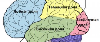
Each hemisphere consists of five lobes: frontal, parietal, occipital, temporal and insular, or insula, located deep in the lateral sulcus (Fig. 116). The boundary between the frontal and parietal lobes is the central sulcus, and between the parietal and occipital lobes is the parieto-occipital sulcus. The temporal lobe is separated from the rest by the lateral sulcus. On the superolateral surface of the hemisphere in the frontal lobe, there is a precentral sulcus, separating the precentral gyrus, and two frontal sulci: superior and inferior, dividing the rest of the frontal lobe into the superior, middle and inferior frontal gyri. The parietal lobe contains the postcentral sulcus, which separates the postcentral gyrus, and the intraparietal sulcus, which divides the rest of the parietal lobe into the superior and inferior parietal lobes. In the lower lobule, the marginal and angular gyri are distinguished. Two parallel sulci, the superior and inferior temporal, divide the temporal lobe into the superior, middle and inferior temporal gyri. In the occipital lobe, transverse occipital sulci and gyri are distinguished. On the medial surface of the hemisphere, the sulcus of the corpus callosum and the cingulate sulcus are clearly visible, between which the cingulate gyrus is located. Above it, surrounding the central sulcus, lies the paracentral lobule. The area between the parieto-occipital sulcus and the calcarine sulcus passing behind it is called the wedge, and the area in front of it is called the precuneus. At the point of transition to the lower surface of the hemisphere, the medial occipitotemporal, or lingual, gyrus stands out. On the lower surface, separating the hemisphere from the brain stem, there is a deep groove of the hippocampus, lateral to which is the parahippocampal gyrus. Laterally, it is separated by a collateral groove from the lateral occipitotemporal gyrus. The insula, located deep in the lateral sulcus, is also covered with grooves and convolutions.
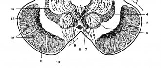
Rice. 116. Convolutions and sulci of the cerebral hemispheres. I - central sulcus; II - lateral groove; III - transverse fissure of the cerebrum; 1 - precentral gyrus; 2 - superior frontal gyrus; 3 - middle frontal gyrus; 4 - inferior frontal gyrus; 5 - superior temporal gyrus; 6 - middle temporal gyrus; 7 - inferior temporal gyrus; 8 - cerebellum; 9 - occipital lobe; 10 - inferior parietal lobule; 11 - superior parietal lobule
The cerebral cortex (cortex cerebri) is a layer of gray matter up to 4 mm thick, covering the surface of the hemispheres and lying deep in the grooves. The cortex is formed by layers of nerve cells and fibers arranged in a certain order (Fig. 117). The most typically structured areas of the phylogenetically newer cortex consist of six layers of cells; the old and ancient cortex has fewer layers and is simpler in structure. Different areas of the cortex have different cellular and fibrous structures. In this regard, there is a doctrine about the cellular (cytoarchitectonics) and fibrous (myeloarchitectonics) structure of the cerebral cortex.
Rice. 117. Structure of the cerebral cortex (diagram), a - location of layers (1 - 6) of nerve cells; b - location of nerve fibers
The olfactory brain in humans is represented by rudimentary formations, well expressed in animals. It makes up the oldest parts of the cerebral cortex.
The basal ganglia are clusters of gray matter within the hemispheres. These include the striatum, consisting of the caudate and lenticular nuclei, interconnected. The lenticular nucleus is divided into two parts: the shell located on the outside and the globus pallidus lying inside. They are subcortical motor centers. Outside the lenticular nucleus there is a thin plate of gray matter - the fence; in the anterior part of the temporal lobe lies the amygdala. Between the basal ganglia and the thalamus there are layers of white matter, the internal, external and outermost capsules. Conducting pathways pass through the internal capsule.
The lateral ventricles (right and left) are cavities of the telencephalon, lie below the level of the corpus callosum in both hemispheres and communicate through the interventricular foramina with the third ventricle. They are irregular in shape and consist of anterior, posterior and lower horns and a central part connecting them. The anterior horn lies in the frontal lobe; posteriorly it continues into the central part, which corresponds to the parietal lobe. At the back, the central part passes into the posterior and inferior horns, located in the occipital and temporal lobes. In the lower horn there is a cushion - the hippocampus. From the medial side, the choroid plexus is invaginated into the central part of the lateral ventricles, continuing into the lower horn. The walls of the lateral ventricles are formed by the white matter of the hemispheres and the caudate nuclei. The thalamus is adjacent to the central part below.
The white matter of the hemispheres occupies the space between the cortex and the basal ganglia. It consists of a large number of nerve fibers running in different directions. There are three systems of hemispheric fibers: associative, connecting parts of the same hemisphere; commissural (commissural) connecting parts of the right and left hemispheres, which include the corpus callosum, anterior commissure and commissure of the fornix, and projection fibers, or pathways connecting the hemispheres with the underlying parts of the brain and spinal cord.
content .. 102 103 104 105 106 107 ..
Functions of the cerebrum
Brain structure
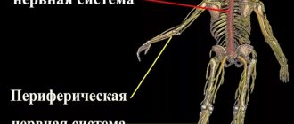
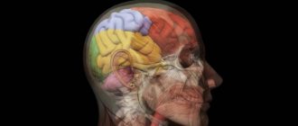
The brain consists of 5 main sections: the telencephalon, diencephalon, midbrain, hindbrain and medulla oblongata. The telencephalon consists of two hemispheres, in which there are many grooves and convolutions. It is divided into several lobes (frontal, parietal, temporal and occipital). There is a distinction between the subcortex and the cerebral cortex. The subcortex consists of subcortical nuclei that regulate various functions of the body. The brain is located in three cranial fossae. The cerebral hemispheres occupy the anterior and middle fossae, and the posterior fossa is the cerebellum, under which the medulla oblongata is located.
Brain functions
Diencephalon
The diencephalon is divided into ventral (hypothalamus) and dorsal (thalamus, metathalamus, epithalamus) parts. The thalamus is a mediator in which all irritations received from the outside world converge and are sent to the cerebral hemispheres so that the body can adequately adapt to the constantly changing environment. The metathalamus is the subcortical center of the auditory analyzer and the subcortical center of the visual analyzer. The epithalamus connects the limbic with other parts of the brain and performs some hormonal functions. The hypothalamus is the main subcortical center for regulating the autonomic functions of the body.
Midbrain
Consists of the cerebrum and quadrigeminal peduncles. All ascending pathways to the cerebral cortex and cerebellum and descending pathways carrying impulses to the medulla oblongata and spinal cord pass through the midbrain. It is important for processing nerve impulses coming from visual and auditory receptors.
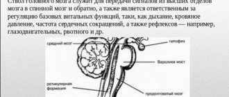
Cerebellum and pons
The cerebellum is located in the occipital part of the brain behind the medulla oblongata and the pons. It consists of two hemispheres and a worm between them. The cerebellum is involved in the coordination of complex motor acts. All sensory and motor pathways of the central nervous system pass through the bridge; it regulates autonomic reactions (lacrimation, salivation, chewing, swallowing, etc.), and is involved in the formation of the voice.
Ventricles of the brain
The cerebrospinal fluid located in the ventricles also circulates in the arachnoid mater.
Functions of the cerebrum
Thanks to the work of the brain, a person can think, feel, hear, see, touch, and move. The large (terminal) brain controls all vital processes, as well as the ability to think. The brain regulates and coordinates the work of all human muscles. The left hemisphere of the brain controls the right half of the body, and the right hemisphere controls the left. The two hemispheres complement each other.
The brain is divided into three large sections - the brainstem, the subcortical section and the cerebral cortex. The total surface of the cerebral cortex increases due to numerous grooves that divide the entire surface of the hemisphere into convex convolutions and lobes. Three main sulci - central, lateral and parieto-occipital - divide each hemisphere into four lobes: frontal, parietal, occipital and temporal. The cerebral cortex receives impulses from receptor formations. Each peripheral receptor apparatus in the cortex corresponds to an area called the cortical nucleus of the analyzer. An analyzer is an anatomical and physiological formation that provides perception and analysis of information about phenomena occurring in the environment and (or) inside the human body, and generates sensations specific to a particular analyzer (for example, pain, visual, auditory analyzer). The areas of the cortex where the cortical nuclei of the analyzers are located are called sensory areas. The motor zone of the cerebral cortex interacts with sensory zones; when it is irritated, movement occurs. This can be shown with a simple example: when a candle flame approaches, the pain and heat receptors of the fingers begin to send signals, then the neurons of the corresponding analyzer identify these signals as pain caused by a burn, and the muscles are “given the order” to withdraw the hand.
Association zones - connect incoming sensory information with previously received and stored in memory, and also compare information received from different receptors. Sensory signals are comprehended, interpreted and, if necessary, transmitted to the associated motor area. Associative zones are involved in the processes of thinking, remembering and learning.