The human body in every way strives for energy intensity and plasticity. A small organ that performs a specific function is better than a large organ that performs the same function. On the road of evolution, the brain (as a multifunctional system) progressed in this way: it was formed and enlarged thanks to a complex system of convolutions and grooves. Thus, being inside the cranium, which was limited in volume, the telencephalon increased its area, while maintaining the entire set of functions.
What it is
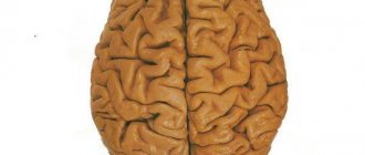
The convolutions of the brain are small elevations above its surface, bounded by grooves. These folds are located throughout the entire telencephalon, and their area averages 1200 cm3. The fact that the functional surface increases due to specific folds is indicated by the numbers: most of the cortex (2/3) is located between the folds in the depths of the depressions. There is an explanation for such a phenomenon as the formation of convolutions: during intrauterine development, the baby’s brain develops unevenly in different places, and, as a result, the tension of the surfaces in different parts is different.
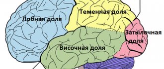
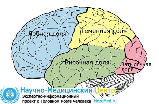
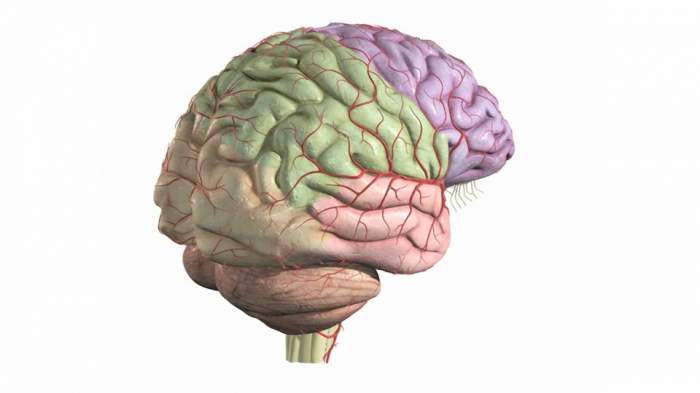
The sulci of the brain are peculiar grooves that separate the convolutions from each other. These formations are classified: primary, secondary and tertiary. The first type of depressions are formed very first in the process of fetal formation. Secondary grooves appear later and are permanent. Tertiary grooves are variable: grooves can change their shape, direction and even size. These depressions divide the surface of the cerebral hemispheres into main lobes: parietal, temporal, frontal, insular and occipital.
Bottom surface
The lower, or basal, surface of the brain is formed by parts of the frontal, temporal and occipital lobes. However, in addition to these structures, the so-called olfactory brain is also located on the basal surface. It consists of the olfactory sulcus, surrounded by the straight gyrus and orbital sulci.
The temporal lobe, based on the brain, contains the inferior temporal and occipitotemporal sulci, between which the gyrus of the same name is located. The lingular gyrus is also detailed nearby.
Structure
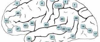
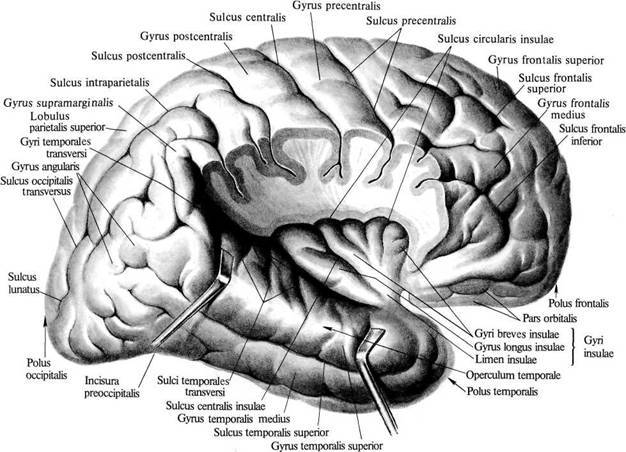
The pattern of convolutions and sulci of the brain is best seen in schematic images. The depressions dividing the cortex into two parts (hemispheres) are called primary. In addition, there are other fundamental limitations of the cortex, namely:
- Sylvian fissure (lateral, lateral): separates the temporal and frontal cortex.
- Roland's fossa (central): separates the parietal from the frontal.
- Parieto-occipital fossa: separates the occipital and parietal lobes of the brain.
- The cingulate recess, which passes into the hippocampal recess: separates the surface of the olfactory brain from other parts.
These structures also have another name: first-order sulci of the brain.
Each part of the telencephalon contains several convolutions, separated by secondary cavities. Tertiary depressions develop purely individually: their presence depends on the personal characteristics of a person and his mental abilities. The third type of notches gives individual relief to the folds.
This area of the telencephalon is limited by three sulci: the lateral, part of the occipital and central. The lateral cavity originates from the lateral fossa. Developing slightly upward and backward, the formation ends on the superolateral surface.
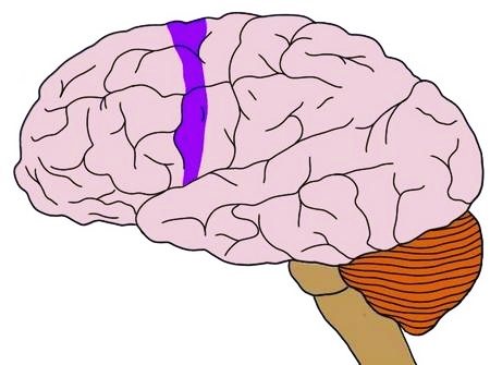
The central sulcus begins at the upper edge of one of the hemispheres. From its middle it goes backward and partially forward. In front of this notch is the frontal lobe of the brain, and behind it is the parietal cortex.
The end of the occipital region serves as the edge of the parietal region. This groove does not have a clear boundary, so the separation is carried out artificially.
This part of the hemispheres has permanent deep grooves. When talking about the formations of the medial surface, first of all, as a rule, one thinks of the groove of the corpus callosum (1). Above this groove there is a belt cavity (2), forming a knee and subsequently a branch. Also in this area is the hippocampal sulcus (3) or seahorse sulcus. Closer to the occipital lobe is the collateral groove (4). On the territory of the posterior part of the median surface there is a calcarine groove (5).
Between the first two formations is the encircling gyrus. And the hippocampal and collateral groove limits the gyrus belonging to the temporal cortex of the hemisphere.
This part of the brain is distributed in different parts of the cortex - temporal, occipital and frontal. The lower surface includes the following grooves:
- Olfactory (1)
- Orbital (2)
- Straight (3)
- Inferior temporal (4)
This area of the hemisphere does not have prominent gyri, however, one should still be noted - this is the lingular gyrus (5).
Medial surface
The sulcus of the corpus callosum is located most medially, which then passes into the sulcus of the hippocampus, which borders the hippocampus itself. Next to the callosal sulcus are the subparietal and callosal-marginal sulci. The rhinal sulcus runs parallel to the hippocampus.
The recesses of the brain listed above limit a specific system, which is called limbic. It, in turn, consists of the cingulate and hippocampal gyri.
In addition to the limbic system itself, on the inner surface of the brain there are also structures that continue their course from the outer part of the cerebral cortex. In this way, the parieto-occipital groove extends, behind which the precuneus is located (a gyrus resembling a trapezoid in shape). Next to this depression there is also a calcarine groove, which extends from the back of the head and forward all the way to the corpus callosum. Between the two recesses mentioned above is the sphenoid gyrus.
Functions of sulci and gyri
The brain is the bearer of various functions. But how was it possible to create such an organ that performs a huge number of tasks and, in general, controls all the vital functions of a complex organism? Nature has made the grooves increase the surface area of the cerebral cortex. Thus, the main sulci and convolutions of the brain perform the function of potentiating cortical tasks and increasing the number of goals performed per unit area of the hemispheres. As mentioned above, the predominant surface of the gray matter is hidden in the grooves between the gyri.
The functions of the convolutions of the brain partially repeat the purpose of the grooves. However, the gyri, in addition to increasing their area, perform specific functions, for example:
- the temporal gyri are responsible for the perception and comprehension of sound and written speech;
- the inferior frontal gyrus formulates sound speech;
- the anterior central gyrus forms conscious motor functions;
- the posterior central gyrus is responsible for general somatic perception (tactile, pain, temperature sensations).
Lobes of the brain and their functions
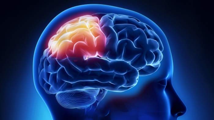
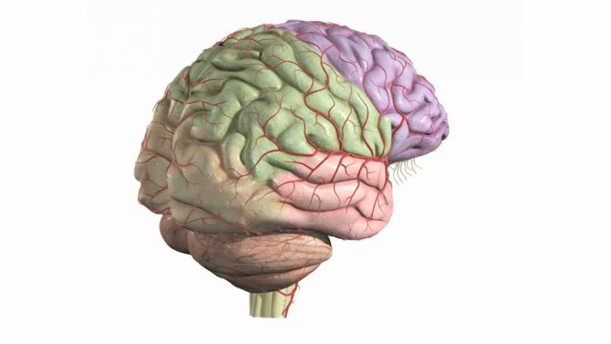
Thanks to the grooves and convolutions, the organ inside the cranium is divided into several zones with different purposes. Thus, the frontal part of the brain, which is located in the anterior cortex, is associated with the ability to express and regulate emotions, make plans, reason and solve problems.
The degree of its development determines the intellectual and mental level of a person.
The parietal lobe is responsible for sensory information. It also allows you to separate contacts made by multiple objects. The temporal region contains everything necessary to process the visual and auditory information received. The medial zone is associated with learning, emotional perception and memory.
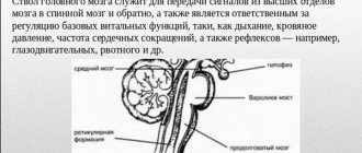
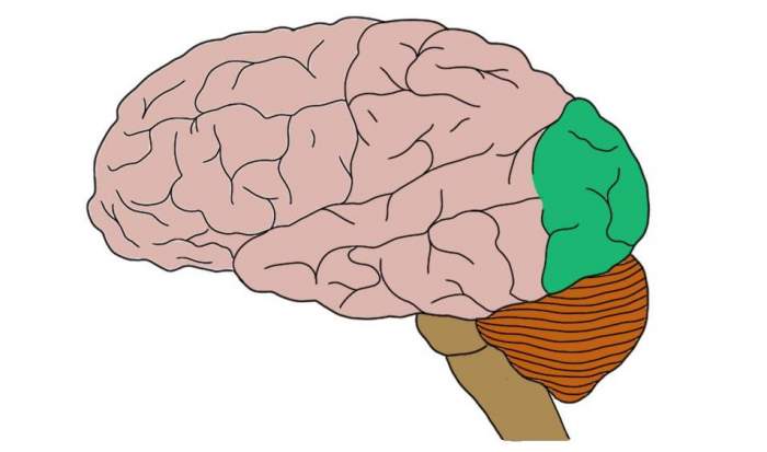
The midbrain allows you to maintain muscle tone and response to sound and visual stimuli. The posterior part of the organ is divided into the medulla oblongata, the pons and the cerebellum. The dorsolateral lobe is responsible for regulating breathing, digestion, chewing, swallowing and protective reflexes.
The bridge connects all parts of the brain with the spinal cord. The function of the cerebellum is to maintain motor coordination and balance.
Let's summarize: for a long time the structure of the contents of the cranium remained a mystery to scientists. Today, thanks to modern research methods, they were able to penetrate this mystery by in-depth studying the structure of the organ. There are many diagrams and tables that allow you to highlight the main areas of the brain and their purposes. And also to identify serious pathologies based on thickenings or damage that are uncharacteristic for a healthy person.
Parts of the brain
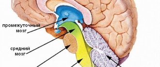
During intrauterine development, a complex brain was formed from an ordinary neural tube. This happened due to the bulging of five brain vesicles, which gave rise to the corresponding parts of the brain:
- telencephalon, or forebrain, from which the cerebral cortex, basal ganglia, and anterior part of the hypothalamus were formed;
- diencephalon, or diencephalon, which gave rise to the thalamus, epithalamus, and posterior part of the hypothalamus;
- mesencephalon, or midbrain, from which the quadrigeminal peduncle and cerebral peduncles subsequently formed;
- the metencephalon, or hindbrain, which gave rise to the cerebellum and pons;
- myelencephalon, or medulla oblongata.
Later in the article we will talk in more detail about the telencephalon, or forebrain. After all, the relief of the cerebral cortex belongs specifically to this part of the central nervous system.
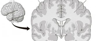
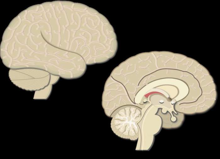
Cross section of the brain
If you conventionally cut the brain in the frontal plane, you can see that part of the brain is dark in color, and part is light. The dark part is the gray matter, which is a collection of nerve cell bodies (neurons). It is represented by the cerebellum and the cerebral cortex, which is located along the perimeter. However, there are areas of gray matter inside the brain, they are called the basal ganglia, or extrapyramidal system.
While the cortex, together with the grooves and convolutions of the brain, performs the functions of coordinating higher nervous activity (speech, writing, thinking, memory, attention, emotions), the gray matter of the extrapyramidal system is necessary for the implementation of high-precision coordinated movements.
The basal ganglia include the following structures:
- striopallidal system, which consists of the caudate nucleus and the lentiform nucleus (the putamen together with the globus pallidus);
- the limbic system, which includes the fence and the amygdala.
White matter, in turn, is a collection of processes of nerve cells that ensure the interaction of the overlying parts of the brain with the underlying ones, as well as the interaction of different neurons within the same structure.
Structure of the cortex
Thanks to the presence of the cortex, a person is able to experience emotions, navigate himself and the surrounding space. What is noteworthy is that the structure of the bark is unique. The grooves and convolutions of the cerebral cortex of one person have a different shape and size than that of another. But the general plan of the building is the same.
What is the difference between the sulci and convolutions of the brain? Fissures are depressions in the cerebral cortex that look like slits. They are the ones who divide the bark into shares. There are four lobes of the cerebral hemispheres:
- frontal;
- parietal;
- temporal;
- occipital
Gyri are convex areas of the cortex that are located between the furrows.
Cellular mechanisms leading to expansion and folding of the cerebral cortex
The structure of the human brain distinguishes it from other mammals, and for this reason may explain its unique mental abilities compared to other animals. The number of folds in the cortex may correlate with some specific cognitive, sensory, and motor abilities. Although there is no clear explanation of how the unique division of the human brain into sulci and convolutions occurs. Today there is progress in understanding the extremely complex processes in the brain, the cortex of which is built with so many grooves and convolutions. Even though all cells have the same DNA, different neural stem cells are formed. It is their work with various properties that creates the basic structure of the brain, consisting of neurons and glial cells.
Telencephalic neuroepithelium
Brain growth occurs through two types of stem cells—neural stem cells and neural progenitors. Both of these forms form neurons, which become permanent in the brain, as well as intermediate cells that create the building material for building the brain. Four different types of stem cells determine the structure of the cortex.
During early embryonic development, expansion of the rostral domain of the neural tube leads to the appearance of two telencephalic vesicles. The dorsal half of these vesicles is molecularly defined as the primordium of the cerebral cortex. At this stage, the cortical primordium consists exclusively of a monolayer of neuroepithelial progenitor cells. They are highly polarized and attached to each other by tight junctions at the level of the apical domain (inner surface of the telencephalic vesicle) and move the cell nucleus between the apical (apical) and basal (lower) side of the neuroepithelium in coordination with the cell cycle.
- basal-directed movement during G1 phase;
- basal position during S phase;
- apically directed movement during G2 phase;
- mitosis on the apical surface.
The cycling movement is known as interkinetic nuclear migration and is completely asynchronous between neuroepithelial cells, giving the neuroepithelium a pseudostratified appearance. Cells undergo only symmetrical self-aggressive divisions, with each division generating two daughter cells, hence exponentially increasing their number. Because they are the fundamental progenitor cells of the cerebral cortex, the size of their association determines the number of derived neurogenic progenitor cells and the final number of cortical neurons, and therefore it has a fundamental influence on the size of the mature cerebral cortex. An increase in quantity leads to an expansion of surface area and the formation of neuroepithelium.
Spread and neurogenesis
Immediately before the onset of neurogenesis, neuroepithelial progenitor cells begin to lose tight junctions and acquire features typical of glial cells (including expression of brain lipid-binding protein, vimentin, and Pax6), thereby becoming apical radial glial cells (ARGCs). They also undergo interkinetic nuclear migration, divide at the apical surface of the developing cortex, and at this early stage also undergo self-reinforcing divisions.
However, they gradually begin to divide asymmetrically to generate one similar cell plus another cell. These new cells accumulate in the basal part of the cortical primordium, while the cell bodies of the ARGC remain on the apical side, forming the ventricular zone (VZ). With the accumulation of cells above the GC, the ARGK process is prolonged, remaining attached to the basal plate, and is now called radial glia. Asymmetric ARGK divisions generate one ARGK plus one neuron or one intermediate progenitor cell. Intermediate progenitor cells (secondary progenitor cells without apical-basal polarity) do not undergo interkinetic nuclear migration, divide in a layer located in the ventricular zone, the subventricular zone (SVZ), and all express a transcription factor (Tbr2).
However, due to the fact that each neuron itself consumes during mitosis, their relative quantity compared to ARGK is quite low. Intermediate progenitor cells in the cerebral cortex generate the majority of cortical excitatory neurons. As neurogenesis progresses, the need for ARGK expansion/renewal decreases and the production of neurons increases. In addition to the expanded ventricular zone, the subventricular zone thickens, populated in abundance by basal precursors, especially in the later stages of neurogenesis. As a result, the SVZ splits into internal and external parts. The outer portion contains a wide variety of progenitor cell types with high developmental potential, which is a key factor for the expansion and formation of the cortex.
Formation of the cortex in embryogenesis
Embryogenesis is the intrauterine development of the fetus from conception to birth. First, uneven depressions form on the cerebral cortex, which give rise to furrows. The primary grooves are formed first. This occurs around the 10th week of intrauterine development. After this, secondary and tertiary depressions are formed.
The deepest groove is the lateral one; it is one of the first to form. It is followed in depth by the central one, which separates the motor (motor) and sensory (sensitive) zones of the cerebral cortex.
Most of the cortical relief develops from 24 to 38 weeks of gestation, and some of it continues to develop after the baby is born.
Brain convolutions and sulci: structure and functions - Lifestyle for good health
In fact, there are a huge number of functions of the human brain, and more than one article can be written about them. In the list below, all functions are combined into separate groups:
- processing information coming from outside;
- planning and decision making;
- carrying out movements;
- emotions;
- memorization and memory;
- attention;
- speech;
- intelligence and thinking.
The brain is the most advanced, and therefore one of the most difficult to study, part of the human body. And its most highly organized component is the cerebral cortex. More details about the anatomy of this formation, the structure of the grooves and convolutions of the brain later in the article.
During intrauterine development, a complex brain was formed from an ordinary neural tube. This happened due to the bulging of five brain vesicles, which gave rise to the corresponding parts of the brain:
- telencephalon, or forebrain, from which the cerebral cortex, basal ganglia, and anterior part of the hypothalamus were formed;
- diencephalon, or diencephalon, which gave rise to the thalamus, epithalamus, and posterior part of the hypothalamus;
- mesencephalon, or midbrain, from which the quadrigeminal peduncle and cerebral peduncles subsequently formed;
- the metencephalon, or hindbrain, which gave rise to the cerebellum and pons;
- myelencephalon, or medulla oblongata.
Later in the article we will talk in more detail about the telencephalon, or forebrain. After all, the relief of the cerebral cortex belongs specifically to this part of the central nervous system.
Structure of the cortex
Embryogenesis is the intrauterine development of the fetus from conception to birth. First, uneven depressions form on the cerebral cortex, which give rise to furrows. The primary grooves are formed first. This occurs around the 10th week of intrauterine development. After this, secondary and tertiary depressions are formed.
The deepest groove is the lateral one; it is one of the first to form. It is followed in depth by the central one, which separates the motor (motor) and sensory (sensitive) zones of the cerebral cortex.
Most of the cortical relief develops from 24 to 38 weeks of gestation, and some of it continues to develop after the baby is born.
The grooves are classified according to the function they perform. The following types are distinguished:
- primary formed - the deepest in the brain, they divide the cortex into separate lobes;
- secondary - more superficial, they perform the function of forming convolutions of the cerebral cortex;
- additional, or tertiary - the most superficial of all types, their function is to provide an individual relief of the bark, increasing its surface.
- Although the shape and size of some of the sulci and convolutions of the cerebral hemispheres differ from individual to individual, their number is normally unchanged. Every person, regardless of age and gender, has the following grooves:
- Sylvian fissure - separates the frontal lobe from the temporal lobe;
- lateral sulcus - separates the temporal, parietal and frontal lobes, and is also one of the deepest in the brain;
- Roland's fissure - separates the frontal lobe of the brain from the parietal lobe;
- parieto-occipital sulcus - separates the occipital region from the parietal;
- cingulate sulcus - located on the medial surface of the brain;
- circular - is the boundary for the insular part on the basal surface of the cerebral hemispheres;
- The hippocampal sulcus is a continuation of the cingulate sulcus.
Main convolutions
The relief of the cerebral cortex is very complex. It consists of numerous convolutions of different shapes and sizes. But we can highlight the most important of them, which perform the most important functions. The main convolutions of the brain are presented below:
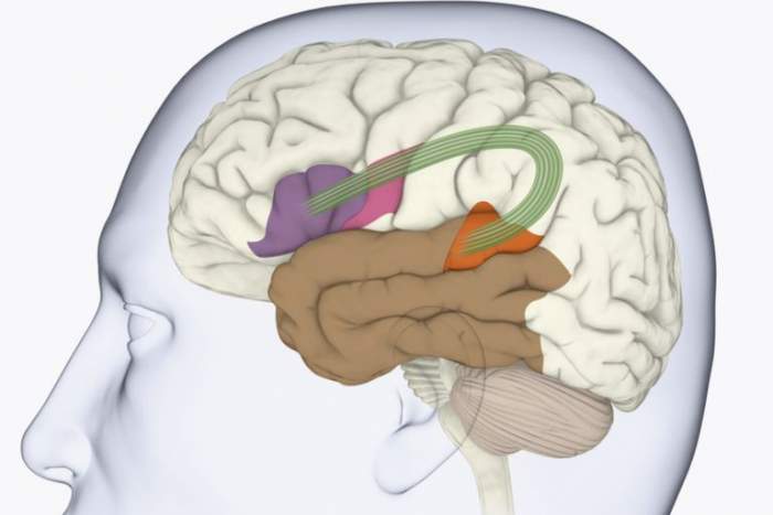
- angular gyrus - located in the parietal lobe, is involved in recognizing objects through vision and hearing;
- Broca's center - the posterior part of the inferior frontal gyrus on the left (in right-handers) or on the right (in left-handers), which is necessary for correct speech reproduction;
- Wernicke's center - located in the posterior part of the superior temporal gyrus on the left or right (similar to Broca's area), is involved in the understanding of oral and written speech;
- cingulate gyrus - located on the medial part of the brain, takes part in the formation of emotions;
- hippocampal gyrus - located in the temporal region of the brain, on its inner surface, necessary for normal memorization;
- fusiform gyrus - located in the temporal and occipital regions of the cerebral cortex, is involved in face recognition;
- lingual gyrus - located in the occipital lobe, plays an important role in processing information coming from the retina;
- precentral gyrus - located in the frontal lobe in front of the central sulcus, necessary for processing sensitive information entering the brain;
- postcentral gyrus - located in the parietal lobe behind the central sulcus, necessary for voluntary movements.
The anatomy of the cerebral convolutions and sulci is best studied in sections. Let's start with the outer surface. It is on the outer surface of the brain that the deepest groove is located - the lateral one.
It begins in the basal (lower) part of the cerebral hemispheres and moves to the outer surface. Here it branches into three more recesses: the ascending and anterior horizontal, which are shorter, and the posterior horizontal, which is much longer.
The bottom of the lateral groove is called the insula. It then continues as the transverse gyrus. The insula is divided into anterior and posterior lobes. These two formations are separated from each other by a central groove.
Parietal lobe
The boundaries of this part of the brain are outlined by the following grooves:
- central;
- parieto-occipital;
- transverse occipital;
- central.
Behind the central sulcus is the postcentral gyrus of the brain. At the back it is bounded by a groove with the appropriate name - postcentral. In some literary publications, the latter is further divided into two parts: upper and lower.
The parietal lobe, using the interparietal sulcus, is divided into two regions, or lobules: superior and inferior. The latter contains the supramarginal and angular gyri of the cerebral hemispheres.
In the postcentral, or posterior central, gyrus there are centers that receive sensory (sensitive) information. It is worth noting that the projection of different parts of the body in the posterior central gyrus is unevenly located. So, most of this formation is occupied by the face and hand - the lower and middle third, respectively. The last third is occupied by projections of the torso and legs.
Praxis centers are located in the lower part of the parietal lobe. It implies the development of automatic movements throughout life. This includes, for example, walking, writing, tying shoelaces, etc.
Frontal lobe
The frontal part of the cerebral hemispheres is located in front of all other structures of the brain. Posteriorly, this area is limited from the parietal lobe by the central sulcus, and laterally, by the lateral sulcus, from the temporal region.
In front of the central sulcus is the precentral gyrus of the brain. The latter, in turn, is limited from other formations of the frontal lobe cortex by means of the precentral recess.
The precentral gyrus, together with the adjacent posterior parts of the frontal lobe, plays an important role. These structures are necessary for the implementation of voluntary movements, that is, those that are under the control of consciousness.
These neurons have a very long process (axon), the endings of which reach the corresponding segment of the spinal cord. This pathway is called the corticospinal pathway.
The relief of the frontal region of the brain is formed by three large convolutions:
- superior frontal;
- average;
- bottom.
These formations are delimited from one another by the superior and inferior frontal grooves.
In the posterior part of the superior frontal gyrus there is an extrapyramidal center, which is also involved in movements. This system is historically more ancient than the pyramidal one. It is necessary for accuracy and smoothness of movements, for automatic correction of motor acts that are already normal for humans.
In the posterior part of the inferior frontal gyrus there is Broca's motor center, which was already mentioned earlier in the article.
Occipital lobe
The boundaries of the occipital region of the brain are outlined by the following formations: it is separated from the parietal lobe by the parieto-occipital recess, from below the occipital part smoothly flows into the basal surface of the brain.
It is in this area of the brain that the most unstable structures are located. But the posterior occipital gyrus of the brain is present in almost all individuals. Moving closer to the parietal region, transitional gyri are formed from it.
On the inner surface of this area there is a calcarine groove. It separates three convolutions from each other:
- wedge;
- lingular gyrus;
- occipitotemporal gyrus.
There are also polar grooves that have a vertical direction.
The function of the most posterior lobe of the brain is the perception and processing of visual information. It is noteworthy that the projection of the upper half of the retina of the eyeball is in the wedge, but it perceives the lower part of the visual field. And the lower half of the retina, which receives light from the upper visual field, is projected in the region of the lingual gyrus.
Temporal lobe
- This structure of the brain is limited by the following grooves: the lateral one from above, a conventional line between the lateral and posterior occipital grooves at the back.
- The temporal lobe, by analogy with the frontal lobe, consists of three large convolutions:
- superior temporal;
- average;
- lower
- The name of the depressions corresponds to the convolutions.
- On the lower surface of the temporal region of the brain, the hippocampal gyrus and the lateral occipitotemporal gyrus are also distinguished.
Wernicke's speech center is located in the temporal lobe, which was already mentioned earlier in the article.
In addition, this area of the brain performs the functions of perception of taste and olfactory sensations. It provides hearing, memory, and synthesis of sounds. Specifically, the superior temporal gyrus, as well as the inner surface of the temporal region, is responsible for hearing.
Main grooves
Although the shape and size of some of the sulci and convolutions of the cerebral hemispheres differ from individual to individual, their number is normally unchanged. Every person, regardless of age and gender, has the following grooves:
- Sylvian fissure - separates the frontal lobe from the temporal lobe;
- lateral sulcus - separates the temporal, parietal and frontal lobes, and is also one of the deepest in the brain;
- Roland's fissure - separates the frontal lobe of the brain from the parietal lobe;
- parieto-occipital sulcus - separates the occipital region from the parietal;
- cingulate sulcus - located on the medial surface of the brain;
- circular - is the boundary for the insular part on the basal surface of the cerebral hemispheres;
- The hippocampal sulcus is a continuation of the cingulate sulcus.
Finite brain
The telencephalon (cerebrum)
consists of the right and left hemispheres and the fibers connecting them, forming the corpus callosum and other commissures.
Under the corpus callosum there is a fornix
in the form of two curved cords connected to each other by adhesions.
The front part of the vault, pointing downward, forms the pillars
.
The posterior part, diverging to the sides, is called the legs of the arch.
Anterior to the trunks of the fornix there is a transverse bundle of fibers -
the anterior (white) commissure.
In front of the fornix in the sagittal plane there is a transparent septum,
consisting of two parallel plates. Anteriorly and superiorly, these plates connect to the anterior part of the corpus callosum. Between the plates there is a narrow slit-like cavity containing a small amount of liquid. Each plate forms the medial wall of the anterior horn of the lateral ventricle.
Each cerebral hemisphere is formed by gray and white matter. The peripheral part of the hemisphere, covered with grooves and convolutions, forms a cloak
, covered with a thin plate of gray matter -
the cerebral cortex.
The surface area of the bark is about 220,000 mm2.
Under the cerebral cortex there is white matter,
in the depths of which there are large accumulations of gray matter -
subcortical nuclei - basal ganglia .
The cavities of the cerebral hemispheres are
the lateral ventricles.
Each hemisphere has three surfaces: the superior lateral
(convex),
medial
(flat), facing the adjacent hemisphere, and
lower,
which has a complex relief corresponding to the unevenness of the internal base of the skull.
On the surfaces of the hemispheres, numerous depressions are visible - grooves
and elevations between the grooves -
gyri.
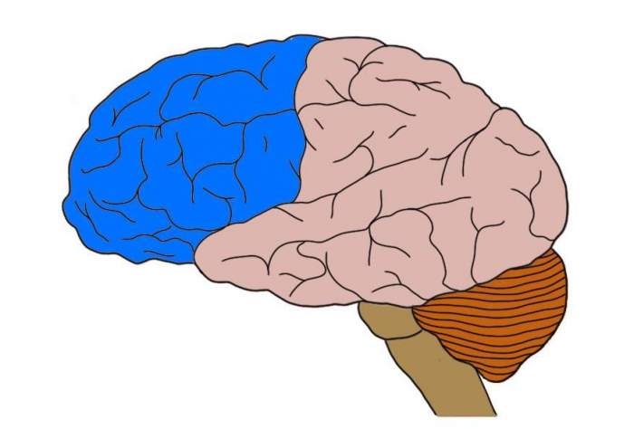
Each hemisphere has five lobes
:
frontal, parietal, occipital, temporal and
insular (
insula).
Main convolutions
The relief of the cerebral cortex is very complex. It consists of numerous convolutions of different shapes and sizes. But we can highlight the most important of them, which perform the most important functions. The main convolutions of the brain are presented below:
- angular gyrus - located in the parietal lobe, is involved in recognizing objects through vision and hearing;
- Broca's center - the posterior part of the inferior frontal gyrus on the left (in right-handers) or on the right (in left-handers), which is necessary for correct speech reproduction;
- Wernicke's center - located in the posterior part of the superior temporal gyrus on the left or right (similar to Broca's area), is involved in the understanding of oral and written speech;
- cingulate gyrus - located on the medial part of the brain, takes part in the formation of emotions;
- hippocampal gyrus - located in the temporal region of the brain, on its inner surface, necessary for normal memorization;
- fusiform gyrus - located in the temporal and occipital regions of the cerebral cortex, is involved in face recognition;
- lingual gyrus - located in the occipital lobe, plays an important role in processing information coming from the retina;
- precentral gyrus - located in the frontal lobe in front of the central sulcus, necessary for processing sensitive information entering the brain;
- postcentral gyrus - located in the parietal lobe behind the central sulcus, necessary for voluntary movements.
Parietal part
This lobe of the brain is limited from other structures by four sulci: central, lateral, parieto-occipital and transverse occipital. Behind the central, by analogy with the frontal lobe, there is a postcentral sulcus, which in some textbooks is further divided into two parts: upper and lower. The two recesses listed above limit the postcentral gyrus.
The parietal part of the brain is divided into two lobes (upper and lower) by the interparietal groove. The inferior lobule includes the supramarginal and angular gyri.
Outside surface
The anatomy of the cerebral convolutions and sulci is best studied in sections. Let's start with the outer surface. It is on the outer surface of the brain that the deepest groove is located - the lateral one. It begins in the basal (lower) part of the cerebral hemispheres and moves to the outer surface. Here it branches into three more recesses: the ascending and anterior horizontal, which are shorter, and the posterior horizontal, which is much longer. The last branch has an upward direction. It is further divided into two parts: descending and ascending.
The bottom of the lateral groove is called the insula. It then continues as the transverse gyrus. The insula is divided into anterior and posterior lobes. These two formations are separated from each other by a central groove.
Frontal part
The most anterior part of the brain is called the frontal lobe. Its boundaries are delineated by two grooves: the central one at the back, separating it from the parietal lobe (this groove is also called the Rolandic groove), the lateral one at the bottom, the structure of which is described in detail above. Anterior to the central recess are the precentral grooves. One is located above, and the second is below. These grooves limit the central gyrus.
The frontal lobe is divided into three frontal gyri: superior, middle and inferior. They are delimited from each other by the superior and inferior frontal grooves. We can say that it is in the frontal lobe that the largest grooves and convolutions of the brain are located.
Parietal lobe
The boundaries of this part of the brain are outlined by the following grooves:
- central;
- parieto-occipital;
- transverse occipital;
- central.
Behind the central sulcus is the postcentral gyrus of the brain. At the back it is bounded by a groove with the appropriate name - postcentral. In some literary publications, the latter is further divided into two parts: upper and lower.
The parietal lobe, using the interparietal sulcus, is divided into two regions, or lobules: superior and inferior. The latter contains the supramarginal and angular gyri of the cerebral hemispheres.
In the postcentral, or posterior central, gyrus there are centers that receive sensory (sensitive) information. It is worth noting that the projection of different parts of the body in the posterior central gyrus is unevenly located. So, most of this formation is occupied by the face and hand - the lower and middle third, respectively. The last third is occupied by projections of the torso and legs.
Praxis centers are located in the lower part of the parietal lobe. It implies the development of automatic movements throughout life. This includes, for example, walking, writing, tying shoelaces, etc.
Occipital lobe
The occipital lobe occupies the posterior parts of the hemispheres.
The function of the occipital lobe is associated with the perception and processing of visual stimuli, the organization of complex processes of visual perception.
1. The central part of the occipital region is the cortical end of the visual analyzer. When damaged, elementary visual impairment occurs in the form of scotomas.
2. The upper part of the occipital region (parieto-occipital cortex) provides the functions of optical-spatial gnosis. If affected, there is optical-spatial agnosia. In mild cases, the patient is not oriented left-to-right, in severe cases - up-down.
3. The middle and lower areas of the occipital lobe provide the functions of visual-object gnosis. If affected, there is visual-object agnosia, the patient sees, but does not recognize what he sees.
The boundaries of the occipital region of the brain are outlined by the following formations: it is separated from the parietal lobe by the parieto-occipital recess, from below the occipital part smoothly flows into the basal surface of the brain.
It is in this area of the brain that the most unstable structures are located. But the posterior occipital gyrus of the brain is present in almost all individuals. Moving closer to the parietal region, transitional gyri are formed from it.
On the inner surface of this area there is a calcarine groove. It separates three convolutions from each other:
- wedge;
- lingular gyrus;
- occipitotemporal gyrus.
There are also polar grooves that have a vertical direction.
The function of the most posterior lobe of the brain is the perception and processing of visual information. It is noteworthy that the projection of the upper half of the retina of the eyeball is in the wedge, but it perceives the lower part of the visual field. And the lower half of the retina, which receives light from the upper visual field, is projected in the region of the lingual gyrus.
Frontal lobe
The frontal part of the cerebral hemispheres is located in front of all other structures of the brain. Posteriorly, this area is limited from the parietal lobe by the central sulcus, and laterally, by the lateral sulcus, from the temporal region.
In front of the central sulcus is the precentral gyrus of the brain. The latter, in turn, is limited from other formations of the frontal lobe cortex by means of the precentral recess.
The precentral gyrus, together with the adjacent posterior parts of the frontal lobe, plays an important role. These structures are necessary for the implementation of voluntary movements, that is, those that are under the control of consciousness. In the fifth layer of the cortex of the precentral gyrus there are giant motor neurons, which are called pyramidal cells, or Betz cells. These neurons have a very long process (axon), the endings of which reach the corresponding segment of the spinal cord. This pathway is called the corticospinal pathway.
The relief of the frontal region of the brain is formed by three large convolutions:
- superior frontal;
- average;
- bottom.
These formations are delimited from one another by the superior and inferior frontal grooves.
In the posterior part of the superior frontal gyrus there is an extrapyramidal center, which is also involved in movements. This system is historically more ancient than the pyramidal one. It is necessary for accuracy and smoothness of movements, for automatic correction of motor acts that are already normal for humans.
In the posterior part of the inferior frontal gyrus there is Broca's motor center, which was already mentioned earlier in the article.
Treatment of Huntington's chorea
The purpose of the operation is to block impulses that can cause an attack of neuralgia, or to eliminate the very cause of neuralgia (vascular compression of the root), if any.
Usually they start with simpler interventions - blockades of individual branches of the V nerve, and lastly (especially in older people) they resort to more complex interventions.
Operations on peripheral branches - novocaine or alcohol blockade of the main peripheral branches.
Blockades or exeresis (excision) of peripheral branches usually give a temporary effect (6-12 months).
Blockade of the gasserian node is carried out by effective and low-traumatic puncture injection of phenol, boiling water into the gasserian node or using radiofrequency coagulation.
Retrogasseral transection of the V nerve root with an approach from the middle cranial fossa (Spiller-Freger operation) or with access from the posterior cranial fossa (Dandy operation) is very traumatic and is rarely used today.
Vascular decompression of the V nerve root.
One of the main causes of trigeminal neuralgia is compression of the V nerve root by an atypically located vessel. In old age, sclerosis and lengthening of the vessels occur, as a result of which they can compress the nerve at the point where it enters the bridge.
The purpose of the operation, which is performed through a small burr hole in the squama of the occipital bone near the pyramid, is to locate this vessel (most often the superior cerebellar artery) and separate it from the nerve using a Teflon sponge or a piece of muscle.
Temporal lobe
This structure of the brain is limited by the following grooves: the lateral one from above, a conventional line between the lateral and posterior occipital grooves at the back.
The name of the depressions corresponds to the convolutions.
On the lower surface of the temporal region of the brain, the hippocampal gyrus and the lateral occipitotemporal gyrus are also distinguished.
Wernicke's speech center is located in the temporal lobe, which was already mentioned earlier in the article. In addition, this area of the brain performs the functions of perception of taste and olfactory sensations. It provides hearing, memory, and synthesis of sounds. Specifically, the superior temporal gyrus, as well as the inner surface of the temporal region, is responsible for hearing.
Thus, the lobes and convolutions of the brain are a complex and multifaceted topic to understand. In addition to the parts discussed in the article, there is also the limbic cortex with its own relief, a structure called the insula. There is a cerebellum, which also has a cortex with its own characteristics. But the anatomy of the brain should be studied gradually, so this article provides only basic information.
The temporal part of the cerebral hemispheres is limited by the lateral sulcus from above, and from behind by a conditional line drawn from this sulcus to the posterior occipital one. The structure of this lobe of the brain is easy to remember: three parallel convolutions are separated by three parallel grooves. The grooves and convolutions of the brain in the temporal part are called the same name: superior, middle and inferior temporal.