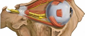
Ischemic neuropathy of the optic nerve is a pathological condition of the optic nerve, which appears as a result of circulatory disorders in the intraorbital and intrabulbar regions.
As the disease progresses, visual acuity is lost quite quickly, the field of vision becomes narrower, and completely invisible areas appear. To diagnose this disease, visometry and ophthalmoscopy are used. To clarify the diagnosis and origin, ultrasound, MRI, angiography, etc. are performed.
Treatment is carried out immediately; until the diagnosis is confirmed, decongestants, anti-spasm drugs and thrombolytics are used. An indispensable element of complex treatment will be physiotherapeutic procedures with stimulation of the optic nerve using a laser or other influence, and eye exercises.
At risk are older people over 40 years of age, mostly men. This complex disease does not tolerate delays in treatment, because it threatens not just loss of visual acuity, but also complete blindness and disability.
Optical pathology cannot be called an independent disease, since it manifests itself only in the complex of the systemic process of development of the disease. This applies not only to the visual system, but also to all other parts of the body. Therefore, not only ophthalmologists work on this problem, but also conduct examinations with the following doctors: endocrinologists, cardiologists, neurologists and other specialists as necessary.
Causes
Anterior ischemic neuropathy occurs as a result of ischemia of the retinal, choroidal (prelaminar) and scleral (laminar) layers of the optic disc against the background of circulatory disorders in the posterior short ciliary arteries. Posterior ischemic neuropathy is associated with impaired blood flow in the carotid and vertebral arteries.
Additionally, acute circulatory disorders of the optic nerve are provoked by arterial spasms, atherosclerotic lesions, and thromboembolism. In addition, the occurrence of the disease is associated with various systemic pathologies, which are the causes of hemodynamic disorders, disorders in the vascular system, and microcirculatory problems.
Quite often, ischemic neuropathy is detected in systemic atherosclerosis, arterial hypertension, temporal arteritis, periarteritis nodosa, obliterating arteritis and atherosclerosis, diabetes mellitus, disorders of the cervical spine, and thrombosis of the great vessels. Sometimes this disease can develop after massive acute gastrointestinal bleeding, trauma, surgery, anemia, arterial hypotension, blood diseases, after anesthesia, after hemodialysis.
Therapeutic methods
In some cases, the cause of retinal ischemia can be eliminated only through surgery. Modern medicine makes it possible to treat retinal ischemia using conservative and surgical methods. The conservative course includes the prescription of antispasmodics, anticoagulants, vitamins and drugs with anti-sclerotizing properties.
Surgery is performed using laser and microinvasive methods. In complex forms of pathology, it is the only way to restore vision. It is also recommended if conservative methods are ineffective.
As an auxiliary therapy, therapeutic courses can be prescribed to treat concomitant diseases that provoke the development of retinal ischemia. Usually this is a complex that includes taking medications and undergoing physiotherapeutic procedures. In some cases, surgery is recommended to eliminate such pathologies.
Clinical features of ischemic neuropathy
In most cases, the lesion is unilateral, but one third of patients may exhibit bilateral changes. Sometimes the second eye is affected after some time (after several days or several years), more often within 2-5 years. Also quite often, anterior and posterior neuropathies are combined with occlusion of the central retinal artery.
Optical ischemic neuropathy usually develops acutely and can occur after sleep, exercise, or taking a hot shower or bath. Against this background, vision drops sharply, sometimes to the point of blindness. This condition develops over a period of several minutes to several hours. In this case, the patient, as a rule, clearly notes the time of onset of vision deterioration. Sometimes this condition is preceded by warning symptoms: pain behind the eye, periodic fog in front of the eyes, intense headache.
This condition is usually accompanied by impaired peripheral vision (in the form of scotomas, loss in the lower part of the visual field, loss of the nasal and temporal halves of the visual field, concentric narrowing of the visual fields).
During the first 4-5 weeks, a period of acute ischemia develops. Then, over time, swelling of the optic disc decreases, hemorrhages resolve, and optic nerve atrophy forms. As a rule, visual field defects remain, but may become much smaller.
Symptoms
The most important symptom of all types of disease is considered to be progressive deterioration of vision, which cannot be corrected with glasses and lenses. Often the rate of disease is so high that blindness occurs within a few weeks. With incomplete nerve atrophy, vision is also not completely lost, since the nervous tissue is affected only in a certain area.
The disease is accompanied not only by a decrease, but also by a narrowing of the visual field, part of the picture may disappear from the field of view, the perception of colors is impaired, and tunnel vision develops.
Often, darkened areas and blind spots appear in the view; the pathology is accompanied by an afferent pupillary defect, i.e., a pathological change in the response to the light source. Symptoms can appear on one or both sides.
Symptoms of hereditary neuropathy
Most patients do not have associated neuralgic abnormalities, although cases of hearing loss and nystagmus have been reported. The only symptom is bilateral loss of vision, paleness of the temporal part is observed, and the perception of yellow-blue hues is impaired. During diagnosis, a molecular genetic study is performed.
Symptoms of nutritional neuropathy
The patient may notice changes in color perception, the red color is washed out, the process occurs simultaneously in both eyes, there is no pain. In the early stages, the images are blurry and foggy, after which a gradual decrease in vision occurs.
With rapid loss of vision, blind spots appear only in the center; in the periphery, pictures are displayed quite clearly, and the pupils react to light as usual.
A lack of nutrients can negatively affect the entire body; pain and loss of sensation in the extremities manifests itself in patients with nutritional neuropathies. The disease epidemic occurred during the Second World War in Japan, when soldiers began to go blind after several months of starvation.
Symptoms of toxic neuropathy
In the early stages, nausea and vomiting are observed, followed by headache, symptoms of respiratory distress syndrome, loss of vision is diagnosed 18-48 hours after toxicity. Without taking appropriate measures, complete blindness may occur; the pupils dilate and stop responding to light.
Diagnosis of ischemic optic neuropathy
All patients with such a diagnosis should be consulted with related specialists: cardiac rheumatologist, endocrinologist, neurologist, hematologist.
In this condition, consultation with an ophthalmologist is carried out in full: an examination, a number of functional tests, ultrasound, X-ray, and electrophysiological studies are carried out.
- The visual acuity test reveals its decrease from minimal to the level of light perception. Visual field defects are also detected, characterizing damage to various parts of the optic nerve.
- Ophthalmoscopy reveals pallor, an increase in size due to ischemic edema of the optic disc, and its protrusion into the vitreous body. Retinal edema around the disc is also detected, and a “star figure” is detected in the macula. In the area of compression by edema, the veins are narrow, but on the periphery, on the contrary, they are dilated. Sometimes focal hemorrhages and exudation are detected.
- Angiography of retinal vessels reveals retinal angiosclerosis, age-related fibrosis, uneven caliber of arteries and veins, occlusion of cilioretinal arteries.
- In case of posterior ischemic optic neuropathy in the acute period, ophthalmoscopy does not reveal any features of the optic disc. However, when performing Dopplerography of the ophthalmic, supratrochlear, carotid, and vertebral arteries, blood flow disturbances in these vessels are often detected.
- Electrophysiological tests reveal a decrease in the functional parameters of the optic nerve.
- In the blood coagulation system, the predominance of coagulation processes is determined. The lipid profile shows hypercholesterolemia and increased low and very low density lipoproteins.
Ischemic optic neuropathy must be differentiated from retrobulbar neuritis, orbital and central nervous system tumors.
Notes[edit | edit code]
- ↑ Sadun, A.A., 1986. Neuroanatomy of the human visual system: Part I. Retinal projections to the LGN and pretectum as demonstrated with a new stain. Neuroophthalmology 6, 353–361.
- ↑ Neil R. Miller, Nancy J. Newman, Valérie Biousse, John B. Kerrison. Walsh & Hoyt's Clinical Neuro-Ophthalmology: The Essentials. Lippincott Williams & Wilkins, 2007.
- ↑ Berg KT, Nelson B, Harrison AR, McLoon LK, Lee MS. "Pegylated interferon alpha-associated optic neuropathy." Journal of Neuroophthalmology
. 2010 Jun;30(2):117-22. - ↑ Petzold, Plant, Diagnosis and classification of autoimmune optic neuropathy, Autoimmun Rev. 2014 Jan 12. pii: S1568-9972(14)00021-4. doi: 10.1016/j.autrev.2014.01.009, PMID 24424177
- ↑ Carelli V, Ross-Cisneros FN, Sadun AA. Mitochondrial dysfunction as a cause of optic neuropathies. Progress in Retinal and Eye Research.
23 (2004) 53-89. - ↑ Oostra, RJ, Bolhuis, PA, Wijburg, FA, Zorn-Ende, G., Bleeker-Wagemakers, EM, 1994. Leber's hereditary optic neuropathy: correlations between mitochondrial genotype and visual outcome. J. Med.
Genet . 31, 280-286. - ↑ Genetic and Rare Diseases Information Center (GARD)
Berk-Tabatznik syndrome. Retrieved September 28, 2013.
Treatment of ischemic optic neuropathy
Assistance should be provided in the first hours from the onset of the disease to prevent the death of nerve cells. As an emergency aid, intravenous aminophylline, sublingual nitroglycerin, and inhalation of ammonia vapor are recommended. Subsequently, it is recommended to undergo a course of inpatient treatment.
Therapy for this disease is aimed at eliminating edema and restoring adequate trophism of the optic nerve, creating collateral blood supply pathways. An important point is the treatment of the underlying disease, restoration of adequate indicators of the blood coagulation system and lipid profile indicators, as well as normalization of blood pressure numbers.
It is recommended to prescribe diuretics (diacarb, lasix), vascular drugs and brain metabolites (cavinton, trental), thrombolytics (phenylin, heparin), glucocorticoids, vitamins B, C, E. Subsequently, magnetic therapy, electrical stimulation, and laser stimulation have proven themselves well.
Drug therapy
In acute retinal ischemia, treatment must be started immediately. For this, ophthalmologists recommend taking the following medications:
- Nitroglycerin tablets: one tablet under the tongue. This drug has an antianginal effect. Its average price is 37 rubles.
- Atropine in the form of a 0.1% solution. It is used for retrobulbar administration. Single dose 0.5 ml. The average cost of the drug is 70 rubles.
- Aminophylline solution 2.4%. Used for infusion administration. Single dosage of the drug is 10 ml. It is an affordable medicine in the mid-price category. Its cost does not exceed 120 rubles.
- Papaverine in solution form. Used for intramuscular injections and intravenous administration. Single dosage 1-2 ml. The price of the drug is within 570 rubles.
- To reduce cholesterol levels and improve tissue nutrition, systematic intake of vitamin complexes is prescribed. They should include retinol, ascorbic acid, vitamins B6 and B12. The average cost of such drugs varies from 150 rubles to 370 rubles.
- Fibrinolysin. The solution is used for retrobulbar administration of 5-10 units. The price of the drug is within 270 rubles.
The selection of the drug, its dosage and regimen of use are prescribed only by an ophthalmologist after a thorough examination and examination. Self-medication of this pathology is categorically unacceptable.
Prognosis and prevention of ischemic optic neuropathy
Unfortunately, quite often, despite the therapy, the prognosis for ischemic neuropathy remains unfavorable: decreased vision and peripheral vision defects that have developed as a result of optic nerve atrophy persist. If both eyes are affected, low vision or complete blindness may occur.
To prevent the formation of the disease, adequate and timely treatment of vascular and systemic pathologies is necessary.
Patients with a history of ischemic optic neuropathy are subject to medical examination by an ophthalmologist.


