Main symptoms:
- Headache
- Lethargy
- Slowness
- Visual impairment
- Menstrual irregularities
- Memory impairment
- Adrenal dysfunction
- Small growth
- Urinary incontinence
- Obesity
- Problems with sex life in men
- Nausea
- Endocrine disorders
- Epileptic seizures
Craniopharyngioma is a benign brain tumor that is prone to degeneration into a malignant neoplasm. In the international classification of diseases (according to ICD-10) it is coded D44.4. A pathological neoplasm arises from embryonic cells, regardless of the age and gender of the patient.
Online consultation on the disease “Craniopharyngioma”.
Ask a question to the specialists for free: Neurologist, Surgeon.
- Etiology
- Classification
- Symptoms
- Diagnostics
- Treatment
- Possible complications
A tumor may not manifest itself in the initial stages of development; as it grows, the tumor will affect intracranial pressure, which will negatively affect a person’s vision, hormonal levels and mental behavior.
Craniopharyngioma is diagnosed after a computed tomography or magnetic resonance imaging scan. Treatment will include surgical removal of the tumor and radiation therapy. If the tumor is eliminated in time, the prognosis is positive. Pregnancy is possible, but it all depends on individual indicators
Where does education come from?
Craniopharyngioma is a congenital benign tumor that develops from overgrown epithelial cells of the embryonic pharyngeal meatus (called Rathke's pouch).
Here, at the end of the first month of embryogenesis, the fetal pituitary gland is formed. If for some reason, after the formation of this part of the brain, the division of cells in the epithelium of Rathke's pouch does not stop, then a craniopharyngioma tumor is formed. Why might this happen?
The role of hereditary predisposition to the development of this pathology and various mutations has been reliably established. Adverse factors operating in the first three months of pregnancy (the period of organ formation) have a direct impact on the formation of pathology:
- intrauterine infections caused by viruses (influenza, herpes, hepatitis, rubella), bacteria (syphilis, tuberculosis, sepsis), as well as fungi, chlamydia, toxoplasma;
- radiation exposure;
- the effects of drugs, toxins and poisons;
- chronic maternal diseases (diabetes mellitus, kidney failure or others).
Craniopharyngioma is a benign neoplasm of the pituitary gland, therefore it is characterized by slow but infiltrative growth with separate phases of development, which is reflected in the diversity of the structure of these tumors.
Diagnosis
The diagnosis is not difficult when there is petrification above the sella turcica. To detect K., lumbar and ventricular punctures are performed (see Ventriculopuncture, Spinal puncture), pneumoencephalography (see), ventriculography (see) and angiography (see Vertebral angiography, Carotid angiography). Valuable information about the size and location of the tumor and its relationship to the cerebrospinal fluid circulation pathways can be obtained by pneumocisternography (pneumoencephalography). The accuracy of the method increases if it is combined with tomography (see). If the cerebrospinal fluid circulation pathways are patent, the third ventricle can be filled with air, which further clarifies the size and location of the tumor.
In case of occlusion of the cerebrospinal fluid circulation pathways, ventriculography should be used through the anterior horns of the lateral ventricles of the brain. In order to detect cysts, the needle is directed to the midline to a depth of 8-10 cm. If the needle gets into a cyst, then after removing its contents, the cavity is filled with air or a contrast agent, after which its image can be obtained on radiographs. In case of retrosellar tumor growth, in addition to carotid angiography, contrasting of the vertebral and basilar arteries is advisable.
For topical diagnosis of K., isotope scanning is used (see) and the so-called. axial computed tomography (see).
What does it look like?
Craniopharyngioma can be of two types in its structure: a neoplasm made of dense tissue and a cystic one, consisting of cavities with yellow or amber liquid. The diameter of this tumor is on average 2 - 3 cm; larger sizes are rare. Over time, the tumor may undergo structural changes.
In dense layers, necrosis, liquefaction and melting of the tissue begins. As a result, cysts appear with fluid containing large amounts of protein, cholesterol crystals, fatty acids and salts. Histological studies show that the tumor contains accumulations of cells of varying degrees of differentiation - epithelium of embryonic and epidermal origin in the form of papillomas or adamantines.
The tumor originates from the remnants of the pituitary tract, so it is usually located directly in the pituitary gland (usually above it) and can grow in various directions:
- forward to the site of the optic chiasm, located in the anterior wall of the third ventricle of the brain;
- upward to the region of the third and lateral ventricles;
- up and along the sides of the sella turcica (it contains the pituitary gland);
- into the cavity of the sella turcica;
- behind the sella turcica.
This division is of practical importance for choosing an operative approach during surgical treatment of a patient.
Diagnosis of craniopharyngioma
Diagnosis of craniopharyngioma begins with a consultation with a neurologist and oncologist. Next, an X-ray of the brain is prescribed, which reveals the structure and erosion of its walls characteristic of the tumor, as well as corresponding changes in the structure of the sella turcica. Calcified tumors are clearly identified by such studies.
Laboratory tests are also performed to determine the concentration of hormones in the blood.
The tumor is diagnosed using MRI and CT scans of the pituitary gland of the brain, which allow one to see the layers of brain structures and determine not only the location of the craniopharyngioma, but also its size, which is extremely important.
Sometimes Dopplerography of cerebral vessels, as well as angiography of the brain and spinal cord, are prescribed.
To make an adequate diagnosis, craniopharyngioma should be differentiated from a number of other tumors with similar symptoms, for example, pituitary adenoma, glioma, colloid cyst of the third ventricle.
...and how does it manifest itself?
This tumor may not manifest itself for quite a long time; the first signs most often appear closer to 10 years or later. The process usually proceeds slowly with the appearance of neurological symptoms during the formation of cysts and gradually acquires the character of a sluggish chronic disease.
Neurological symptoms depend on the size, growth pattern of the tumor and the effect on certain areas of the brain. Main clinical manifestations:
- attacks of headache , which are not relieved by analgesics and are accompanied by vomiting (cause - impaired outflow of fluid from the left ventricle compressed by the tumor and increased intracranial pressure);
- visual disturbances (compression of the chiasm causes various types of narrowing and loss of visual fields, rarely - one-sided blindness);
- pituitary insufficiency (underdevelopment and dysfunction of the genital organs, retardation in physical development, short stature);
- endocrine disorders (obesity, diabetes insipidus with diabetes up to 10 liters per day, galactorrhea - secretion of milk from the mammary glands);
- sleep disorders;
- increased blood pressure;
- sometimes psychopathic manifestations - schizophrenia-like states, depression, epileptic seizures;
- lesions of the cranial nerves - trigeminal neuralgia, disturbances of smell or other manifestations.
Complications
During the operation, damage to the brain structures adjacent to the tumor, the bottom of the third ventricle, and large vessels at the base of the brain, which are often included in the tumor, is possible. The use of microsurgical techniques during surgery can significantly reduce the risk of such complications. Phenomena of aseptic meningitis may be observed.
When the brain is dislocated as a result of the emptying of large cysts, hyperthermia, symptoms of impaired consciousness, subcortical symptoms, and endocrine disorders may be observed. Diabetes insipidus is a common complication. Changes in mineral and carbohydrate metabolism are possible.
In order to prevent and treat postoperative complications in the postoperative period, adrenal cortex preparations (hydrocortisone, prednisolone, etc.) are used. For diabetes insipidus, adiurecrine is indicated; for hyperthermia, antipyretics (amidopyrine, analgin) and physical cooling methods are indicated. Drugs that reduce cerebral edema (Lasix, Diacarb, mannitol, etc.) are usually prescribed.
For faster sanitation of cerebrospinal fluid and reduction of intracranial hypertension, repeated lumbar and ventricular punctures are performed.
Diagnosis and tumor removal
The presence of cerebral craniopharyngioma can be suspected in children with growth retardation, obesity, and bone age lagging behind biological age. With these complaints, the child is often referred to an endocrinologist.
If headaches, vomiting and visual disturbances come to the fore, the patient first turns to a neurologist and ophthalmologist. To clarify the diagnosis, an X-ray examination of the skull is performed, which allows one to detect an increase in volume and changes in the walls of the sella turcica.
Additional research methods for diagnosing a tumor are MRI, which makes it possible to establish the exact location, size and structure of the tumor. In addition, the patient is prescribed tests to study hormonal levels.
The gold standard treatment for craniopharyngioma is surgery, the goal of which is radical surgical removal of the tumor to stop compression of the pituitary gland and restore its functions. It is not always possible to perform the operation in full, which is associated with topographic difficulties of access for a certain localization of the tumor.
When a craniopharyngioma is partially removed, courses of radiation therapy are performed to suppress the growth of remaining cells. In children, the irradiation method is considered the most dangerous and is rarely used due to complications and negative effects on the child’s neuropsychic development. In childhood, preference is given to repeated surgery.
The surgical method has its own risks and complications, including bleeding, infection, nerve and soft tissue damage.
Currently, an innovative non-invasive method is radiosurgery - under CT control, a radiation beam is directed to the tumor with high precision, using a special device (gamma knife, cyber knife). This alternative treatment has virtually no contraindications and is used in all cases where radical surgery is not possible.
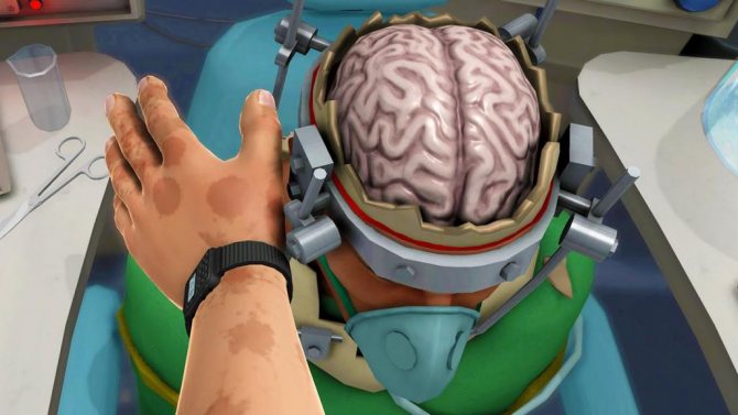
Be careful, video of the operation! Click to open
Treatment
According to the WHO classification, Craniopharyngioma belongs to tumors of the first degree of malignancy, therefore, if it is detected, it is necessary to take timely measures.
Treatment of craniopharyngioma should be comprehensive, including surgical and conservative measures.
Before surgery, to reduce the risk of complications, the patient is prescribed:
- short course of corticosteroid hormones;
- performing shunting in the presence of severe obstructive hydrocephalus to relieve the brain from high intracranial pressure.
The operation itself is performed using stereotactic methods or by craniotomy. Rathke's pouch is cleared of its contents stereotactically. To remove the remainder of the tumor and exclude recurrence of the disease, the neurosurgeon injects bleomycin into the capsule cavity, which accelerates the process of sclerosis of the tumor. Endoscopic intervention methods cause less damage to the brain and are easier to tolerate by patients. Radiosurgical methods of exposure with the introduction of radioactive isotopes are also used.
If craniopharyngioma is removed using the classical method, then specialists use osteoplastic trepanation with subsequent access to the area of the sella turcica.
In the postoperative period, the patient is prescribed:
- corticosteroids to reduce inflammation and swelling of damaged brain tissue;
- diuretics;
- a course of radiation therapy, since the benign nature of the tumor does not exclude the possibility of malignancy and relapse.
In the long-term postoperative period, hormone replacement therapy is prescribed if necessary. Residual effects of the removed tumor include the following disorders:
- poor vision, up to blindness;
- menstrual irregularities and decreased potency;
- diabetes insipidus (increased daily volume of urine with low specific gravity);
- Signs of infantilism, hypothyroidism and adrenal insufficiency may remain.
Forecast
Depends on the size, location, time of detection of the tumor, and the patient’s health condition. In 40-80% of patients, survival exceeds ten years with timely surgery. But even successful surgery does not save from the dangers of the disease returning; relapses are observed in every fifth operated patient. Radiation therapy gives good results even with incomplete tumor removal.
Considering such serious consequences of craniopharyngioma as visual and mental disorders, hormonal disorders, and the formation of persistent intracranial hypertension, timely consultation with a specialist and examination are necessary. A timely operation will prevent the development of complications, eliminate the possibility of relapses and help maintain health for many years.
What's next?
The prognosis for the life of a patient with craniopharyngioma is determined by the size and location of the tumor, as well as the quality of the treatment provided.
Mortality in the postoperative period is no more than 10% and is associated with damage to the hypothalamus region. The five-year postoperative survival rate varies according to various sources at the level of 50-85%. Relapses are possible throughout this period; those that occur in the first year after tumor removal are especially dangerous.
In the postoperative period, almost all patients require long-term hormonal replacement therapy and observation by an endocrinologist.
Preventive measures in this situation come down to preventing harmful effects on a pregnant woman, especially in the first trimester.
This is the exclusion of all types of intoxication and exposure to penetrating radiation. Timely registration of a woman with a medical institution and further proper management of pregnancy and childbirth are of great importance. To prevent tumor recurrence in the postoperative period, early diagnosis, examination at the first signs of the disease and timely surgical treatment are important.
basic information
Craniopharyngioma is a benign neoplasm in a part of the brain, which occurs from the embryonic period, most often located in the hypothalamic-pituitary region. During the development of a tumor, cysts filled with fluid with an impressive composition of proteins and cholesterol can form in its tissues. The disease is more common in children, but can occur at different ages.
The range of occurrence of the disease is approximately 2-3% of all types of brain tumors. The most common is neuroepithelial craniopharyngioma (about 10% of cases), with the peak of disease progression occurring in the age period from 5 to 15 years. Another form of craniopharyngioma disease, papillary, most likely occurs after 40 years.
Symptoms: pay attention
A severe variant of the pathology is the spread of the tumor process to the ventricles of the brain. This development of the situation affects a person’s ability to sleep, and the disturbances are very persistent.
In pathology, a general cerebral syndrome is observed. Craniopharyngioma can be suspected by increased pressure inside the skull and hydrocephalus. Hypertension is indicated by headaches. The patient describes the syndrome as bursting. Many people vomit, but after that it doesn’t get better.
Reasons and factors
The tumor is formed by fairly dense tissue. The diameter of the neoplasm is from a couple to five centimeters. At the moment, scientists do not know the causes of the disease. It is assumed that any pathological factor due to which the development of the embryo is disrupted can provoke cancer. If the mother smoked and drank alcohol, took medications or faced increased stress, the child born to her may develop such a pathology. It can be provoked by overload of the mother's body and malnutrition. The risk of developing a benign neoplasm is higher for those who were exposed to an infectious agent during intrauterine development. Additional risks are associated with toxicosis in the early period of gestation.