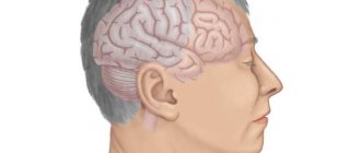
Brain examination is always a relevant area of medicine. This body exercises control, coordination, and optimization of the activities of all systems of the human body. A slight deviation can disrupt this complexly organized communication link, causing serious illness and death of a person.
Today, there are several types of brain research. Below are the most effective of them. It should be said that each of the methods makes it possible to implement specific medical tasks, so one type of research may require clarification using a different technique. However, there are fairly universal ways of examining the brain. In this regard, the following brain research methods are divided into two types:
Group 1 of brain research methods – rather “narrow”, specialized methods that do not give an idea of the entire system of brain functioning;
Group 2 is a combination of methods that allow us to obtain a more complete understanding of the brain and its interaction with other structures of the body.
It should be understood that this division is arbitrary, adopted exclusively in this article in order to optimize perception.
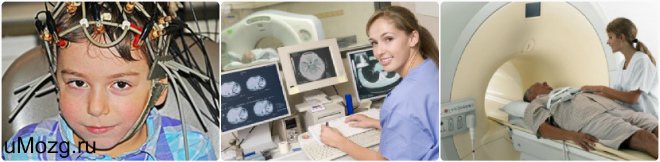
Commonly Used Methods
There are rare signs of health problems, the appearance of which a person does not pay attention to.
In hot weather, I lost my balance and had a severe headache - it’s okay, it will pass, it’s because of the heat. I stood up sharply, “spots” flew in front of my eyes - that’s okay too, I had to get up more slowly. The pressure has risen - of course it will rise, the weather is changing so quickly. There are explanations and justifications for everything, but any of these signs may indicate a serious pathology. If you experience the following symptoms, you should immediately consult a doctor:
- The headache is constant and getting worse.
- Vision, speech, hearing deteriorate, and there are incidents with complete loss of these functions.
- Thought processes, memory, and attention are disrupted.
- Coordination of movements changes, loss of balance and the appearance of a shaky gait are possible.
- Convulsions appear.
- The person loses consciousness or experiences a faint state.
- Intracranial and blood pressure changes.
- “Floaters” appear before the eyes.
These symptoms indicate pathology of the cerebral vessels. To exclude it, a hardware medical examination may be prescribed. It includes both the basic procedure for checking vessels for patency (called angiography), as well as ultrasound, rheoencephalography, and electroencephalography.
These methods are used to clarify traumatic brain injuries, neck injuries, stroke, neoplasms, diseases accompanied by inflammation of the brain, thrombosis, and atherosclerosis. A head examination is performed before or after vascular surgery.
Depending on the pathology, either one type of study or several may be prescribed. Many methods have limitations, so they are prescribed when absolutely necessary.
Diagnosis of brain diseases using CT is based on calculating the intensity of penetration of X-rays through brain tissue. Thus, it is possible to obtain their detailed cross-sectional image. Modern devices for computer diagnostics have a low level of radiation, which does not affect the accuracy of the results.
Such a brain examination may be prescribed if the patient has a history of:
- dizziness;
- headache;
- loss of consciousness;
- convulsions;
- strokes;
- speech and memory disorders;
- auditory and visual impairments.
This method of diagnosing brain pathologies is contraindicated for children and pregnant women. If intravenous administration of a contrast agent is necessary, the following are added to the list of contraindications: renal and liver failure, diabetes mellitus, heart disease, asthma, thyroid pathologies, allergic reaction to iodine.
CT scan does not require special preparation of the patient. Unless before the procedure with contrast, you should not consume food or liquid for 4 hours. The examination lasts from 15 minutes to half an hour. At this time, the person is on a movable table, which moves into the tomograph. The patient is not allowed to move, and sometimes will need to hold his breath at the command of the medical staff.
It’s not for nothing that in foreign films about the work of medical institutions, doctors constantly refer patients to MRI – today it is one of the leaders among brain research methods. Visualization of the state of the organ occurs thanks to the magnetic field constantly maintained in the tomograph. Electromagnetic waves are passed through it, the energy flow from which is repelled by hydrogen atoms, which are present in all cells of the human body. Computer equipment converts the data into images of brain tissue.
MRI is effective for diagnosing a wide range of diseases: from vascular pathologies to tumors.
Examination with a tomograph is contraindicated in the following cases:
- the patient is mentally unstable, has acute pain syndrome or is in a coma;
- The patient’s body contains metal and ferromagnetic implants, pins, clips on blood vessels, and permanent crowns on teeth.
- The patient's skin has tattoos made with paint containing metal particles.
As with a CT scan, for the examination a person must lie down on a movable table, where his body will be secured with special belts, and sensors that send and read a signal are attached to his head. Afterwards the table moves into the tomograph. Depending on the number of programs used for scanning, the duration of the procedure is 15-40 minutes. All this time the person must lie motionless.
The principle of using a low-frequency electromagnetic field makes MRI completely safe for children and adults.
This technique for studying the brain is based on the same principles as MRI, but its main task is to identify pathologies of the vascular bed. Modern equipment is designed to obtain a three-dimensional image of the entire network of brain vessels, as well as to isolate thin sections of individual vessels and nerve trunks.
Using this method, you can examine the brain in order to record in a three-dimensional projection all the functional processes occurring in it. Analysis of the metabolism of brain structures occurs at the cellular level, therefore PET is the best way to distinguish benign from malignant neoplasms in the early stages.
You should not eat 4-6 hours before the procedure. It is recommended to have a protein-free dinner the night before. Part of the examination is the intravenous administration of a radiopharmaceutical. The scanning itself lasts 30-75 minutes.
Indications for examination are:
- frequently recurring headaches, migraines, dizziness;
- sharp deterioration of vision, hearing, tinnitus;
- nosebleeds in the absence of injury;
- frequent fainting with short loss of consciousness;
- tremors of the hands and head in Parkinson's disease;
- previous cerebral infarction, signs of impaired arterial blood flow (chronic ischemia);
- head and cervical spine injuries;
- symptoms indicating the presence of neoplasms, vessel aneurysm;
- encephalopathy (reduction in the volume of nervous tissue and impaired brain function against the background of another pathology);
- arterial and intracranial pressure.
Today, there are several ways to check blood vessels, but the most accurate and informative are magnetic resonance imaging and computed tomography. However, due to the complexity of the equipment and the lack of highly qualified doctors in small towns, the examination is carried out only in a few specialized clinics and is quite expensive. The demand for MRI and CT scans not only in the head and neck area, but also in the entire body is steadily increasing, and the cost is gradually decreasing.
In addition to magnetic resonance and computed tomography, Doppler ultrasound and rheoencephalography are most often used as accessible but informative methods. Checking the blood vessels of the brain and neck is carried out free of charge upon doctor's prescription in all clinics of large cities.
Examinations are:
- Non-invasive (without penetration into the body).
- Invasive (angiographic), when a contrast agent is injected into the patient’s artery to scrupulously examine the smallest details and structures in the affected area. Angiography is used for X-ray examination, ultrasound, MRI and CT.
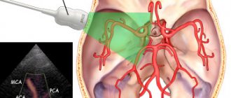
Doppler ultrasound is a technique based on the Doppler effect. Its essence is that high-frequency ultrasonic waves directed at the study area are reflected from moving blood elements and converted on the device screen into a two-dimensional image that characterizes the condition of the blood vessels.
Doppler ultrasound allows you to check the main arteries (vertebral, carotid, basilar, subclavian) and veins (anterior and internal jugular, subclavian), which are located in the neck and at the base of the skull. Ultrasound ultrasound determines:
- patency (lumen diameter), degree of narrowing (stenosis), blockage, elasticity and damage to the walls of blood vessels;
- aneurysms;
- speed and state of blood flow (hemodynamics);
- the presence of a change in direction (tortuosity) caused by damage to the intervertebral discs and other spinal tissues.
Doppler ultrasound is widely used because it is informative, harmless and does not require patient preparation. Contraindications include severe health conditions and the patient’s inability to lie down.
A method of checking the vessels of the head, based on recording changes in the degree of resistance of blood and brain tissue when a weak high-frequency electric current flows through them, is called rheoencephalography. Metal electrodes are placed on the patient's head, secured with rubber bands, and current is passed through them.
REG informs about tension, elasticity, blood filling of blood vessels, viscosity and speed of blood movement through the arteries and veins of the brain. It is used for traumatic brain injuries, concussions, ischemia, strokes, and encephalopathies. The method is especially often used to determine the severity of atherosclerosis in older people, to control cerebral circulation after surgery, and for headaches and dizziness.
The method is based on the interaction of a magnetic field and radio frequency pulses, which results in electromagnetic oscillations. Reflecting from the internal organs of a patient placed in a closed tomograph chamber, they form an image on the monitor screen. The device scans the area under study layer by layer and forms a three-dimensional image. The method allows you to clearly and in detail consider:
- the structure of the circulatory network, all large and small vessels;
- inflammation of brain tissue, nerves;
- aneurysms, blood clots, neoplasms, areas of hemorrhage during a stroke;
- intervertebral hernias and other lesions of the spine and spinal cord.
Indications
There are certain symptoms that require you to be diagnosed by a doctor. If a person notices a number of manifestations, then he should immediately go to the hospital so that the disease can be identified. Even if there is no disease, in any case it will be useful to check the head so that, if necessary, certain abnormalities can be identified.
Alarming symptoms:
- Constant headaches. However, they are poorly controlled by conventional medications and occur regardless of external factors.
- Pressure surges. A person can suffer from both hypertension and hypotension. In any case, such a condition cannot be considered normal.
- Numbness of the extremities, which is accompanied by tingling of the legs and arms. This often indicates that there are problems with blood vessels.
- The occurrence of seizures for unknown reasons.
- Fainting. In such a situation, it is recommended to consult a doctor and undergo an examination so that you can understand what caused the loss of consciousness.
- Problems with memory, concentration, and coordination. As a rule, this does not occur without a good reason, especially if the person is young or middle-aged.
- Emotional disorders. A person may suffer from depression, be too aggressive, or indifferent to surrounding events. However, such conditions are not typical for him, and they have not been observed before.
- Problems with hearing and vision that have arisen for unknown reasons.
- Noise in the ears, as well as pulsation.
There are also a number of situations in which it is necessary to check the condition of the brain. In particular, it is necessary to undergo diagnostics after an injury. At first, you may not even experience negative symptoms, but this does not mean that complications will not arise. It is imperative to make sure that the injury will not lead to dangerous consequences.
For migraine and arterial hypertension, it is also necessary to undergo examination so that the patient’s condition can be monitored. If a person has suffered a heart attack or stroke, it is important for him to seek medical help. Because such conditions require mandatory monitoring to ensure that there are no negative consequences.
Vegetative-vascular dystonia, although not considered a dangerous disease, also requires control. Because it can lead to various complications, especially if a person leads an incorrect lifestyle. Diabetes mellitus, neck osteochondrosis and encephalopathy also need diagnosis.
It is important to understand the state of a person’s brain so that negative consequences can be prevented.
Electroencephalography
EEG is a non-invasive way to study the electrical activity of the brain, including under the influence of certain stimuli: light, sound, movement. Indirectly indicates a change in blood circulation, therefore it is not the main way to diagnose vascular disorders.
The brain examination is carried out using a special device - an encephalograph, capable of recording the frequency of electrical oscillations from 0.5 to 100 Hz. Electrodes are placed on the head to pick up minimal brain signals. The signals enter the amplifier, are magnified millions of times, and are transmitted to the computer monitor in the form of a graph - an encephalogram. In children under 3 years of age, the study is performed only during sleep or under light anesthesia.
The recording reflects the fluctuations and rhythms of the electrical process occurring in the nerve cells of the head:
- Alpha rhythm with a frequency of 8-14 Hz, characterizing a state of rest.
- Beta rhythm 13-30 Hz, indicating depression and anxiety.
- Delta rhythm 0.5-3 Hz, typical for sleep.
- Theta rhythm is 4-7 Hz, characteristic of an adult and a child in sleep.
The predominance of alpha and beta rhythms, the same electrical activity in both hemispheres and the occurrence of only a local reaction to the stimulus are signs of a normal encephalogram. EEG does not provide an anatomical picture of the structure of blood vessels.
Before performing an encephalography, you need to prepare. You should not eat 2 hours before the procedure, you should not drink coffee or energy drinks 12 hours before, and you should not smoke. Do not use hair styling products. The time the procedure may take is 45-120 minutes.
Electroencephalography is not used during periods of exacerbation of mental disorders, mental trauma, infectious diseases, injuries or wounds on the scalp. Unless absolutely necessary, it is not performed in children under 7 years of age.
This procedure is quite useful in terms of collecting anamnesis for headaches. With it you can see exactly where there are abnormalities in the functioning of the brain, where there are vascular lesions, as well as the slightest signs of just beginning tumors.
What pathologies can be identified?
Even if a person only has a headache, it will be useful for him to check the condition of the cranium. In this case, it will be possible to understand exactly what pathologies we are dealing with, as well as how advanced they are. There are many diseases that can be diagnosed using MRI, CT, angiography and other examinations. It is worth considering the most common deviations that can be identified using such examinations.
In the photographs you will be able to see the neoplasms, as well as determine their nature. It will also be possible to detect cysts, hematomas and abscesses. From vascular pathologies, thrombosis, narrowing of the lumen, and also rupture of the vessel can be determined. It is also possible to diagnose aneurysm, underdevelopment of vascular canals and many other abnormalities.
With the help of head examinations, you can not only see the existing pathology, but also determine its size, boundaries, degree of development and nature. This is exactly what is needed so that the correct treatment can be prescribed and a prognosis made.
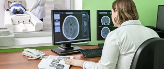
Magnetic resonance imaging
This study is considered the most popular, which is excellent for checking the head. With its help, you can identify various deviations from the norm that affected a person. The procedure takes place in a certain way. The person will need to lie down on a sliding table, after which the patient will be placed inside a special device. After this, you need to lie still, because otherwise the pictures will be distorted.
As a result, after the procedure it will be possible to see the brain from different angles. The specialist will be able to examine the various pathologies that affected a particular person. A contrast agent may be used to obtain more detailed information. However, it is not used in cases where a person has an allergic reaction.
Certain citizens are not allowed to participate in the research due to their characteristics. People weighing more than 120 kg will not be able to undergo diagnostics. They will not fit inside the device, because the equipment has a certain diameter. Also, the procedure is not recommended for citizens with mental disorders, especially claustrophobia. During the procedure you will have to be inside the device, which creates the feeling of a closed space. This may provoke panic among certain citizens.
If the diagnosis needs to be carried out for a small child, then anesthesia may be used for him. As already mentioned, a person will have to lie still all the time, and not all children are capable of this. Pregnant women are not allowed to participate in the study during the first trimester. An exception may be made if there is a clear need to check the head. Separately, it is worth noting that MRI is not suitable for those citizens who have metal implants. They will not be able to undergo diagnostics, because magnetic waves do not interact well with metal.
Otherwise, the procedure is considered harmless and can be used even once a week. With its help, it is convenient to determine not only existing pathologies, but also to control treatment. It is convenient to observe the changes that occur in the human body. If a specific treatment regimen is not suitable, then another option will be prescribed.
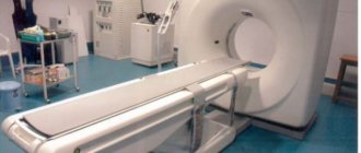
CT head
Computed tomography is also considered an informative study of the head. The procedure is performed if tumors, inflammatory lesions, hemorrhages and other abnormalities are suspected. An examination is often prescribed if there has been a head injury.
In the photographs you will be able to see various brain structures, its membranes, cranial bones, large vessels, as well as the nasal sinuses. The diagnostic method is considered safe, although the person is exposed to radiation. The effectiveness of the research will largely depend on how high-quality the equipment is. The procedure itself will take no more than 30 minutes, and no special preparation is required.
In a situation where it is necessary to use a contrast agent, a person will first have to consult an allergist. It will be useful to perform special tests so that you can understand whether the contrast can be used for a particular person. It is worth noting that this product is iodine with additional components. It allows you to visualize brain areas and blood vessels. A strict contraindication is during pregnancy. In such a situation, a woman cannot use a contrast agent; the procedure can be carried out without it.
Vascular ultrasound
Ultrasound examination is often used in cases where there is suspicion of vascular pathology in the head and neck. In most cases, the examination is carried out using Doppler. The procedure is harmless, has no contraindications or any restrictions. It also does not cause side effects in people.
Before the session, a person is not recommended to drink alcohol, smoke or drink coffee. It is also worth giving up medications that affect the condition of blood vessels. Otherwise, the results will be distorted, and it will not be possible to understand exactly what is happening to the person.
The person will need to lie on a couch during the test. The specialist will apply a special gel to the areas of interest and then use an ultrasound probe. With its help, you will be able to see the condition of the vessels, as well as assess the quality of blood flow. The procedure is inexpensive and at the same time useful; its cost starts from about 1,000 rubles.
Electroencephalography
This procedure allows you to assess brain activity using special sensors. They are attached to a person’s head, after which electrical impulses will be recorded. All information is transferred to the computer, and the doctor will be able to see if there are any deviations from the norm. The procedure is used for people of all ages, including infants.
It is often necessary to perform additional steps in order to obtain more accurate information about brain activity. For example, a person may need to hold their breath, remain still, respond to light, and take deep breaths.
Based on the results of the studies, it will not be possible to understand whether there is pathology in the vessels. In this case, it will be possible to determine the state of a person’s brain, as well as how well it functions. In particular, the study is useful for seizures, as well as after a person has suffered a stroke.
Radiography
This procedure cannot be considered harmless, which is why it is performed quite rarely. X-rays of the brain show general pathologies such as neoplasms, skull fractures and hematomas. In this case, a contrast agent can be used if it is necessary to assess the condition of the vessels.
The procedure is not recommended for children, because the study may have a negative impact on a fragile body. That is why examination is used only in extreme cases when there are no other options. For example, it may be prescribed for a serious head injury, when it is important to ensure that there is no fracture.
A person does not have to prepare specially for the examination. Before entering the office, you will need to leave all metal objects, including jewelry, and also put out your phone. In the room itself, you will need to put on a protective gown, and then take the necessary position, which the doctor will advise. The specialist will take the picture from another room, and it will be important for the person not to move at this time. The procedure itself does not take much time; the description of the image takes the longest, which takes up to 15 minutes.
Head testing often involves complex procedures that will be called MRI, CT, X-ray, etc. All of them allow you to check the state of the brain and surrounding structures, as well as draw conclusions about human health.
Rheoencephalography of cerebral vessels
REG (rheoencephalography) is a non-invasive method for diagnosing cerebral vessels, recording changes in the electrical resistance of vascular walls when a high-frequency pulse passes through them. Provides an opportunity to check the vessels of the head, their reactivity, tone, elasticity, level of resistance of vascular tissue, possible blockage and characteristics of blood supply.
REG is indicated for suspected diseases:
- vascular pathologies;
- circulatory disorders;
- atherosclerosis;
- stroke;
- vascular stenosis;
- clarification of the effect of drugs;
- traumatic brain injuries.
To perform rheoencephalography, rheographs with a number of channels from 2 to 6 are used, recording the corresponding number of vessels. The patient is sitting or lying down. Electrodes are installed on his head using rubber bands. A special paste can be applied underneath them to improve contact with the skin and reduce resistance.
Some electrodes send electrical signals, others receive them and transmit them to the rheograph. The latter processes and transmits to a monitor or print in the form of a graph - a rheoencephalogram. The procedure lasts several minutes.
During the examination, the patient may be given a small dose of nitroglycerin under the tongue, Papaverine, or asked to change body position or turn his head. These measures will allow you to see a change in tone and blood supply.
The advantage of rheoencephalography is its simplicity, safety for the patient, and the ability to monitor blood flow for a long time. Disadvantages include the inability to measure cerebral blood flow and determine the exact cause of circulatory disorders.
Before the study, you should not smoke, drink alcohol, coffee, or energy drinks. If the patient is taking medications, the doctor must be informed about this. Some may need to be stopped for a while.
Rheoencephalography is not prescribed for children under 7 years of age, as well as for wounds and injuries at the places where the electrodes are attached.
Which doctor should I contact?
To find out how to check the blood vessels in the brain, you need to make an appointment with a doctor. A referral for examination of vessels located in the brain and neck is given by a therapist. Among the doctors who check the condition of the vascular system and determine which research method is suitable in a particular case are highly specialized doctors - neurologist, cardiologist, phlebologist, angiologist. Consultation with a psychiatrist is necessary if malfunctions in the cerebral blood flow system are accompanied by disturbances in mental activity.
Timely detection of pathological processes occurring in the vascular system of the brain contributes to successful treatment. Modern methods for diagnosing vascular diseases affecting the brain make it possible to detect disorders at an early stage of development.
Angiography of cerebral vessels
Angiography is the most accurate and most informative examination of cerebral vessels, accompanied by the administration of a contrast agent. It will allow you to learn about the functional state, characteristics of blood flow, changes in vascular patency, and localization of the disorder.
This study checks for the following pathologies:
- aneurysm;
- arteriovenous malformation;
- vascular stenosis;
- blockage of blood vessels;
- neoplasms.
Angiography is also performed in the postoperative period to control the location of clips installed on the vessels.
There are 3 methods of angiography: classical, CT angiography, magnetic resonance imaging.
Classical and CT angiography is not performed in cases of increased sensitivity to contrast agents and anesthetics, impaired hemostasis, pathology of internal organs, acute infectious diseases, exacerbation of mental disorders, changes in thyroid function, pregnancy, lactation.
The procedure examines the aorta, large vessels (carotid arteries), and small arteries.
Preparation includes giving up alcohol 2 weeks in advance. It is forbidden to buy or drink 4 hours before the procedure. In some cases, hydration is given to facilitate the removal of the contrast agent from the body. They do fluorography and electrocardiogram. It is possible to prescribe antihistamines.
During the procedure, the patient is placed on a special table, fixed, and connected to a cardiac monitor. The femoral (possibly carotid or vertebral) artery is punctured, a catheter is installed into it, through which a contrast agent (mainly iodine) is injected, and an examination is carried out. X-ray television records everything that happens in the vessel. Upon completion of the study, a bandage is applied to the puncture site for a day. In the absence of contraindications, it is recommended to drink as much water as possible.
Duration: 60-180 minutes, performed only in a hospital. The advantage is the accuracy of the results. Disadvantages: invasiveness and radiation exposure.
CT angiography
This study is a type of computed tomography of the brain, but to obtain a clearer picture, a contrast agent is injected through a catheter into the cubital vein. It is performed on an outpatient basis using a computed tomograph. A week before the procedure, a creatinine test is taken.
The tomograph takes images layer by layer with a distance between layers of about 1 mm in different projections. As a result, the doctor receives many images, which are later combined into three-dimensional images of the structure of the blood vessels. The study allows you to combine images of arterial and venous blood flow.
Advantages of CT angiography: absence of shadows, high information content. Disadvantages: invasiveness and x-ray exposure.
An additional contraindication is third degree obesity.
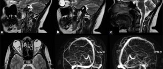
This type of angiography is a type of MRI. Magnetic fields are used instead of X-rays. With 4D angiography, not only a three-dimensional image is obtained, but also the dynamics of blood flow are visualized. In rare cases, a contrast agent may be administered.
After the patient has taken off his jewelry and glasses, he is placed on the table and moved into a closed capsule. It is important to ensure immobility, so if the examination is performed on young children, they are given light anesthesia.
Duration: 30 minutes. The procedure is performed on an outpatient basis.
Advantages: no radiation, accuracy of information, possibility of carrying out in case of sensitivity to iodine-containing substances.
Contraindications include installed pacemakers and metal-containing implants, heart failure, pregnancy, mental disorders, third-degree obesity, claustrophobia.
Who to go to with varicose veins
The leading place among vascular diseases is occupied by varicose veins on the legs. Most often the problem is provoked by the following factors:
- Weak tone of the venous walls.
- Standing for a long time without moving.
- Violation of the functions of vein valves.
- Pathologies of the thyroid gland.
- Genetic predisposition.
A number of symptoms have been identified that indicate the formation of varicose veins:
- Swelling of veins.
- Pain.
- Swelling.
- Vascular drawing.
- Ulcers that most often appear on the legs.
Venous blood stagnates, which becomes the result of thrombophlebitis - blocking of a vessel with a blood clot, followed by an inflammatory process.
The phlebologist will determine a number of studies:
- Dopplerography of veins, which evaluates blood flow in the vessels.
- CT scan to detect blood clots.
- Blood test for clotting.
- Phlebomanometry (measurement of pressure in the veins).
The disease is associated with pressure in the blood vessels. In the early stages of development, drug therapy is prescribed; in advanced forms, only surgical intervention can help.
Differential diagnosis
When a patient experiences ailments in the neck and head, headaches, sleep disturbances, and gait instability, he consults a neurologist who, in the absence of contraindications, prescribes a study using one of the listed methods. After scanning and taking instrument readings, the specialist who performed the procedure interprets the images and gives the result to the patient.
Based on the information received, the neurologist diagnoses brain diseases and prescribes treatment. If difficulties arise in making a diagnosis, he will refer you for a repeat examination of a higher level - contrast CT or MRI, which will allow you to see the affected area structurally and clearly.
Due to the fact that headache can have a large number of symptoms, it is imperative to carry out a differential diagnosis. For each doctor, the main task is to determine the connection between a specific headache and a specific disease. You need to understand that there will be no benefit in treating an exceptional symptom.
We present to you the sequence of all necessary examinations:
- The first step is to interview the patient. It is important for the doctor to find out the duration of pain, their sequence, nature and location of pain. It is also important to find out the patient’s body’s reaction to taking analgesics. Often, most people cannot describe such pain when they first see a doctor with such a problem;
- After the general survey is completed, doctors prescribe certain tests that need to be completed in the near future. But it is important to choose the right diagnostic measures so that the patient receives his course of treatment faster.
Treatment
When vascular disease in the brain and neck occurs, medications are first prescribed. Only a specialist can say how long the treatment will last. Proper lifestyle and nutrition play an important role. It is necessary to eat foods that contain low amounts of cholesterol. You need to add fresh fruits and vegetables to your diet. Once and for all, you should forget about foods that contain rich animal fats. This includes smoked and fried foods, including fast food.
It is useful to eat seafood for people who have problems with blood vessels. Such food contains biologically active substances, and with their help, cholesterol in the blood is reduced. The metabolism in the human body also becomes normal. If you adhere to proper nutrition, it will only bring positive effects.
As soon as a disease associated with blood vessels has been found, you must immediately stop drinking alcohol and quit smoking. If a patient is overweight, then he needs not only to switch to proper nutrition, but also to do physical exercise.
In order not to encounter diseases of the vascular system, you need to follow preventive measures. Breathing exercises, as well as swimming and cycling give good results. You can take a contrast shower to harden your body and blood vessels.
It is important to avoid stressful situations, as well as emotional and physical stress. The main thing is not to overstrain your body and completely give up alcohol and tobacco products. Proper nutrition plays an important role, because if you follow all the recommendations, you can avoid many diseases. It is important to examine the vessels of the head and neck in a timely manner and not turn a blind eye to various symptoms. If the disease is found in a timely manner, it will be easier to undergo treatment. As soon as unpleasant symptoms appear, such as dizziness, fainting or severe headaches, it is better to immediately seek help from a medical facility.
Causes
Pain in the head can have a completely different onset, from banal fatigue to infection with some kind of infection. But, despite the initial cause, the mechanism for creating pain is the same. Let's look at it in more detail.
First, the inflammatory process begins, which affects areas of the human central nervous system. Next, irritation of the membranes of the brain occurs.
This happens for two reasons, first of all, there is an increase in cerebrospinal fluid pressure, and the second reason is poisoning of the body. At the end of the process, a spasm occurs and the blood vessels dilate.
Some experts highlight a theory about the mental source of headaches. At first glance, the cause of this type of pain is obvious. But in fact there can be a very large number of reasons. Let's look at the most common of them:
- diseases that were caused by infection;
- various tumors in the head area;
- increased blood pressure;
- diseases in the sinus area;
- disruption of hormones;
- human sensitivity to weather conditions.
Examination of young children
Neurosonography is intended for examining newborns. Its essence is to study the brain using ultrasound scanning through the fontanel.
The study can be prescribed both in case of suspected pathology and for preventive purposes (for example, during difficult childbirth). Using ultrasound, information is obtained about the state of blood flow and its possible disturbances.
It must be remembered that an effective solution to health problems is possible only if the disease is detected and correctly diagnosed in a timely manner.
An examination of the cerebral vessels will enable the attending physician to establish a diagnosis and prescribe appropriate treatment. In turn, the progression and development of a disease associated with cerebral vessels can cause significant harm to health.
Which MRI should I do?
If the pain in the head is incredibly severe and lasts for quite a long period, and also has many symptoms that are associated with irritation, poor sleep, upset stomach, vomiting and nausea, then this indicates some serious diseases in the brain area.
If an MRI of the brain is prescribed during diagnosis, this should be done to check for vascular pathologies.
Migraine occurs due to the fact that the blood vessels in the human brain contract at a certain moment.
Due to this, a spasm occurs in the brain. A tomograph of this type allows you to learn about blood flow disorders, as well as reflections of various ischemic types.
Some patients encounter a problem after an MRI - their headaches begin to hurt more severely. The nature of this phenomenon has not yet been revealed - the construction of the image is carried out due to electromagnetic influence, and it is harmless to the body.
In any case, the various examinations must be complete. This will allow the doctor to see the whole picture of the disease and prescribe the most accurate treatment. Through a complete diagnosis, you will be able to find out the cause of the pain in your head. As a rule, specialists first do an MRI of the brain.
Such procedures are carried out regardless of whether there are headaches at the moment. A complete medical history will provide complete information for the doctor.
Cardiac Vascular Specialist
A cardiologist deals with the treatment of heart pathologies. Patients come to him with complaints about:
- Pain in the chest area.
- Shortness of breath.
- Increased work of sweat glands.
- Violation of heart contractions.
- Heart disease.
- Heart attack.
- Angina pectoris.
In addition, a cardiologist deals with the vessels that nourish the heart muscle.
It is recommended to visit a cardiologist in a number of cases:
- Age after 35 years.
- Planned pregnancy.
- Genetic predisposition to heart pathologies.
- Overweight, obesity.
If a person follows these rules, then the risk of serious diseases of the cardiovascular system is reduced significantly . If a certain problem is detected, the patient receives timely treatment without risking the development of complications.
The person himself is responsible for the blood vessels and their health, and the doctor only helps to cope with the problems that arise.