Structure and functions of nerve fibers, their classification
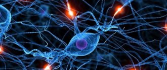
Nerve fibers are processes of nerve cells (neurons) that have a membrane and are capable of conducting nerve impulses.
The main component of the nerve fiber is the process of the neuron, which forms, as it were, the axis of the fiber. Mostly this is an axon. The nerve process is surrounded by a membrane of complex structure, together with which it forms a fiber.
Nerve fibers are divided into myelinated and unmyelinated. The former have a myelin sheath covering the axon, the latter lack a myelin sheath.
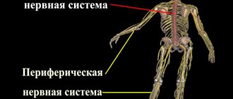
Myelin fibers predominate in both the peripheral and central nervous systems. Nerve fibers lacking myelin are located predominantly in the sympathetic division of the autonomic nervous system.
Structure of nerve fibers:
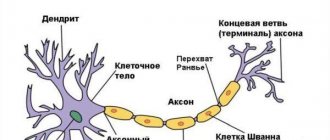
The myelinated nerve fiber contains the following elements (structures): 1) an axial cylinder located in the very center of the nerve fiber,
2) the myelin sheath covering the axial cylinder,
3) Schwann shell.
The axial cylinder consists of neurofibrils. The pulpy membrane contains a large amount of lipoid substances known as myelin. Myelin ensures the speed of nerve impulses. The myelin sheath does not cover the entire axial cylinder, forming gaps called nodes of Ranvier. In the area of the nodes of Ranvier, the axial cylinder of the nerve fiber is adjacent to the superior Schwann membrane.
The fiber space located between two nodes of Ranvier is called a fiber segment. In each such segment, the nucleus of the Schwann membrane can be seen on stained preparations. It lies approximately in the middle of the segment and is surrounded by the protoplasm of the Schwann cell, the loops of which contain myelin. Between the nodes of Ranvier, the myelin sheath is also not continuous. In its thickness, so-called Schmidt-Lanterman notches are found, running in an oblique direction.
Schwann membrane cells, as well as neurons with processes, develop from the ectoderm. They cover the axial cylinder of the nerve fiber of the peripheral nervous system, similar to how glial cells cover the nerve fiber in the central nervous system. As a result, they may be called peripheral glial cells.
In the central nervous system, nerve fibers do not have Schwann sheaths. The role of Schwann cells here is performed by elements of oligodendroglia. An unmyelinated (unmyelinated) nerve fiber is devoid of a myelin sheath and consists only of an axial cylinder and a Schwann sheath.
Function of nerve fibers.
The main function of nerve fibers is the transmission of nerve impulses. Currently, two types of nerve transmission have been studied: pulsed and non-pulse. Impulse transmission is provided by electrolyte and neurotransmitter mechanisms. The speed of nerve impulse transmission in myelinated fibers is much higher than in nonmyelinated fibers. In its implementation, the most important role is played by myelin. This substance is capable of isolating a nerve impulse, as a result of which signal transmission along the nerve fiber occurs spasmodically, from one node of Ranvier to another. Pulseless transmission is carried out by axoplasmic current along special axon microtubules containing trophogens - substances that have a trophic effect on the innervated organ.
Structural classification.
Based on the number and arrangement of dendrites and axons, neurons are divided into axonless neurons, unipolar neurons, pseudounipolar neurons, bipolar neurons, and multipolar (many dendritic arbors, usually efferent) neurons.
Axonless neurons are small cells grouped near the spinal cord in the intervertebral ganglia, which do not have anatomical signs of division of processes into dendrites and axons. All processes of the cell are very similar. The functional purpose of axonless neurons is poorly understood. Unipolar neurons are neurons with one process, present, for example, in the sensory nucleus of the trigeminal nerve in the midbrain.
Bipolar neurons are neurons that have one axon and one dendrite, located in specialized sensory organs - the retina, olfactory epithelium and bulb, auditory and vestibular ganglia;
Multipolar neurons - Neurons with one axon and several dendrites. This type of nerve cells predominates in the central nervous system. Pseudounipolar neurons are unique in their kind. One tip extends from the body, which immediately divides in a T-shape. This entire single tract is covered with a myelin sheath and is structurally an axon, although along one of the branches the excitation goes not from, but to the body of the neuron. Structurally, dendrites are branches at the end of this (peripheral) process. The trigger zone is the beginning of this branching (i.e., it is located outside the cell body).
Nerve fibers -
processes of nerve cells covered with membranes.
Some nerve fibers have a sheath made of a fat-like substance called myelin
. This substance performs trophic, protective and electrical insulating functions.
In the early stages of ontogenesis, the myelin sheath is absent; it develops in the first 2–3 years of life, its formation depends on the child’s living conditions. Under unfavorable conditions, the process of myelination can slow down for several years, which complicates the control and regulatory activity of the nervous system.
Nerve fibers combine with each other to form nerves
, which in the form of white threads are visible to the naked eye. Nerves connect all parts of our body with the central parts of the nervous system. The main function of nerve fibers and nerves is to conduct nerve impulses.
There are three types of nerves:
1. sensitive or centripetal - conduct nerve impulses to the central nervous system;
2. motor or centrifugal – conduct nerve impulses from the central nervous system to peripheral organs;
3. mixed - consist of sensory and motor fibers.
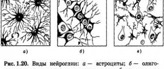
Glial cells (neuroglia)
more numerous than neurons, accounting for half the volume of the central nervous system. They are capable of division throughout their lives. Glial cells perform supporting, protective, insulating, and metabolic (supplying neurons with nutrients) functions.
During human development, the ratio between glial and nerve cells changes significantly. In a newborn, the number of neurons is higher than glial cells; by the age of 20–30, their ratio becomes equal; after 30 years, the number of glial cells increases.
Basic properties of nervous tissue.
1. Excitability
– the ability of nerve tissue cells to quickly respond to irritation by changing the electrical properties of the cell membrane and their metabolism.
A quantitative measure of excitability is the irritation threshold
– the minimum amount of stimulus that can cause a tissue response.
The most common and natural stimulus for all cells of the body is the nerve impulse
.
A stimulus of lesser strength is called subthreshold
, and a greater one is
suprathreshold.
The latter cause more significant response changes in the vital activity of the tissue or organism.
2. Conductivity –
the ability of living tissue to conduct excitation. Excitation occurs due to the spread of a nerve impulse, which passes through the synapse to neighboring cells and can be transmitted to any part of the nervous system.
Having arisen at the site of excitation, an action potential (a change in the electrical charge of the membrane) causes a change in electrical charges in the adjacent area, and those, in turn, in the next one, etc.
3. Lability –
the ability of excitable tissue to reproduce the maximum number of action potentials per unit time.
The functional state of nervous tissue depends on its lability. Pathological processes and fatigue lead to a decrease in lability, and systematic special training leads to its increase.
Anatomical and physiological features of the development of the central nervous system.
Spinal cord.
CM. of an adult is located in the spinal canal and is a cylindrical cord 40-45 cm long, with a total weight of 34 - 38 g. The spinal cord of a newborn is the most mature part of the central nervous system, but its final development ends only by the age of 20. During this period, the brain mass increases 8 times.
CM. has segmental
.
(31 segments). Two pairs of anterior and posterior roots extend from each segment. Each two pairs correspond to one vertebra. The dorsal roots are formed by sensory (afferent)
neurons.
Neurons located at the front of the spinal cord are motor neurons
, which control the functioning of skeletal muscles.
CM. conditionally divided into 4 departments:
1. cervical;
2. chest;
3. lumbar;
4. sacral,
Each of which contains several segments, a pair of spinal nerves extends from any segment. Each pair of nerves innervates a specific area of the body. For example: the nerves of the cervical and lumbar regions innervate the muscles of the limbs.
Building S.M. – cross section:
Gray matter located centrally
(nerve cells), which borders
the white matter
(nerve fibers). Sensitive roots s.m. consist of afferent neurons, and motor neurons - of efferent ones. Switching the signal from afferent to efferent neurons is carried out using intercalary or directly.
A huge number of reflex arcs are closed in the spinal cord, thanks to which it is able to regulate many functions of the body - such as flexion and extension of limbs, maintaining a certain posture...
Cm. human contains two thickenings: cervical and lumbar. They begin to develop in the first years of a child's life.
The cervical thickening regulates the movement of the upper limbs, the lumbar thickening regulates the movement of the lower limbs. The formation of cervical and lumbar thickenings depends on the child’s motor activity.
Nervous impulses from motor centers S.M. provides constant, slightly slower tension of the entire skeletal muscles, called muscle tone
, which allows a person to conduct normal motor activity.
Nerve fibers. Classification. Structure.
Nerve fibers are extensions of nerve cells that are usually covered with sheaths.
Depending on the structure of the shell, they are divided into two main groups:
1. Myelin. 2. Unmyelinated.
Both consist of a nerve cell process, which is located in the center of the fiber and is therefore called the axial cylinder
and a membrane formed by oligodendroglial cells called neurolemmocytes (Schwann cells).
Myelinated nerve fibers
These are fibers consisting of an axial cylinder
,
myelin sheath
,
neurolemma and basement membrane.
The diameter of the cross section is from 1 to 20 microns.
Localization - central nervous system, peripheral nervous system.
The axial cylinder is a process of a nerve cell (axon or dendrite). The axial cylinder consists of neuroplasm covered with a membrane - the axolemma.
Neuroplasma
is the cytoplasm of a nerve cell, which contains longitudinally oriented neurofilaments and neurotubules. The neuroplasm contains mitochondria, which are more numerous in the immediate vicinity of the interceptions and are especially numerous in the terminal apparatus of the fibers.
Axolemma
- represents a continuation of the cell membrane of the neurocyte, which ensures the conduction of the nerve impulse. The speed of nerve impulse transmission along thick myelin fiber ranges from 5 to 120 m/s.
The myelin sheath is a tube with a thickness of 0.3 to 20 microns, which covers the entire length of the axial cylinder. There is no myelin sheath at the sites where the process exits the perikaryon, at the sites of terminal branches of the axon and at the sites of nodal interceptions. The interceptions correspond to the border of adjacent neurolemmocytes. The fiber segment located between adjacent interceptions is called the internodal segment, and its membrane is represented by one glial cell. The length of the internodal segment ranges from several micrometers to several millimeters. The nodal interception has dimensions from 0.25 to 1 µm.
Due to the fact that the myelin sheath contains lipids, when the fiber is treated with osmic acid, it becomes intensely stained dark brown. In this case, the entire fiber has the appearance of a homogeneous cylinder, in which light lines—myelin notches—are located at a certain distance from each other.
During the development of the myelin fiber, the axial cylinder, plunging into the neurolemmocyte, bends its membrane and forms a deep fold. This double fold of the plasma membrane of the neutrolemmocyte is called mesoxon. As the Schwann cell develops, it slowly turns around the axial cylinder, as a result of which the mesaxon repeatedly envelops it. Under an electron microscope, each mesaxon curl is visible as a light layer, about 8-12 nm wide, which corresponds to the lipid layers of the two layers of the plasma membrane of the neurolemmocyte. In the middle and along its surface, thin dark lines formed by protein molecules are visible. The myelin notches correspond to those places where the mesaxon curls are pushed apart by the cytoplasm of the Schwann cell.
The sheath of one nerve fiber is formed by many neurolemmocytes. They contact each other at the sites of nodal interceptions. The internodal segment corresponds to one glial cell.
In a longitudinal section near the interception, a region is visible in which the mesaxon curls sequentially contact the axial cylinder. The attachment sites of the deepest mesaxon curls are farthest from the interception, and all subsequent ones are naturally located closer to it. This is explained by the fact that during the growth of the axial cylinder and neurolemmocytes, mesaxon is layered, so its first layers are shorter than the subsequent ones. The edges of two adjacent lemmocytes in the interception area form ring-shaped processes with a diameter of 50 nm; the length of these processes is different.
Neurolemma is a peripheral zone of a nerve fiber containing the cytoplasm of neurolemmocytes and their nuclei pushed here.
Basement membrane - covers the outside of the myelin fiber. It is associated with dense strands of collagen fibrils, which are oriented longitudinally and are not interrupted at the interception.
Previous27282930313233343536373839404142Next
Date added: 2017-01-29; ;
SEE MORE:
Nerve fibers (meaty and non-meaty) - structure and functions. Conduction of excitation along a nerve
Previous12345678910111213141516Next
The function of rapid transmission of excitation to and from a nerve cell is performed by its processes - dendrites and axons, i.e. nerve fibers . Depending on their structure, they are divided into pulpy , having a myelin sheath , and non-pulpy . This membrane is formed by Schwann cells, which are modified glial cells. They contain myelin , which is mainly composed of lipids. It performs insulating and trophic functions . One Schwann cell forms the sheath per 1 mm of nerve fiber. Areas where the shell is discontinuous , i.e. not covered with myelin, called nodes of Ranvier . Interception width 1 µm.
Functionally, all nerve fibers are divided into 3 groups:
1. Type A fibers are thick fibers that have a myelin sheath .
This group includes 4 subtypes:
• And alpha - motor fibers of skeletal muscles and afferent nerves coming from muscle spindles - stretch receptors. Conduction speed 70-120 m/s.
• And beta - afferent fibers coming from pressure and touch receptors of the skin . Speed 30-70 m/s.
• And gamma – efferent fibers going to muscle spindles (15-30 m/s).
• And delta – afferent fibers from temperature and pain receptors of the skin (12-30 m/s).
2. Group B fibers are thin myelinated fibers, which are preganglionic fibers of the autonomic efferent pathways. Conduction speed 3-18 m/s.
3. Group C fibers are non-myelinated postganglionic fibers of the autonomic nervous system. Speed 0.5-3 m/s.
The conduction of excitation along the nerves is subject to the following laws:
1. The law of anatomical and physiological integrity of nerves , i.e. the nerve is able to perform its function only under both of these conditions. The first violations occur during cutting, the second - due to the action of substances that block conduction, for example, novocaine.
2. The law of two-way conduction of excitation . It spreads in both directions from the site of irritation. In the body, excitation most often goes through afferent pathways to the neuron, and through efferent pathways from the neuron. This type of distribution is called orthodromic. Very rarely, reverse or antidromic propagation of excitation occurs.
3. Law of isolated conduction . Excitation is transmitted from one nerve fiber to another fiber, which is part of the same nerve trunk.
4. Law without decremental implementation . Excitation is carried out along the nerves without decrement, i.e. without attenuation. Consequently, nerve impulses are not weakened as they pass through the nerves .
5. The speed of conduction is directly proportional to the diameter of the nerves.
Nerve fibers have the properties of an electrical cable, which is not very well insulated. The mechanism of excitation is based on the occurrence of local current. As a result of the generation of an action potential in the axon hillock and reversal of the membrane potential, the axon membrane acquires a positive charge. Outside it becomes negative, inside positive. The membrane of the underlying unexcited axon is charged in the opposite way. Therefore, local currents begin to pass between these areas along the outer and inner surfaces of the membranes. These currents depolarize the membrane of the underlying unexcited portion of the nerve to a critical level, and an action potential is also generated in it. Then the process is repeated and a more distant part of the nerve is excited, etc.
Since local currents flow through the membrane of the pulpless fiber without interruption, such conduction is called continuous. With continuous conduction, local currents cover a large surface area of the fiber, so they require a long time to pass through a section of the fiber. As a result, the range and speed of transmission along the non-pulp fiber is small.
In the pulp fibers, areas covered with myelin have high electrical resistance. Therefore, continuous conduction of the action potential is impossible. When an action potential is generated, local currents flow only between adjacent nodes. According to the “all or nothing” law, the node of Ranvier closest to the axon hillock is excited, then the adjacent underlying node, etc.
This is called a saltatory jump. With this mechanism, local currents do not weaken, and nerve impulses travel over a greater distance and at high speed.
Previous12345678910111213141516Next
Date: 2015-07-02; view: 551; Copyright infringement
| Did you like the page? Like for friends: |
Normal physiology: lecture notesSvetlana Sergeevna Firsova
2. Mechanisms for conducting excitation along the nerve fiber. Laws for the conduction of excitation along nerve fibers
The mechanism for conducting excitation along nerve fibers depends on their type. There are two types of nerve fibers: myelinated and unmyelinated.
Metabolic processes in unmyelinated fibers do not provide rapid compensation for energy expenditure. The spread of excitation will occur with gradual attenuation - with decrement. Decremental behavior of excitation is characteristic of a low-organized nervous system. Excitation propagates due to small circular currents that arise into the fiber or into the surrounding liquid. A potential difference arises between excited and unexcited areas, which contributes to the emergence of circular currents. The current will spread from the “+” charge to the “-”. At the point where the circular current exits, the permeability of the plasma membrane for Na ions increases, resulting in depolarization of the membrane. A potential difference again arises between the newly excited area and the neighboring unexcited one, which leads to the emergence of circular currents. The excitation gradually covers neighboring areas of the axial cylinder and thus spreads to the end of the axon.
In myelin fibers, thanks to the perfection of metabolism, excitation passes without fading, without decrement. Due to the large radius of the nerve fiber due to the myelin sheath, electric current can enter and exit the fiber only in the area of interception. When stimulation is applied, depolarization occurs in the area of interception A, and the neighboring interception B is polarized at this time. Between the interceptions, a potential difference arises, and circular currents appear. Due to circular currents, other interceptions are excited, while the excitation spreads saltatory, jumpwise from one interception to another. The saltatory method of propagation of excitation is economical, and the speed of propagation of excitation is much higher (70–120 m/s) than along unmyelinated nerve fibers (0.5–2 m/s).
There are three laws for the conduction of stimulation along a nerve fiber.
Law of anatomical and physiological integrity.
Conduction of impulses along a nerve fiber is possible only if its integrity is not compromised. If the physiological properties of the nerve fiber are disrupted by cooling, the use of various drugs, compression, as well as cuts and damage to the anatomical integrity, it will be impossible to conduct a nerve impulse through it.
Law of isolated conduction of excitation.
There are a number of features of the spread of excitation in peripheral, pulpal and non-pulpate nerve fibers.
In peripheral nerve fibers, excitation is transmitted only along the nerve fiber, but is not transmitted to neighboring ones, which are located in the same nerve trunk.
In the pulpy nerve fibers, the myelin sheath plays the role of an insulator. Due to myelin, the resistivity increases and the electrical capacitance of the sheath decreases.
In non-pulp nerve fibers, excitation is transmitted in isolation. This is explained by the fact that the resistance of the fluid that fills the intercellular gaps is significantly lower than the resistance of the nerve fiber membrane. Therefore, the current that arises between the depolarized area and the unpolarized one passes through the intercellular gaps and does not enter neighboring nerve fibers.
The law of two-way conduction of excitation.
The nerve fiber conducts nerve impulses in two directions - centripetal and centrifugal.
In a living organism, excitation is carried out only in one direction. Bilateral conductivity of the nerve fiber is limited in the body by the place where the impulse originates and the valve property of synapses, which consists in the possibility of excitation in only one direction.
Nerve fibers. Classification. Structure.
Nerve fibers are extensions of nerve cells that are usually covered with sheaths.
Depending on the structure of the shell, they are divided into two main groups:
1. Myelin. 2. Unmyelinated.
Both consist of a nerve cell process, which is located in the center of the fiber and is therefore called the axial cylinder
and a membrane formed by oligodendroglial cells called neurolemmocytes (Schwann cells).
Myelinated nerve fibers
These are fibers consisting of an axial cylinder
,
myelin sheath
,
neurolemma and basement membrane.
The diameter of the cross section is from 1 to 20 microns.
Localization - central nervous system, peripheral nervous system.
The axial cylinder is a process of a nerve cell (axon or dendrite). The axial cylinder consists of neuroplasm covered with a membrane - the axolemma.
Neuroplasma
is the cytoplasm of a nerve cell, which contains longitudinally oriented neurofilaments and neurotubules. The neuroplasm contains mitochondria, which are more numerous in the immediate vicinity of the interceptions and are especially numerous in the terminal apparatus of the fibers.
Axolemma
- represents a continuation of the cell membrane of the neurocyte, which ensures the conduction of the nerve impulse. The speed of nerve impulse transmission along thick myelin fiber ranges from 5 to 120 m/s.
The myelin sheath is a tube with a thickness of 0.3 to 20 microns, which covers the entire length of the axial cylinder. There is no myelin sheath at the sites where the process exits the perikaryon, at the sites of terminal branches of the axon and at the sites of nodal interceptions. The interceptions correspond to the border of adjacent neurolemmocytes. The fiber segment located between adjacent interceptions is called the internodal segment, and its membrane is represented by one glial cell. The length of the internodal segment ranges from several micrometers to several millimeters. The nodal interception has dimensions from 0.25 to 1 µm.
Due to the fact that the myelin sheath contains lipids, when the fiber is treated with osmic acid, it becomes intensely stained dark brown.
In this case, the entire fiber has the appearance of a homogeneous cylinder, in which light lines—myelin notches—are located at a certain distance from each other.
During the development of the myelin fiber, the axial cylinder, plunging into the neurolemmocyte, bends its membrane and forms a deep fold. This double fold of the plasma membrane of the neutrolemmocyte is called mesoxon. As the Schwann cell develops, it slowly turns around the axial cylinder, as a result of which the mesaxon repeatedly envelops it. Under an electron microscope, each mesaxon curl is visible as a light layer, about 8-12 nm wide, which corresponds to the lipid layers of the two layers of the plasma membrane of the neurolemmocyte. In the middle and along its surface, thin dark lines formed by protein molecules are visible. The myelin notches correspond to those places where the mesaxon curls are pushed apart by the cytoplasm of the Schwann cell.
The sheath of one nerve fiber is formed by many neurolemmocytes. They contact each other at the sites of nodal interceptions. The internodal segment corresponds to one glial cell.
In a longitudinal section near the interception, a region is visible in which the mesaxon curls sequentially contact the axial cylinder. The attachment sites of the deepest mesaxon curls are farthest from the interception, and all subsequent ones are naturally located closer to it. This is explained by the fact that during the growth of the axial cylinder and neurolemmocytes, mesaxon is layered, so its first layers are shorter than the subsequent ones. The edges of two adjacent lemmocytes in the interception area form ring-shaped processes with a diameter of 50 nm; the length of these processes is different.
Neurolemma is a peripheral zone of a nerve fiber containing the cytoplasm of neurolemmocytes and their nuclei pushed here.
Basement membrane - covers the outside of the myelin fiber. It is associated with dense strands of collagen fibrils, which are oriented longitudinally and are not interrupted at the interception.
Previous27282930313233343536373839404142Next
Date added: 2017-01-29; ;
Erlanger-Gasser classification
It is the most complete classification of nerve fibers based on the speed of nerve impulse conduction.
| Fiber type | Function | Diameter, microns | Conduction speed, m/s | Myelination |
| Aα | Afferent - muscle spindles, tendon organs; efferent - skeletal muscles | 10-20 | 60-120 | |
| Aβ | Afferent - tactile sense; collaterals of Aα fibers to intrafusal muscle fibers | 7-15 | 40-90 | |
| Aγ | Efferent - muscle spindles | 4-8 | 15-30 | |
| Aδ | Afferent - temperature, rapid pain transmission | 3-5 | 5-25 | |
| B | Sympathetic, preganglionic; postganglionic fibers of the ciliary ganglion | 1-3 | 3-15 | intermittent |
| C | Sympathetic, postganglionic; afferent - slow pain conduction | 0,3-1 | 0,5-2 | — |
Classifies only afferent neurons.
| Fiber type | Function | Diameter, microns | Conduction speed, m/s | Myelination |
| Ia | Muscle spindles | 18-22 | 90-120 | |
| Ib | Tendon receptors | 15-18 | 60-90 | — |
| II | Skin mechanoreceptors, secondary muscle spindles | 7-15 | 40-90 | |
| III | Ligament receptors | 1-5 | 3-25 | intermittent |
| IV | Pain receptors, connective tissue receptors | 0,1-1 | 0,5-2 | — |