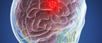
A brain tumor is a type of oncology that occurs most often in children and older people. The neoplasm can be localized in different parts and have different definitions (medical names). In order to understand how advanced the process is, it was decided to create a unified international classification that would allow, by reading the letter values, to understand what we are talking about.
We are talking about the TNM system, where the values correspond to the following notations:
- T – stands for tumor or tumor in Russian. If there is this letter, it means that the doctor wants to write about the stage of the disease, therefore, the size of the tumor, as well as the advanced stage of the malignant process.
- T1 - this value may indicate that in case of cancer of the subcerebellar region the tumor has reached a size of up to 3 cm, and in case of supracerebellar cancer - up to 5 cm.
- T2 - this stage indicates that the malignant tumors have exceeded the permissible size of 3 cm and 5 cm; if the tumor has not gone beyond the limits, then even with sizes over 7 cm it will be classified as T2.
- T3 – at this stage, the tumor extends beyond the original zone and grows into the ventricles and begins to put pressure on neighboring areas, which directly affects the symptoms.
- T4 – the formation greatly increases and spreads to the second part of the brain.
- N – this letter has the meaning nodes, which means nodes. We are talking about the lymph nodes involved in the oncological process.
- N0 – no atypical cells are observed in the lymph nodes.
- N1 – at this stage, those lymph nodes that are located near the tumor (in the cervical area, for example) are affected.
- N2 – cancer cells are found even in distant lymph nodes.
- M – metastasis or metastases.
- M0 – no metastases.
- M1 – metastatic process noticed.
Considering that we are talking about the brain, doctors do not particularly evaluate the significance of the condition of the lymph nodes and metastases. The reason is that the skull has certain dimensions beyond which the tumor cannot extend. Therefore, special attention is paid to exactly what zone the formation is located in, whether it is one or several, as well as how much it has managed to grow.

Cancer treatment in Israel is selected based on all the nuances described above. This is important, since the task of doctors is not only to eliminate the formation, but not to harm the body. Doing this is sometimes problematic, because even at the initial stage the tumor can affect important areas responsible for speech or motor activity. Any wrong action will result in disability. Knowing this, doctors are developing a whole range of measures aimed at stopping the growth of a malignant tumor, but at the same time preserving a normal and fulfilling human life. Thanks to this, the medicine of this country is famous, which shows better results every year.
Short description
Brain tumors are a heterogeneous group of neoplasms for which a common feature is location or secondary penetration into the cranial cavity. Histogenesis varies and is reflected in the WHO histological classification (see below). There are 9 main types of tumors of the central nervous system • A: neuroepithelial tumors • B: tumors of the membranes • C: tumors of the cranial and spinal nerves • D: tumors of the hematopoietic series • E: germinal cell tumors • F: cysts and tumor-like formations • G: tumors of the region sella turcica • H: local spread of tumors from adjacent anatomical regions • I: Metastatic tumors.
Code according to the international classification of diseases ICD-10:
- C71 Malignant neoplasm of the brain
- D33 Benign neoplasm of the brain and other parts of the central nervous system
Epidemiology. Given the heterogeneity of the concept of “brain tumor,” precise generalized statistical data are not available. It is known that central nervous system tumors in children occupy the second place among all malignant neoplasms (after leukemia) and the first place in the group of solid tumors.
Classification. The main working classification used to develop treatment tactics and determine prognosis is the WHO Classification for tumors of the central nervous system • Tumors of neuroepithelial tissue •• Astrocytic tumors: astrocytoma (fibrillary, protoplasmic, gemistocytic [mast cell], or large cell), anaplastic (malignant) astrocytoma, glioblastoma (giant cell glioblastoma and gliosarcoma), pilocytic astrocytoma, pleomorphic xanthoastrocytoma, subependymal giant cell astrocytoma (tuberous sclerosis) •• Oligodendroglial tumors (oligodendroglioma, anaplastic [malignant] oligodendroglioma) •• Ependymal tumors: ependymoma (cellular, with papillary, clear cell), anaplastic (malignant ) ependymoma, myxopapillary ependymoma, subependymoma •• Mixed gliomas: oligoastrocytoma, anaplastic (malignant) oligoastrocytoma, etc. •• Choroid plexus tumors: papilloma and choroid plexus cancer •• Neuroepithelial tumors of unknown origin: astroblastoma, polar spongioblastoma, glioma brain omatosis •• Neuronal and mixed neuronal glial tumors: gangliocytoma, dysplastic gangliocytoma of the cerebellum (Lhermitte Duclos), desmoplastic ganglioglioma in children (infantile), dysembryoplastic neuroepithelial tumor, ganglioglioma, anaplastic (malignant) ganglioglioma, central neurocytoma, paraganglioma of the filum terminale, olfactory neuroblast oma (esthesioneuroblastoma), variant : olfactory neuroepithelioma •• Parenchymal tumors of the pineal gland: pineocytoma, pineoblastoma, mixed/transitional tumors of the pineal gland •• Embryonic tumors: medulloepithelioma, neuroblastoma (option: ganglioneuroblastoma), ependymoblastoma, primitive neuroectodermal tumors (medulloblastoma [option: desmoplastic honey Ulloblastoma], medullomyoblastoma, melanin-containing medulloblastoma) • Tumors of the cranial and spinal nerves •• Schwannoma (neurilemoma, neuroma); options: cellular, plexiform, melanin-containing •• Neurofibroma (neurofibroma): limited (solitary), plexiform (mesh) •• Malignant tumor of the peripheral nerve trunk (neurogenic sarcoma, anaplastic neurofibroma, “malignant schwannoma”); variants: epithelioid, malignant tumor of the peripheral nerve trunk with divergence of mesenchymal and/or epithelial differentiation, melanin-containing • Tumors of the meninges •• Tumors of meningothelial cells: meningioma (meningothelial, fibrous [fibroblastic], transitional [mixed], psammomatous, angiomatous, microcystic, secretory, clear cell, chordoid, rich in lymphoplasmacytic cells, metaplastic), atypical meningioma, papillary meningioma, anaplastic (malignant) meningioma •• Mesenchymal non-meningothelial tumors: benign (osteochondral tumors, lipoma, fibrous histiocytoma, etc.) and malignant (heme angiopericytoma, chondrosarcoma [ option: mesenchymal chondrosarcoma] malignant fibrous histiocytoma, rhabdomyosarcoma, meningeal sarcomatosis, etc.) tumors •• Primary melanocytic lesions: diffuse melanosis, melanocytoma, malignant melanoma (option: meningeal melanomatosis) •• Tumors of unknown histogenesis: hemangioblastoma ( capillary hemangioblastoma) • Lymphomas and tumors of hematopoietic tissue •• Malignant lymphomas •• Plasmacytoma •• Granulocellular sarcoma •• Others • Tumors of germ cells (germ cell) •• Germinoma •• Embryonic carcinoma •• Yolk sac tumor (endodermal sinus tumor) •• Choriocarcinoma •• Teratoma: immature, mature, teratoma with malignancy •• Mixed germ cell tumors • Cysts and tumor-like lesions •• Rathke's pouch cyst •• Epidermoid cyst •• Dermoid cyst •• Colloid cyst of the third ventricle •• Enterogenous cyst •• Neuroglial cyst •• Granular cell tumor ( choristoma, pituicytoma) •• Neuronal hamartoma of the hypothalamus •• Nasal glial heterotopia •• Plasmacytic granuloma • Tumors of the sella turcica •• Pituitary adenoma •• Pituitary cancer •• Craniopharyngioma: adamantinoma-like, papillary • Tumors growing into the cranial cavity •• Paraganglioma (chemodectoma) •• Chordoma •• Chondroma •• Chondrosarcoma •• Cancer • Metastatic tumors • Unclassified tumors
Symptoms (signs)
Clinical picture. The most common symptoms of brain tumors are progressive neurological deficit (68%), headaches (50%), and seizures (26%). The clinical picture mainly depends on the location of the tumor and, to a lesser extent, on its histological characteristics • Supratentorial hemispheric tumors •• Signs of increased ICP due to mass effect and edema (headaches, congestive optic discs, disturbances of consciousness) •• Epileptiform seizures • • Focal neurological deficit (depending on location) •• Personality changes (most typical for frontal lobe tumors) • Midline supratentorial tumors •• Hydrocephalic syndrome (headache, nausea/vomiting, disturbances of consciousness, Parinaud's syndrome, congestive optic discs) •• Diencephalic disorders (obesity/emaciation, thermoregulation disorders, diabetes insipidus) •• Visual and endocrine disorders in tumors of the chiasmatic-sellar region • Subtentorial tumors •• Hydrocephalic syndrome (headache, nausea/vomiting, disturbances of consciousness, congestive optic discs) •• Cerebellar disorders •• Diplopia, severe nystagmus, dizziness •• Isolated vomiting as a sign of an effect on the medulla oblongata • Tumors of the base of the skull •• Often asymptomatic for a long time and only in the later stages cause neuropathy of the cranial nerves, conduction disorders (hemiparesis, hemihypesthesia) and hydrocephalus.
Reasons for the appearance of the tumor
A single cause for the development of cancer has not yet been identified, although active searches are being conducted in this direction. For now, the multifactor theory prevails. It states that several factors can simultaneously take part in the occurrence of a tumor. Most often this is:
- genetic predisposition (if close relatives had cancer).
- belonging to the age category (usually over forty-five years, with the exception of medulloblastoma).
- exposure to harmful production factors, especially chemicals.
- exposure to radiation.
- race (oncological diseases are more common in people belonging to the Caucasian race, with the exception of meningioma, which is typical for Negroids).
Diagnostics
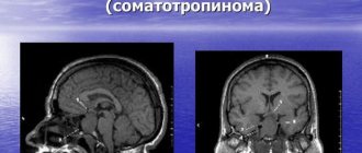
Diagnostics. Using CT and/or MRI at the preoperative stage, it is possible to confirm the diagnosis of a brain tumor, its exact location and extent, as well as the presumptive histological structure. For tumors of the posterior cranial fossa and base of the skull, MRI is more preferable due to the absence of artifacts from the bones of the base (the so-called beam-harding artifacts). Angiography (both direct and MR and CT angiography) is performed in rare cases to clarify the characteristics of the blood supply to the tumor.
Classification of brain tumors by location.
In relation to brain tissue.
- Intracerebral (intracerebral) brain tumors are located inside the brain tissue, from which they do not have a clear boundary. As these tumors grow, they replace and destroy the brain matter. These include glial and metastatic tumors. In addition, you need to understand that all intracerebral tumors are malignant, but have varying degrees of malignancy. In adults, intracerebral tumors occur in approximately 50% of cases, in children - in 80-90%.
- Extracerebral (extracerebral) brain tumors are located outside the medulla. As they grow, they compress and push away brain tissue. These tumors include tumors of the meninges and cranial nerves. They are often benign.
In relation to the tentorium of the cerebellum.
- Supratentorial brain tumors are localized above the tentorium of the cerebellum, occurring in adults in 65-70% of cases, in children - in 20-45% of cases.
- Subtentorial brain tumors are located in the posterior cranial fossa under the tentorium of the cerebellum and are more common in children - up to 80% of cases.
Treatment
Treatment . The treatment strategy depends on the exact histological diagnosis, the following options are possible: • observation • surgical resection • resection in combination with radiation and/or chemotherapy • biopsy (usually stereotactic) in combination with radiation and/or chemotherapy • biopsy and observation • radiation and/or chemotherapy without tissue verification based on CT/MRI results and tumor marker studies.
The prognosis depends mainly on the histological structure of the tumor. Without exception, all patients operated on for brain tumors require regular MRI/CT control studies due to the risk of relapse or continued tumor growth (even in cases of radically removed benign tumors).
ICD-10 • C71 Malignant neoplasm of the brain • D33 Benign neoplasm of the brain and other parts of the central nervous system
Diagnostic methods
A neurological examination is very important, as it provides information about the dysfunction of the brain and data on the location of the lesion. A neurologist studies:
- muscle strength of the limbs
- reflexes, including knee, palm, Achilles
- quality of hearing and vision
- skin sensitivity
- ability to maintain balance, coordination
- memory and mental abilities are tested using questionnaires.
If any pathological signs are detected, instrumental examinations are carried out:
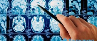
- Computed tomography (CT) of the brain. In this study, a detailed picture of the brain is obtained in the form of multiple x-rays.
- Magnetic resonance imaging (MRI), which uses a strong magnetic field and radio waves.
- Electroencephalography (EEG). The method records the electrical activity of brain cells.
- Biopsy, i.e. taking a small fragment to examine under a microscope. The manipulation is carried out in stationary conditions. After anesthesia, a small hole is made in the skull bone and a tissue sample is taken with a thin needle to determine the type of tumor.