Brain: structure and functions, general description
The brain is the main regulator of all functions of a living organism. It is one of the elements of the central nervous system. The structure and functions of the brain are still the subject of study by doctors.
general description
The human brain consists of 25 billion neurons. These cells are the gray matter. The brain is covered with membranes:
- hard;
- soft;
- arachnoid (the so-called cerebrospinal fluid, which is cerebrospinal fluid, circulates through its channels). Liquor is a shock absorber that protects the brain from shock.
Despite the fact that the brains of women and men are equally developed, they have different masses. So, among representatives of the stronger sex, its weight is on average 1375 g, and among women - 1245 g. The weight of the brain is about 2% of the weight of a person of normal build. It has been established that the level of a person’s mental development is in no way related to his weight. It depends on the number of connections created by the brain.
Brain cells are neurons that generate and transmit impulses and glia that perform additional functions. Inside the brain there are cavities called ventricles. Paired cranial nerves (12 pairs) depart from it to different parts of the body. The functions of the parts of the brain are very different. The vital functions of the body completely depend on them.
Structure
The structure of the brain, pictures of which are presented below, can be considered in several aspects. So there are 5 main parts of the brain:
- final (80% of the total mass);
- intermediate;
- posterior (cerebellum and pons);
- average;
- oblong.
The brain is also divided into 3 parts:
- cerebral hemispheres;
- brain stem;
- cerebellum.
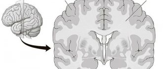
Structure of the brain: drawing with the names of the departments.
Structure of the brain: names of departments
Finite brain
The structure of the brain cannot be briefly described, since without studying its structure it is impossible to understand its functions. The telencephalon extends from the occipital to the frontal bone.
It distinguishes 2 large hemispheres: left and right. It differs from other parts of the brain by the presence of a large number of convolutions and grooves. The structure and development of the brain are closely interrelated.
Experts distinguish 3 types of cerebral cortex:
- ancient, which includes the olfactory tubercle; perforated anterior substance; semilunar, subcallosal and lateral subcallosal gyri;
- old, which includes the hippocambus and dentate gyrus (fascia);
- new, represented by the rest of the cortex.
The structure of the cerebral hemispheres: they are separated by a longitudinal groove, in the depths of which the fornix and the corpus callosum are located. They connect the hemispheres of the brain. The corpus callosum is a new cortex made up of nerve fibers. There is a vault underneath it.
The structure of the cerebral hemispheres is presented as a multi-level system. So they distinguish between lobes (parietal, frontal, occipital, temporal), cortex and subcortex. The cerebral hemispheres perform many functions. The right hemisphere controls the left half of the body, and the left hemisphere controls the right. They complement each other.
Bark
The cerebral cortex is a 3 mm thick superficial layer covering the hemispheres. It consists of vertically oriented nerve cells with processes. It also contains afferent and efferent nerve fibers, neuroglia.
What is the cerebral cortex? This is a complex structure with horizontal layering.
The structure of the cerebral cortex: it has 6 layers (outer granular, molecular, outer pyramidal, inner granular, inner pyramidal, spindle cells), which have different density, width, size and shape of neurons.
Due to the vertical bundles of nerve fibers, neurons and their processes present in the cortex, it has vertical striations. The human cerebral cortex, which contains more than 10 billion neurons, has an area of about 2200 sq.cm.
The cerebral cortex is responsible for several specific functions. Moreover, each of its parts is responsible for something of its own. Functions of the cerebral cortex:
- temporal lobe – hearing and smell;
- occipital – vision;
- parietal – touch and taste;
- frontal – speech, movement, complex thinking.
Each neuron (gray matter) has up to 10 thousand contacts with other neurons. The white matter of the brain is made up of nerve fibers. A certain part of them connects both hemispheres. The white matter of the cerebral hemispheres consists of 3 types of fibers:
- association (connecting different cortical areas in one hemisphere);
- commissural (connecting the hemispheres);
- projection (conducting pathways of analyzers that connect the cerebral cortex with the underlying formations). Inside the hemispheres of the brain there are accumulations of gray matter (basal ganglia). Their function is to transmit information. The white matter of the human brain occupies the space between the basal ganglia and the cerebral cortex. There are 4 parts in it (depending on its location):
- located in the convolutions between the furrows;
- present in the outer parts of the hemispheres;
- part of the internal capsule;
- located in the corpus callosum.
The white matter of the brain is formed by nerve fibers that connect the gyral cortex of both hemispheres and the underlying formations. The subcortex of the brain consists of subcortical nuclei. The telencephalon controls all processes important for human life and our intellectual abilities.
Diencephalon
It consists of a ventral (hypothalamus) and dorsal (metathalamus, thalamus, epithalamus) part. The thalamus is a mediator in which all received stimuli are sent to the cerebral hemispheres. It is often called the optic thalamus. Thanks to it, the body quickly adequately adapts to a changing external environment. The thalamus is connected to the cerebellum by the limbic system.
The hypothalamus is a subcortical center in which the regulation of autonomic functions occurs. Its influence occurs through the endocrine glands and the nervous system.
It is involved in the regulation of the functioning of some endocrine glands and metabolism. Below it is the pituitary gland. Thanks to it, body temperature, digestive and cardiovascular systems are regulated.
The hypothalamus regulates wakefulness and sleep, shapes drinking and eating behavior.
hindbrain
This section consists of the pons located in front and the cerebellum located behind it. The structure of the cerebral pons: its dorsal surface is covered by the cerebellum, and its ventral surface has a fibrous structure. These fibers are directed transversely. On each side of the bridge they pass into the cerebellar middle peduncle. The bridge itself looks like a white thick roller.
It is located above the medulla oblongata. The nerve roots emerge from the bulbar-pontine groove. Hindbrain: structure and functions - on the frontal section of the bridge, it is noticeable that it consists of a large ventral (anterior) and a small dorsal (posterior) part. The border between them is the trapezoidal body. Its thick transverse fibers belong to the auditory tract.
The hindbrain provides the conductive function.
The cerebellum, often called the cerebrum, is located posterior to the pons. It covers the rhomboid fossa and occupies almost the entire posterior fossa of the skull. Its mass is 120-150 g. The cerebral hemispheres hang above the cerebellum, separated from it by a transverse fissure of the brain. The inferior surface of the cerebellum is adjacent to the medulla oblongata.
It distinguishes 2 hemispheres, as well as the upper and lower surfaces and the worm. The boundary between them is called a deep horizontal gap. The surface of the cerebellum is cut by many slits, between which there are thin ridges (gyri) of the medulla.
The groups of gyri located between the deep grooves are lobules, which, in turn, make up the lobes of the cerebellum (anterior, flocnonodular, posterior).
There are 2 types of substance in the cerebellum. Gray is on the periphery. It forms the cortex, which contains the molecular, pyriform neurons and granular layer. The white matter of the brain is always located under the cortex. Likewise, in the cerebellum it forms the brain body.
It penetrates into all convolutions in the form of white stripes covered with gray matter. The white matter of the cerebellum itself contains interspersed gray matter (nuclei). In cross-section, their relationship resembles a tree. Our coordination of movement depends on the functioning of the cerebellum.
Midbrain
This section extends from the anterior edge of the pons to the papillary bodies and optic tracts. It contains a cluster of nuclei, which are called quadrigeminal tubercles. The midbrain is responsible for hidden vision. It also contains the center of the orienting reflex, which ensures the body turns in the direction of a sharp noise.
Medulla
It is a continuation of the spinal cord. The structure of the brain and spinal cord have much in common. This becomes clear upon a detailed examination of the white matter of the medulla oblongata.
The white matter of the brain is represented by long and short nerve fibers. Gray matter is presented in the form of nuclei. This brain is responsible for coordination of movement, balance, regulation of metabolism, blood circulation and breathing.
It is also responsible for coughing and sneezing.
The structure of the brainstem: it is a continuation of the spinal cord, divided into the midbrain and hindbrain. The trunk is called the medulla oblongata, midbrain, diencephalon and pons. The structure of the brain stem consists of ascending and descending pathways that connect it with the brain and spinal cord. It controls articulate speech, breathing and heartbeat.
(55 3,49 of 5) Loading...
Source: https://golmozg.ru/stroenie/golovnoj-mozg-stroenie-i-funkcii-obshhee-opisanie.html
content .. 51 52 55 ..Human brain (anatomy)
The brain is located in the cranial cavity and consists of five sections: medulla oblongata, hindbrain, midbrain, diencephalon and telencephalon.
A number of authors divide the brain into the stem part and the cerebral hemispheres. The stem part includes parts of the brain that are more ancient in origin - the medulla oblongata, posterior, middle and intermediate. The cerebral hemispheres are newer formations in the evolution of the central nervous system. They are most developed in humans.
The weight of the human brain ranges from 1000 to 2200 g, in men it weighs on average 1375 g, and in women - 1245 g. There is no connection between brain weight and mental abilities. For example, Byron’s brain weight was 2238 g, that of I. S. Turgenev was 2012, and that of A. France was 1017 g.
The human brain has a specific structure and differs from the brain of animals in the predominance of the cerebrum over the spinal cord, and the cerebral hemispheres over the stem part, the highest development of the frontal lobes, sulci and gyri, the large relative weight of the brain (to body weight), the presence of a second signaling system, manifested in form of thinking.
Medulla.
The medulla oblongata is a relatively small but important part of the brain. On its front surface, on the sides of the anterior median fissure, longitudinal thickenings are visible - pyramids, which consist of motor - pyramidal (corticospinal) tracts connecting the brain with the spinal cord (Fig. 102 and 103).
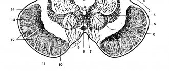
Rice. 102. Brain stem (bottom view): 1 - lateral cord of the spinal cord; 2 - anterior cord; 3, 4 - intersection of pyramidal paths; 5 - olive; 6 - pyramid; 7 - abducens nerve; 8 - middle cerebellar peduncle; 9 - bridge; 10 - trigeminal nerve; 11 - cerebral peduncle; 12 - optic tract; 13 - mastoid body; 14 — gray tubercle with a funnel (cut); 15 - olfactory pathway; 16 - olfactory triangle; 17 - trigeminal node; 18 - motor root of the trigeminal nerve; 19 - facial nerve; 20 - intermediate nerve; 21 - vestibulocochlear nerve; 22 - glossopharyngeal nerve; 23 - vagus nerve; 24 - hypoglossal nerve; 25 - accessory nerve; 26 - anterior root of the first cervical nerve
Rice. 103. Brain stem (top view): 1 - bottom of the fourth ventricle (diamond-shaped fossa); 2 - superior cerebellar peduncles; 3 - average; 4 - lower; 5 - posterior longitudinal groove; 6 - tubercle of the sphenoid nucleus; 7 - tubercle gonogo, nucleus; 8 - rope body; 9 - wedge-shaped bundle; 10 - thin beam; 11 - trochlear nerve; 12 - lower colliculi; 13 - medial geniculate body; 14 - superior colliculi; 15 - pineal body; 16 - brain stripes; 17 - visual thalamus
On the sides of the pyramids there are oval elevations - olives, the function of which is related to maintaining the body in an upright position. On the posterior surface of the medulla oblongata there is a posterior median sulcus, which is a continuation of the sulcus of the same name in the spinal cord. To the side of it lie thin and wedge-shaped tufts. Rising upward, they form thickenings - the tubercle of the thin nucleus and the tubercle of the sphenoid nucleus (the location of the second neurons of the sensitive proprioceptive pathways of the thin and cuneate fasciculi).
The thin and wedge-shaped fasciculi on each side of the medulla oblongata connect to the lateral funiculus to form the inferior cerebellar peduncle, which runs to the cerebellum. The depression located between the inferior cerebellar peduncles forms the lower part of the rhomboid fossa, which is the floor of the fourth ventricle of the brain.
The fourth ventricle of the brain is a residual cavity of the rhombencephalon.
In the area of the rhomboid fossa lie the nuclei of the cranial nerves (from the V to the XII pairs), surrounded by the reticular substance. The roof of the fourth ventricle is made up of thin sheets of white matter running from the cerebellum to the roof of the midbrain (superior medullary velum) and to the medulla oblongata (inferior medullary velum). The fourth ventricle communicates with the midbrain aqueduct, the central canal and the subarachnoid space of the spinal cord.
The medulla oblongata consists of gray and white matter. The gray matter is not butterfly shaped as in the spinal cord, but forms clusters called nuclei or centra. In the region of the medulla oblongata there is an automatically functioning respiratory center, centers that regulate the functioning of the heart and blood vessels, the secretion of digestive juices, reflexes of sneezing, coughing, swallowing, etc., as well as the nuclei of the cranial nerves from the IX to the XII pairs.
Of great importance is the powerfully developed reticular (mesh) substance, consisting of small branched cells that connect various neurons of the medulla oblongata.
The white matter of the medulla oblongata consists of ascending (sensory) and descending (motor) pathways, as well as its own nerve fibers. Thus, the medulla oblongata, like the spinal cord, performs reflex and conduction functions.
Hindbrain.
The medulla oblongata and hindbrain have a common cavity - the fourth ventricle of the brain. The hindbrain includes the pons and cerebellum. The pons is located in front of the medulla oblongata. Its anterior surface is convex, and its posterior surface, forming the upper part of the diamond-shaped fossa, is flat. The ridges extending to the sides of the bridge go to the cerebellum - the middle cerebellar peduncles. The pons, like the medulla oblongata, consists of gray and white matter. The gray matter has the pontine nuclei, the nuclei of the cranial nerves from pairs V to VIII, lying in the upper part of the rhomboid fossa, and the reticular reticular substance, which is a continuation of the formation of the same name in the medulla oblongata. Through the pontine nuclei, the cerebral cortex connects to the cerebellum. The white matter consists of fibers running in the longitudinal and transverse directions. In the longitudinal direction there are ascending (sensitive) and descending (motor) pathways. Transverse fibers connect the pons to the cerebellum.
Cerebellum
adjoins posteriorly to the flat surfaces of the pons and medulla oblongata. It has two hemispheres and a middle part called the vermis. In the cerebellum, two surfaces are distinguished: the upper, somewhat flattened, to which the occipital lobes of the telencephalon hemispheres are adjacent, and the lower, convex. The surfaces of the cerebellum are divided by transverse grooves into small convolutions and lobules. The cerebellum has three pairs of peduncles - superior, middle and inferior.
Inferior cerebellar peduncles
The cerebellum connects to the medulla oblongata, and they contain unconscious proprioceptive pathways from the spinal cord to the cerebellum.
The middle cerebellar peduncles
connect the cerebellum to the pons and consist of fibers that connect the pontine nuclei to the cerebellar cortex.
The pontine nuclei, in turn, are connected to the cerebral cortex, so there is constant control of the cerebral cortex over the functions of the cerebellum. The superior cerebellar peduncles
connect the cerebellum to the roof of the midbrain, in particular to the red nucleus. They also include proprioceptive spinal cerebellar tracts.
Numerous fibers connecting the cerebellum with the spinal cord and parts of the brain form its white matter. The gray matter in the cerebellum is located both on the periphery, forming the cerebellar cortex, and inside the white matter, forming the cerebellar nuclei, of which the largest is the dentate.
If you make a median (sagittal) section of the cerebellum through the vermis, you will see a peculiar distribution of gray and white matter in it, reminiscent of a branching tree and called the “tree of life.”
The functions of the cerebellum are very diverse and represent a single continuous automatic regulatory function, very complex and at the same time precise. The cerebellum receives information about the state of all muscles, the position of the head and, in the case of its rotational movement, its speed. Having received input information, the cerebellum influences the red nucleus of the midbrain and the reticular formation of the brainstem, which, together with the nuclei of the base of the brain, directly regulate muscle tone and ensure coordination of movements.
The cerebellum is also involved in the regulation of autonomic functions, influencing the functioning of the gastrointestinal tract, blood composition, etc.
Midbrain.
The midbrain is formed by the roof of the midbrain, the cerebral peduncles and the narrow cavity between them - the aqueduct.
Roof of the midbrain
consists of four mounds - two upper and two lower, located on the back side of the trunk, above the rhomboid fossa. The superior colliculi contain the subcortical centers of vision, and the inferior colliculi contain the subcortical centers of hearing.
The subcortical centers of vision receive irritations from the retina of the eye, in response to this the size of the pupil is regulated and the eye is accommodated. Impulses from the nuclei of the auditory nerves arrive at the subcortical hearing centers, and in response, indicative auditory reflexes are carried out.
Thus, the nuclei of the midbrain roof provide a “guard” reflex, which prepares the body to react to a new sudden irritation. A person with impairments in this area of the brain is not capable of a quick response to unexpected stimulation.
Brain stems
They are two rollers connecting the bridge with the cerebral hemispheres. A cross-section of the legs shows that they are divided by a black (mesh) substance into a tire and a base. The tegmentum is located directly under the roof of the midbrain and contains the nuclei of the III and IV pairs of cerebral nerves, as well as the red nucleus (subcortical motor center). The red nuclei are approached by nerve pathways from the cerebral cortex, from the subcortical nuclei, and from the cerebellum. As already mentioned, the red nuclei play an important role in regulating skeletal muscle tone. The reflex motor pathway begins from the red nuclei, along which impulses are transmitted to the motor cells (motoneurons) of the spinal cord, and from them to the muscles of the body. At the base of the cerebral peduncles there are fibers of the conductive pathways - sensory and motor.
Water pipes
is the residual cavity of the midbrain. It connects the fourth ventricle of the brain with the third ventricle - the cavity of the diencephalon.
Diencephalon.
The diencephalon includes the optic thalamus, supratuberous, subtubercular, subtubercular areas and the third ventricle of the brain.
Optic thalamus
It is a rather large ovoid-shaped formation lying above the roof of the midbrain. The anterior end of the visual thalamus is narrowed, the posterior end is widened and forms a cushion of the visual thalamus. Its upper surface faces the cavity of the lateral ventricle, and the inner surface faces the cavity of the third ventricle. The visual thalamus consists mainly of gray matter, which is grouped into nuclei - lateral, medial and anterior, separated from each other by thin layers of white matter. The fibers of all sensitive pathways going to the cerebral cortex approach the nuclei of the visual thalamus. Thus, sensory impulses, before reaching the cortex, must certainly reach the visual hillocks. This is like a gate on the way to the cortex, through which all information passes from receptors that perceive irritations from the external and internal environment.
To the supra-tuberculous region
refers to the pineal gland (see organs of internal secretion), or pineal body, a small, unpaired triangular formation located above the upper colliculi of the roof of the midbrain.
Foreign region
represented by the lateral and medial geniculate bodies, connecting with the superior and inferior tubercles of the roof of the midbrain.
To the sub-mountain region
includes formations: optic chiasm, optic tract, gray tubercle with infundibulum and pituitary gland, mastoid bodies. All these formations lie directly under the visual thalamus, forming the floor of the third ventricle of the brain. They are clearly visible on preparations of the base and sagittal section of the brain.
The optic chiasm and the optic tract following the chiasm consist of fibers (conducting pathways) coming from the retina of the eye.
The gray hillock contains vegetative centers that regulate fat, carbohydrate and water-salt metabolism, as well as heat metabolism.
The pituitary gland, connected to the gray tubercle by a funnel, belongs, like the pineal gland, to the organs of internal secretion.
The mammillary bodies, located behind the gray tuberosity, consist of cells that form the subcortical centers of smell.
Third ventricle of the brain
- This is the cavity of the diencephalon, located between the visual thalamus. The bottom of the third ventricle is the subtubercular region, and the roof is the choroid plexus of the third ventricle and the corpus callosum. In front, the third ventricle communicates through the interventricular foramina with the lateral ventricles of the telencephalon, and behind, through the midbrain aqueduct, with the fourth ventricle of the rhombencephalon.
The functions of the brainstem are associated with the reticular (mesh) formation of the brain. It is located along the entire length of the brain stem - in the medulla oblongata, hindbrain, midbrain and diencephalon, occupying a central position there, and consists of numerous neurons, the bodies of which have different sizes, from small to giant, and different shapes - fusiform, oval, round . Cell dendrites branch weakly, and neurites are divided into ascending and descending branches, which, in turn, have branches. The number of contacts of one cell of the reticular formation with other cells can reach 30,000. Since the processes of the cells go in different directions, they create the impression of a network, which was the reason for the name of this substance. The neurons of the reticular formation are scattered in some places and form nuclei in some places (96 nuclei are currently described).
The reticular formation is connected to a number of parts of the brain. These connections can be divided into three groups: 1) going from the reticular formation to the spinal cord, to the nuclei of the cranial nerves, to the cerebellum, to the cerebral cortex; 2) going to the reticular formation from all sensory and motor conductors of the brain stem, from the cerebellum, from the autonomic subcortical centers, from the cerebral cortex; 3) connections between the various nuclei of the reticular formation itself.
There are two types of influence of the reticular formation on the nerve centers: activating and inhibitory. Both influences can spread in an ascending direction - to the cerebral cortex and in a descending direction - to the spinal cord.
The ascending activating influence covers the entire cortex if the impulses come from the reticular formation of the midbrain, or individual areas of the cortex if the impulses come from the reticular formation of the diencephalon. This influence ensures the normal activity of the cerebral cortex - the state of wakefulness, the alternation of wakefulness and sleep, etc.
The inhibitory effect of the reticular formation on the cortex is reflected in the formation of more and less active sections of it or in the diffuse inhibition of cortical centers, which sometimes occurs when performing monotonous work for a long time.
The descending influence of the reticular formation on the neurons of the spinal cord can be activating and suppressive and is expressed, respectively, in an increase or decrease in the excitability of motor neurons. The complex relationships and feedbacks between spinal cord neurons and skeletal muscles are also regulated by the reticular formation, determining muscle tone (with the participation of the cerebellum, the red nuclei of the midbrain and the nuclei of the base of the hemispheres).
The reticular formation also plays an important role in the processes of adaptation of the human body, maintaining spatial relationships between parts of the body.
However, it should be noted that the reticular formation, influencing the cerebral cortex and maintaining the level of its activity, is itself under the constant regulatory influence of impulses from the cortex.
Finite brain.
The telencephalon consists of two hemispheres connected by a commissure - the corpus callosum. Between the hemispheres there is a deep longitudinal fissure of the cerebrum. Between the posterior parts of the hemispheres and the cerebellum there is a transverse fissure of the cerebrum. Each hemisphere has three surfaces: superior-lateral, medial and inferior, and three most prominent parts, or three poles: frontal, occipital and temporal. In addition, the following parts are distinguished in each hemisphere: the cloak, the olfactory brain, the nuclei of the base of the brain and the lateral ventricle.
The telencephalon is made up of gray and white matter. Gray matter is located on the outside, forming the mantle, or cortex of the cerebral hemispheres, followed by white matter, at the base of which lie clusters of gray matter - the nuclei of the base of the brain.
Before talking about the structure and functional significance of the cortex, it is advisable to consider its relief, that is, the grooves and convolutions on the surfaces of the hemisphere.
On the superior lateral surface of the hemisphere there are three main sulci: central, lateral and parieto-occipital, which divide the hemisphere into four main lobes: frontal, parietal, occipital and temporal. In the depths of the lateral sulcus, if you move it apart, you can see an additional, fifth, lobe of the brain, called the insula.
Each lobe of the brain has convolutions separated from each other by grooves.
Frontal lobe
located in front of the central sulcus and has three grooves: the precentral, lying in front of the central sulcus, the superior and inferior frontal. Between the grooves there are four gyri: precentral, superior, middle and inferior frontal (Fig. 104, see color insert).
Rice. 104. Furrows and convolutions of the cerebral hemispheres. A. Superolateral surface of the hemisphere: 1 - central sulcus; 2 - lateral groove; 3 - precentral sulcus; 4 - postcentral sulcus; 5 - superior frontal sulcus; 6 - inferior frontal sulcus; 7 - intraparietal sulcus; 8 - parieto-occipital groove; 9 - superior temporal sulcus; 10 - inferior temporal sulcus; 11 - precentral gyrus; 12 - superior frontal gyrus; 13 - middle frontal gyrus; 14 - inferior frontal gyrus; 15 - postcentral gyrus; 16—superior parietal lobule; 17 - inferior parietal lobule; 18-middle temporal gyrus; 19 - superior temporal gyrus; 20 - inferior temporal gyrus; 21 - cerebellum; 22 - medulla oblongata. B. Medial and lower surfaces of the hemisphere: 1 - corpus callosum; 2 - groove of the corpus callosum; 3 - cingulate groove; 4 - parieto-occipital groove; 5 - calcarine groove; 6 - collateral groove; 7 - hippocampal sulcus; 8 - precuneus; 9 - wedge; 10 - inferior temporal gyrus; 11 - parahippocampal gyrus; 12 - lateral groove; 13 - cingulate gyrus; 14 - beak of the corpus callosum; 15 - brain stem (cut)
Parietal lobe
located behind the central sulcus. It has two main sulci: the postcentral sulcus, which runs behind the central sulcus, and the intraparietal sulcus, which extends transversely from the posterior central sulcus. These two sulci divide the parietal lobe into the postcentral gyrus and two lobules: the superior parietal lobule and the inferior parietal lobule.
Occipital lobe
is located behind the parieto-occipital sulcus and has very variable grooves and convolutions running longitudinally and transversely.
Temporal lobe
lies below the lateral sulcus and two temporal sulci, superior and inferior, and is divided into the superior, middle and inferior gyri. The inferior gyrus is limited from below by the occipitotemporal sulcus, which is located on the border of the lateral and inferior surfaces of the temporal lobe.
Island
has the shape of a triangle. Its surface is covered with short convolutions.
On the medial surface of the hemisphere, above the corpus callosum, which is the commissure connecting the two hemispheres, runs the corpus callosum groove. Above it, maintaining the same direction, there is a cingulate groove. Between them is the cingulate gyrus. On the medial surface of the hemisphere, a groove is clearly visible that separates the parietal lobe from the occipital lobe - the parieto-occipital groove. A calcarine groove extends upward from its lower end at an angle. The area of the brain between these sulci is called the wedge, and the area of the brain lying in front of the parieto-occipital sulcus is called the precuneus (Fig. 105).
Rice. 105. Sagittal section of the brain: 1 - medulla oblongata; 2 - bridge; 3 cerebral peduncle; 4 - visual thalamus; 5 - cover of the midbrain; 6 - pituitary gland; 7 - water supply; 8 - cerebellum; 9 - corpus callosum; 10 - groove of the corpus callosum; 11 cingulate groove; 12 - cingulate gyrus; 13 - parieto-occipital groove; 14 - calcarine groove; 15 - wedge; 16 - precuneus; 17 - fourth ventricle
The inferior surfaces of the frontal, temporal and occipital lobes of the brain are visible on the inferior surface of the hemisphere. On the frontal lobe there are olfactory grooves that run near the longitudinal fissure that separates the hemispheres from each other. In these grooves lie the formations of the peripheral part of the olfactory brain - the olfactory bulbs, the olfactory tract and the olfactory triangle. Between the longitudinal fissure and the olfactory sulcus there is a straight gyrus. The remaining surface of the lower part of the frontal lobe is occupied by the orbital sulci and gyri.
On the lower surface of the temporal lobe runs the already mentioned occipitotemporal sulcus, and inward from it are the collateral sulcus and the hippocampal sulcus. Between the occipitotemporal and collateral sulci is the lateral occipitotemporal gyrus (fusiform), and between the collateral sulcus and the hippocampal sulcus is the parahippocampal gyrus, which ends in an extension called the uncus. The parahippocampal gyrus, together with the cingulate gyrus, makes up the vaulted gyrus.
The cerebral cortex, with a total area of 220,000 mm2, consists of gray matter (up to 4 mm thick) and contains more than 14 billion nerve cells. All cortical cells are interneurons of reflex arcs. They have different shapes - pyramidal, spindle-shaped, stellate, arachnid, etc. Neurites extending from cells are divided into short and long. Short neurites mainly perform an association function, connecting individual areas of the cortex, and long neurites have a projection function, connecting the cortex with the underlying areas of the brain and the spinal cord.
The cells of the cortex, together with their processes, form six to nine layers of the cortex. Since in the last period of the uterine life of the fetus almost all areas of the cortex have six layers, the initial type of structure of the cortex is considered to be six-layer. However, in some areas of the cortex the number of layers varies, for example, in some areas of the occipital lobe there are nine, and in the olfactory area of the cortex there are only five. Different areas of the cortex differ in their functional significance. One of the first scientists in Russia to pay attention to the structural features of individual sections of the cortex was Professor V.A. Bets (1874), who identified 8 characteristic fields in the human cortex (currently there are more than 200 of them).
A new stage in the study of the cortex was the work of I. P. Pavlov. He showed that the cortex is the material substrate of higher nervous activity and the regulator of all body functions.
Based on the relationship between areas of the cerebral cortex and the outside world, I. P. Pavlov created the doctrine of “analyzers,” that is, about nervous mechanisms that begin with the external perceptive apparatuses and end in the brain. According to this teaching, each “analyzer” consists of three parts: peripheral (receptors, sensory organs), conductive (conducting pathways) and central (cortical end of the analyzer). The peripheral part of the analyzer serves to receive information coming from the external world or the internal environment of the body and transform it into nerve impulses. The conductive part of the analyzer transmits impulses from the receptor to the cerebral cortex. In the central part, information processing, analysis and synthesis take place.
The significance of this concept is that the entire cortex is viewed as an area that receives and processes information.
The most important cortical ends of the analyzer include the following.
1. Cortical end of the motor analyzer, located in the precentral gyrus. This area of the cortex was previously considered only a motor area. It is currently considered to be the region containing interneurons and motor neurons. Interneurons perceive stimuli from bones, joints, muscles and tendons.
Motor neurons lie in the 5th and partially in the 6th layers of the central gyrus. They are giant Betz cells, from which the corticospinal, or pyramidal, tract begins. The fibers of this pathway descend to the motor neurons of the anterior horns of the spinal cord segments. Dysfunction of the precentral gyrus leads to muscle paralysis on the opposite side of the body, since most of the fibers of the pyramidal tract intersect in the medulla oblongata.
2. The cortical end of the skin analyzer, which is located in the postcentral gyrus and perceives pain, temperature and tactile sensitivity. Damage to this analyzer results in loss of sensitivity.
3. Cortical end of the auditory analyzer. It is located in the superior temporal gyrus, in its middle part.
4. The cortical end of the visual analyzer, located along the edges of the calcarine groove, which is located on the inner surface of the occipital lobe of the brain.
5. Cortical ends of the olfactory and gustatory analyzers. They are located in the uncus of the parahippocampal gyrus (on the inferior surface of the temporal lobe).
6. The cortical end of the motor analyzer of complex coordinated movements, located in the inferior parietal lobule.
7. Cortical end of the analyzer for recognizing objects by touch. It is located in the superior parietal lobule.
8. Speech analyzers providing speech function. Their cortical ends occupy four areas of the cortex. The cortical end of the motor speech analyzer is located in the posterior part of the inferior frontal gyrus. The cortical end of the motor writing analyzer is located in the posterior part of the middle frontal gyrus. The cortical end of the reading analyzer lies in the inferior parietal lobule. The cortical end of the auditory speech analyzer is located in the same place as the cortical end of the hearing analyzer - in the superior temporal gyrus. When it is impaired, a person hears speech, but does not understand it.
It should be noted that the function is not localized in one specific field, but is only predominantly associated with it and spreads over a larger area.
All the sensations and ideas that we receive constitute, according to I.P. Pavlov, the first signal system of reality, which we have in common with animals, and speech is the second signal system. The second signaling system is human thinking, which is always verbal.
Speech is the most complexly localized function in which the entire cortex is involved. This function is performed by nerve cells with short processes located in the superficial layers of the cortex. In accordance with the development of new experience, speech functions associated with the mentioned analyzers can move to other areas of the cortex (gesticulation of the deaf and dumb, reading of the blind, writing with the foot of the armless, etc.).
The second signaling system functions in close connection with the first. I. P. Pavlov’s teaching about two cortical systems determines the materialistic understanding of human mental activity and forms the basis of V. I. Lenin’s theory of reflection, according to which the objective real world is reflected in our consciousness, existing independently of our consciousness,
Thus, the cerebral cortex receives information, processes it and stores it (memory apparatus). In the process of adaptation (adaptation) of the body to the external environment, complex systems were formed in the cerebral cortex, such as systems of self-regulation, stabilization, providing a certain level of function, self-learning systems with a memory code, control systems (software control based on genetic code and age, optimal control that ensures the optimal level of functions in the body), comparison systems that determine the transition from one form of control to another.
Olfactory brain
The human brain is divided into two sections: peripheral and central. The peripheral section includes: the olfactory bulb, the olfactory tract, the olfactory triangle and the adjacent anterior perforated substance. The central section includes: the vaulted gyrus, consisting of the cingulate gyrus and the parahippocampal gyrus, as well as the hippocampus, a peculiarly shaped formation located in the cavity of the inferior horn of the lateral ventricle.
The olfactory brain is considered the most ancient (in terms of development) part of the telencephalon, which arose in connection with the olfactory receptor.
Studies have shown that this part of the brain, especially the hippocampus, has extensive connections with the cerebral cortex and the subthalamic region of the diencephalon. The hippocampus is involved in the formation of emotional states. In mentally ill patients, all layers of nerve cells in the hippocampus are affected. The cingulate gyrus takes part in reactions of alertness, awakening, and emotional activity. It is connected by fibers to the reticular formation and the autonomic part of the nervous system.
Nuclei of the base of the brain
They are a collection of gray matter in the white matter at the base of the brain. These include the caudate nucleus, the lentiform nucleus, the cervical nucleus, and the amygdala.
Caudate nucleus
located in the frontal lobe and takes part in the formation of the lateral wall of the anterior horn of the lateral ventricle.
Lenticular nucleus
located outward from the caudate nucleus and is separated from it by a layer of white matter called the internal capsule. The lenticular nucleus is divided into two parts - the outer (putamen) and the inner (globus pallidus).
Caudate nucleus
together with the putamen is called the striatum, and the globus pallidus is separated into an independent morpho-functional unit called the pallidum.
The caudate and shell do not have direct motor function. They perform a controlling function in relation to the globus pallidus, somewhat inhibiting its influence. When these nuclei are damaged, a person experiences rhythmic involuntary movements of the limbs (chorea).
Fence
It is a thin strip of gray matter that lies outward from the shell and is separated from it by a layer of white matter - the outer capsule.
Amygdala
located deep in the temporal lobe, closer to its anterior end.
Of all the listed nuclei, the most ancient in development is the globus pallidus, which performs the functions of the motor nucleus. Thanks to its connection with the red nucleus of the midbrain, such friendly movements are carried out as movements of the torso and arms when walking - cross-coordination, a number of auxiliary movements when changing body positions, facial movements, etc.
The nuclei of the base of the brain function in close relationship with the cerebral cortex, diencephalon and other parts of the brain. They take part in the formation of both conditioned and unconditioned reflexes - defensive, food, etc.
Together with the red nucleus, thalamus opticum and substantia nigra of the midbrain, the nuclei of the base of the brain are called the extrapyramidal system.
Lateral ventricles
are the cavities of the cerebral hemispheres. Each ventricle has a central part adjacent to the upper surface of the optic thalamus, and three horns extending from it - anterior, posterior and inferior. The anterior horn is directed from the central part to the frontal lobe (it is limited from the outside by the caudate nucleus), the posterior one - to the occipital lobe, and the lower one - to the depth of the temporal lobe. In the lower horn, forming its internal and partially lower wall, there is an elevation - the hippocampus - a depression of the hippocampal sulcus.
The lateral ventricles, like the rest of the ventricles of the brain, are filled with cerebrospinal fluid. Through the interventricular foramina, which are located in front of the visual thalamus, the lateral ventricles communicate with the third ventricle of the diencephalon. Most of the walls of the lateral ventricles are formed by the white matter of the cerebral hemispheres. It is located between the cortex and the nuclei of the base of the brain, consists of a large number of nerve fibers belonging to the pathways of the nervous system.
The brain, like the spinal cord, is covered with three membranes - dura mater, arachnoid membrane and vascular membrane.
Dura shell
The brain is different from the dura mater of the spinal cord. It is fused with the inner surface of the skull bones (there is no suprahard space); forms channels for the outflow of venous blood from the brain - the sinuses of the dura mater; gives processes that provide fixation of the brain - the falx cerebri (between the right and left hemispheres of the brain), the tentorium cerebellum (between the occipital lobes of the cerebral hemispheres and the cerebellum) and the diaphragm sellae (above the sella turcica, in which the pituitary gland is located).
Arachnoid
The brain is located under the dura mater of the brain. It covers the brain, not going into its grooves and grooves, but spreading over them in the form of bridges. Between the arachnoid and choroid, a subarachnoid space is formed, which, unlike the same space of the spinal cord, is not evenly expressed.
Its most well-defined areas containing cerebrospinal fluid are called cisterns (for example, cisterns between the cerebellum and medulla oblongata, between the cerebral peduncles, in the region of the lateral sulcus, etc.). The subarachnoid space of the brain communicates with the space of the same name in the spinal cord and the fourth ventricle of the brain.
Choroid
the brain is closely fused with the substance of the brain, entering all the cracks and grooves. It is rich in blood vessels that supply blood to the brain. Penetrating into the ventricles of the brain, the choroid forms choroid plexuses there, which produce cerebrospinal fluid.
Age-related changes in the brain.
Starting from the 5th month of uterine life, the hemispheres are covered with furrows; by the 6th month, all lobes of the brain are well defined, including the insula. Then secondary and tertiary grooves of the cerebral cortex appear. At birth, the frontal lobe is less developed than the others, but has the thickest cortex, while the well-developed precentral gyrus and occipital lobes have a thinner cortex.
The differentiation of the cortex proceeds from deep to superficial layers. In newborns, the brain weighs about 370 g, accounting for 1/8 of the body weight. Compared to the weight of the brain in newborns, the weight of the brain doubles at the 8th month, triples at the age of 3, and by the end of puberty it increases 4 times in girls and 31/2 times in boys. The weight of the brain increases until the age of 20-25, then it remains unchanged for some time, and decreases somewhat in old age.
The relative weight of the brain (in relation to body weight) decreases with age: in the 2nd year of life it is 7p of the body weight, in the third year it is 1/18, and in an adult it is 1/33 or 1/50.
content .. 51 52 55 ..
Anatomy of the human brain
The human brain is currently a “black box” for scientists. The brain consists of neurons and structures that ensure their normal functioning and protection.
Neurons control all brain activity. For their normal functioning, they require constant nutrition, as these cells consume large amounts of oxygen and glucose, which are the key source of their energy.
Basic information about the structure of the brain
The structure of the human brain distinguishes the following main sections:
- Large hemispheres
- Intermediate
- Average
- Rear
- Oblong
The brain system is surrounded by 3 membranes. The following types of brain membranes are distinguished:
- The dura mater fuses with the skull bone and plays an additional protective role for the brain.
- Arachnoid. This shell contains cerebrospinal fluid, which provides a shock-absorbing effect.
- Vascular. Characterized by the presence of a dense cluster of choroid plexuses
Each brain section has cavity structures, namely the cerebral ventricles. Ascending upward, the central canal of the spinal cord expands and connects to the 4th ventricle, the bottom of which is a diamond-shaped fossa formed by the medulla oblongata and the pons.
Pairs of cranial nuclei (from 5 to 12 pairs) are localized in the thickness of the bottom of the 4th ventricle. The cerebellum is located above the 4th ventricle.
Outside this ventricle there is its limiter - the cerebral peduncles, and from above the ventricle is limited by the vascular plate, the superior and inferior velum of the brain.
In the upper part, the 4th ventricle begins to narrow and in the midbrain region it passes into the cerebral aqueduct, which envelops the gray matter.
The cerebral aqueduct is directed to the 3rd ventricle, namely to the cavity of the intermediate section. The lateral walls of the 3rd ventricle are the visual tuberosities. In the same way as in the 4th and 3rd ventricles, the choroid plexuses are located in the lateral ones.
These plexuses produce cerebrospinal fluid (CSF), which fills the ventricles and the cavity of the central canal of the spinal cord.
Through the openings of the lower velum, cerebrospinal fluid flows from the cavity of the 4th ventricle into the subarachnoid space and washes the surface of the brain and spinal cord.
If the patency of these holes is impaired, as well as with additional pressure on the structures due to tumor formation, there is a risk of developing occlusive hydrocephalus.
The structure of the cerebral hemispheres
The cerebral cortex is an anatomical layer of gray matter, about 3 mm thick, and covers the cerebral hemispheres of the brain. This part of the brain, developing in the later periods of evolution, played a key role in the implementation of higher nervous activity. Therefore, the cerebral cortex controls and coordinates all functions in the human body.
The white matter of the cerebral hemispheres consists of several types of fibers, namely the following types:
- Association, which connect different cortical areas in one hemisphere
- Projection, are determined by the presence of adductor pathways of analyzers, which communicate the cortical area with the underlying formations
- Commissural, connect the hemispheres to each other
In humans, due to the uneven growth of individual structures of the gray matter, the surface of the cortex becomes folded, covered with grooves and convolutions. They expand the surface area of the cortex without increasing the volume of the skull. Thus, in humans, about 2/3 of the surface of the entire cortex is located deep in the grooves.
Cortical neurons are located in delimited layers. Each layer is characterized by the predominance of one type of cell. The motor cortex has 6 main layers:
- Molecular
- External granular
- Pyramidal
- Internal grainy
- Ganglion (Betz cell layer)
- Multiform
The hemispheres are separated from each other by a longitudinal fissure, which is the corpus callosum - a plate that lies deep and connects the hemispheres of the telencephalon. Below the corpus callosum is the fornix. Anterior commissure is located in front of the pillars of this arch. Between the anterior part of the corpus callosum you can see a vertically stretched plate of brain tissue - a transparent septum.
Both hemispheres are divided into 4 lobes:
- Frontal
- Parietal
- Occipital
- Temporal
The limiting component of the frontal and parietal lobe is the central sulcus. The temporal lobe is separated from the others by the lateral sulcus. On the lateral surface in the frontal lobe, a precentral sulcus is distinguished, which separates the precentral gyrus and two sulci.
Intermediate brain
The intermediate section is located directly under the corpus callosum and fornix, fused with the cerebral hemispheres. This department includes the following parts:
- Thalamus
- Epithalamus
- Hypothalamus
- Metathalamus
The anatomy of the human brain, namely the thalamus, is represented by paired accumulations of gray matter, which are covered with white matter. The structure of the thalamus includes 3 key groups of nuclei, namely:
- Front
- Lateral
- Medial
The function of the lateral nuclei is to switch sensory pathways heading to the cerebral cortex.
The hypothalamus is localized in the area of the visual thalamus and includes the subtubercular area and a number of other formations located at the base of the brain.
Also connected here are the terminal plate, the optic chiasm, the gray tubercle and the infundibulum with the pituitary gland. In the hypothalamic region there are certain groups of nuclei.
The posterior part of the hypothalamus is determined by the presence of nuclei formed by small neurons, which in turn interact with the circulatory system of blood vessels.
Midbrain
The average human brain appears to be the smallest and simplest in structure. It includes 2 main parts: the roof, where the subcortical visual-auditory centers are located, and the cerebral peduncles, where the pathways are localized.
- The roof of the midbrain is hidden under the posterior end of the corpus callosum and is limited by two cross grooves, into 4 small hills
- The cerebral peduncles form pathways that are directed to the anterior part of the brain. The legs themselves are included in the thickness of the cerebral hemispheres
- The cavity, which is the residual part of the brain bladder, appears as a narrow channel - the cerebral aqueduct. This narrow canal (about 2 cm long) is lined with ependyma and connects the fourth ventricle with the third. The canal is limited dorsally by the roof of the midbrain, and ventrally by the tegmentum of the cerebral peduncles.
Functions of the midbrain:
- Implementation of motor reactions to a specific stimulus
- Visual autonomic reactions (reaction to light)
- Maintaining skeletal muscle tone
Medulla oblongata
The medulla oblongata is a direct continuation of the spinal cord. The nuclei located in this section originate from the cranial nerves.
Conductive impulses pass through this section from the spinal cord to the brain and back.
Of particular importance is the pyramidal tract, which connects the motor cortex with the motor neurons of the anterior horns of the spinal cord.
In the bordering area of the medulla oblongata and spinal cord, a crossover of the pyramidal tracts occurs, which is characterized by functional disorders when a certain area of the brain is damaged.
For example, when the pyramidal fasciculus is damaged slightly above the intersection, hemiplegia begins to appear, characterized by the manifestation of symptoms on the opposite side of the body. With simultaneous damage to the cranial nerves, symptoms arise precisely at the site of the lesion.
The oblongata region performs a number of functions such as:
- Regulation of blood pressure and respiration
- Reflex processes (chewing, coughing and a number of others)
The connecting place between the pons, the medulla oblongata and the cerebellum is called the cerebellopontine angle, which is located at the base of the brain. In the presence of a neoplasm in the area of the cerebellopontine angle, a compressive effect of its structures occurs, which is manifested by certain clinical symptoms.
Neurologist Konstantin Olegovich Makheev.
Source: https://umozg.ru/struktura/anatomiya-golovnogo-mozga.html
Third ventricle
The cavity of the diencephalon is the third ventricle. It is a sagittal fissure located in the median plane. Its width is 4–5 mm, its length in the upper section is about 25 mm, and its maximum height is also 25 mm. Posteriorly, the cerebral aqueduct opens into the third ventricle. Through the interventricular foramina, which are located in the anterior part of the lateral walls of the third ventricle, there is communication with the lateral ventricles.
The lateral wall of the third ventricle is formed by the surfaces of the optic hillocks and the subthalamic region itself (see Fig. 3.16). They are separated by the subthalamic groove. Most of the floor of the third ventricle consists of formations related to the hypothalamus, namely: the dorsal surface of the optic chiasm, the gray tubercle and the substance of the brain between the mammillary bodies. Posterior to them is the posterior perforated substance of the midbrain.
At the bottom of the third ventricle there is the previously noted supraoptic recess and infundibulum recess. The posterior wall of the third ventricle is: the posterior commissure of the brain, which is located above the entrance to the midbrain aqueduct, and the base of the epiphysis, into which a small pineal recess is inserted. The dorsal (upper) wall of the neural tube is preserved only in the form of a layer of ependymal cells, which are externally covered with a duplicate of the choroid, represented by the choroid plexus of the third ventricle. The ependymal plate and the choroid are firmly fused to each other.
The anterior wall of the third ventricle in the upper part is formed by the columns of the vault, which look like white ridges located one next to the other. They diverge downwards. Anterior to the columns is the anterior commissure of the brain. On a cross section, the commissure has a round shape with a diameter of about 4 mm. Below the anterior commissure of the brain is stretched the terminal plate, which reaches the bottom of the ventricle.
Posterior to the column of the fornix, between it and the anterior tubercle of the thalamus on each side is the interventricular foramen (foramen of Monro). The upper part of the interventricular foramen is occupied by the choroid plexus, which continues from the third ventricle into the lateral ventricles. The choroid plexus is covered with ependyma.
Structure of the human brain: sections, membranes, functions
The central nervous system is the part of the body responsible for our perception of the outside world and ourselves. It regulates the functioning of the entire body and, in fact, is the physical substrate of what we call “I”. The main organ of this system is the brain. Let's look at how the parts of the brain are structured.
Functions and structure of the human brain
This organ is primarily made up of cells called neurons. These nerve cells produce electrical impulses that enable the nervous system to function.
The work of neurons is ensured by cells called neuroglia - they make up almost half of the total number of cells in the central nervous system.
Neurons, in turn, consist of a body and processes of two types: axons (transmitting impulses) and dendrites (receiving impulses). The bodies of nerve cells form a tissue mass, which is commonly called gray matter, and their axons are woven into nerve fibers and represent white matter.
To protect it, nature has created a whole arsenal of various means. Externally, the parts of the brain are protected by the cranium, under which there are three more membranes of the brain:
- Solid. It is a thin film, one side adjacent to the bone tissue of the skull, and the other directly to the cortex.
- Soft. It consists of loose tissue and tightly envelops the surface of the hemispheres, going into all the cracks and grooves. Its function is to supply blood to the organ.
- Arachnoid. It is located between the first and second membranes and exchanges cerebrospinal fluid (cerebrospinal fluid). Liquor is a natural shock absorber that protects the brain from damage when moving.
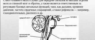
Next, let's take a closer look at how the human brain works. According to morpho-functional characteristics, the brain is also divided into three parts. The lowermost section is called rhomboid. Where the rhomboid part begins, the spinal cord ends - it passes into the medulla oblongata and posterior (pons and cerebellum).
Next comes the midbrain, which unites the lower parts with the main nerve center - the anterior section. The latter includes the telencephalon (cerebral hemispheres) and diencephalon. The key functions of the cerebral hemispheres are the organization of higher and lower nervous activity.
Cortex
This small superficial layer of gray matter (up to 4.5 mm) is the youngest formation in the central nervous system. It is the cerebral cortex that is responsible for the work of higher nervous activity in humans.
Research has made it possible to determine which areas of the cortex were formed relatively recently during evolutionary development, and which were present in our prehistoric ancestors:
- neocortex - the new outer part of the cortex, which is its main part;
- archicortex - an older formation responsible for human instinctive behavior and emotions;
- The paleocortex is the most ancient area involved in the control of autonomic functions. In addition, it helps maintain the internal physiological balance of the body.
This is possible thanks to the convolutions and grooves, which in addition also divide the hemispheres into lobes, each of which has different functions:
Frontal lobes
The largest lobes of the cerebral hemispheres, responsible for complex motor functions. In the frontal lobes of the brain, planning of voluntary movements occurs, and speech centers are also located here. It is in this part of the cortex that volitional control of behavior is exercised. If the frontal lobes are damaged, a person loses control over his actions, behaves antisocially and simply inappropriately.
Occipital lobes
Closely related to visual function, they are responsible for the processing and perception of optical information. That is, they transform the entire set of light signals that enter the retina into meaningful visual images.
Parietal lobes
They carry out spatial analysis and process most sensations (touch, pain, “muscle feeling”). In addition, it promotes the analysis and integration of various information into structured fragments - the ability to feel one’s own body and its sides, the ability to read, count and write.
Temporal lobes
In this department, audio information is analyzed and processed, which ensures the function of hearing and the perception of sounds. The temporal lobes are involved in recognizing the faces of different people, as well as facial expressions and emotions. Here information is structured for permanent storage, and thus long-term memory is realized.
In addition, the temporal lobes contain speech centers, damage to which leads to the inability to perceive spoken language.
Insula
It is considered responsible for the formation of consciousness in a person. In moments of compassion, empathy, listening to music and the sounds of laughter and crying, active work of the insular lobe is observed. This is also where the processing of feelings of aversion to dirt and unpleasant odors, including imaginary stimuli, takes place.
Hindbrain: pons and cerebellum
The structure of the hindbrain includes the pons and the cerebellum. The function of the bridge is very similar to its name, since it consists mainly of nerve fibers. The cerebral pons is essentially a “highway” through which signals travel from the body to the brain and impulses travel from the nerve center to the body. Along the ascending pathways, the brain bridge passes into the midbrain.
The cerebellum has a much wider range of capabilities. The functions of the cerebellum are to coordinate body movements and maintain balance. Moreover, the cerebellum not only regulates complex movements, but also contributes to the adaptation of the motor system to various disorders.
For example, experiments using an invertoscope (special glasses that invert the image of the surrounding world) have shown that it is the functions of the cerebellum that are responsible for the fact that when wearing the device for a long time, a person not only begins to navigate in space, but also sees the world correctly.
Anatomically, the cerebellum follows the structure of the cerebral hemispheres. The outside is covered with a layer of gray matter, under which there is an accumulation of white matter.
Limbic system
The limbic system (from the Latin word limbus - edge) is a set of formations surrounding the upper part of the trunk. The system includes the olfactory centers, hypothalamus, hippocampus and reticular formation.
The main functions of the limbic system are the body's adaptation to changes and the regulation of emotions. This education promotes the creation of lasting memories through associations between memory and sensory experiences. The close connection between the olfactory tract and the emotional centers is why smells evoke such strong and clear memories in us.
If we list the main functions of the limbic system, then it is responsible for the following processes:
- Smell
- Communication
- Memory: short-term and long-term
- Restful sleep
- Performance of departments and bodies
- Emotions and motivational component
- Intellectual activity
- Endocrine and vegetative
- Partially involved in the formation of food and sexual instinct
Source: https://MozgPortal.ru/stroenie-mozga/funktsii-i-stroenie-golovnogo-mozga.html
Hindbrain (human anatomy)
Source: https://zinref.ru/000_uchebniki/03200medecina/000_03_anatomia_i_fiziologia_vorobiova_1981/102.htm
| content .. 100 101 102 103 104 105 .. The hindbrain includes the pons and cerebellum. It develops from the fourth cerebral vesicle (metencephalon). The pons borders the medulla oblongata from below, passes into the cerebral peduncles from above, and its lateral sections form the middle cerebellar peduncles. In the anterior (basilar) part of the pons there are clusters of gray matter - the pons' own nuclei; in the posterior part of the pons tegmentum there are nuclei of the superior olive, reticular formation and V - VIII pairs of cranial nerves. These nerves emerge from the base of the brain lateral to and behind the pons, at the border with the cerebellum and medulla oblongata. The white matter of the pons in its anterior part is represented by transversely running fibers going to the middle cerebellar peduncles. They are penetrated by powerful longitudinal bundles of fibers of the pyramidal tracts, which then form the pyramids of the medulla oblongata and go to the spinal cord. In the rear part of the bridge (pontineum) there are ascending and descending fiber systems (Fig. 113). Rice. 113. Brain stem (front view). 1 – anterior median fissure; 2 – pyramids of the medulla oblongata; 3 – olive; 4 – cerebellum; 5 – decussation of the pyramids (place of transition of the medulla oblongata into the spinal cord); 6 – middle cerebellar peduncle; 7 – bridge; 8 – interpeduncular fossa; 9 – cerebral peduncle; III – XII roots of cranial nerves; C – first spinal nerve Physiology of the medulla oblongata and ponsThe medulla oblongata and the pons perform two functions – reflex and conductive. Through the sensitive fibers of the roots of the cranial nerves, it receives impulses - information from the receptors of the scalp, mucous membranes of the eyes, nose, mouth (including taste buds), from the organ of hearing, the vestibular apparatus (organ of balance), from the receptors of the larynx, trachea, lungs, as well as from interoreceptors of the cardiovascular system and digestive apparatus. Through the medulla oblongata, many simple and complex reflexes are carried out, covering not individual metameres of the body, but organ systems, for example, the digestive, respiratory, and circulatory systems. The reflex activity of the medulla oblongata can be observed in a bulbar cat, that is, a cat in which the brain stem above the medulla oblongata has been cut. The reflex activity of such a cat is complex and diverse. The following reflexes occur through the medulla oblongata: 1) protective: coughing, sneezing, blinking, lacrimation, vomiting; 2) food: sucking, swallowing, secretion of juice from the digestive glands; 3) cardiovascular, regulating the activity of the heart and blood vessels; 4) in the medulla oblongata there is an automatically working respiratory center that provides ventilation to the lungs; 5) the vestibular nuclei are located in the medulla oblongata and the pons.
A bulbar cat can neither stand nor walk, but the medulla oblongata and cervical segments of the spinal cord provide those complex reflexes that are elements of standing and walking. All reflexes associated with the standing function are called positioning reflexes. Thanks to them, the animal holds its body, usually with the crown up. The special importance of this part of the central nervous system is determined by the fact that the medulla oblongata contains vital centers: respiratory, cardiovascular. Therefore, not only removal, but even damage to the medulla oblongata ends in death. In addition to the reflex function, the medulla oblongata performs a conductive function. Conducting pathways pass through it, connecting the cortex, diencephalon, midbrain, cerebellum and spinal cord with a two-way connection. The cerebellum is located dorsal to the pons and medulla oblongata. It has two hemispheres and a middle part - the worm. The surface of the cerebellum is covered with a layer of gray matter (cerebellar cortex) and forms narrow convolutions separated by grooves. With their help, the surface of the cerebellum is divided into lobules. The central part of the cerebellum consists of white matter, which contains accumulations of gray matter - the cerebellar nuclei. The largest of them is the dentate nucleus. The cerebellum is connected to the brain stem by three pairs of peduncles: the upper ones connect it to the midbrain, the middle ones to the pons, and the lower ones to the medulla oblongata. The peduncles contain bundles of fibers connecting the cerebellum to various parts of the brain and spinal cord. During development, the isthmus of the rhombencephalon forms the boundary between the hindbrain and midbrain. From it develop the superior cerebellar peduncles, the superior medullary velum located between them and the triangles of the loop, lying outward from the superior cerebellar peduncles.
At the bottom, the IV ventricle communicates with the central canal of the spinal cord, at the top it passes into the cerebral aqueduct of the midbrain, and in the roof area it is connected by three openings to the subarachnoid space of the brain. Its anterior (ventral) wall—the bottom of the fourth ventricle—is called the rhomboid fossa. The lower part of the rhomboid fossa is formed by the medulla oblongata, and the upper part by the pons and isthmus. The posterior (dorsal) wall - the roof of the IV ventricle - is formed by the superior and inferior medullary sails and is supplemented from behind by a plate of the pia mater lined with ependyma. This area contains a large number of blood vessels that form the choroid plexus of the fourth ventricle. The convergence of the superior and inferior sails protrudes into the cerebellum and forms a tent. The rhomboid fossa is of vital importance, since most of the nuclei of the cranial nerves (V – XII pairs) are located in this area. |