The cerebral hemispheres within the brain are the largest and most anterior sections. Thanks to the structures of the cortical layer covering the hemispheres, a person is able to feel, understand, talk, and navigate the environment. For example, when the frontal regions are damaged, psycho-emotional reactions are disrupted and movement disorders occur. Damage to the parietal regions is accompanied by impaired perception of the size and position of one’s own body in space.
Characteristic
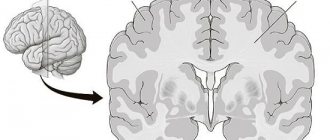
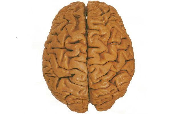
The cerebral hemispheres are the final section of the brain, which has a characteristic structure and performs many functions, the most important of which is ensuring the relationship of the body with the outside world. The terminal section is the largest brain structure in size. The central sulcus divides the terminal part of the brain into the cerebral hemispheres, which are connected by a system of thin cords formed by connective tissue.
The surface of the hemispheres has a complex topography due to the presence of many grooves and convolutions. The cortex, covering the cerebral hemispheres, is represented by gray matter; its complex, differentiated structure determines the variety of functions performed. The subcortical layer consists of white matter, within which the basal ganglia, otherwise called the nerve nuclei, are located.
Inside the white matter are the lateral ventricles. The thickness of the cortex covering the cerebral hemispheres is about 1-5 mm. The mass of the cortex makes up 78% of the weight of the entire brain. The importance of the cortex within the cerebral hemispheres is due to its main function - maintaining all types of conscious activity.
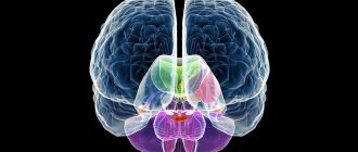
Morpho - functional features of the limbic system
The limbic system is understood as a morpho-functional association of phylogenetically old parts of the forebrain.
The most important structures of the limbic system are:
- cingulate gyrus,
- amygdala complex,
- hippocampus,
- transparent septum (septum).
The limbic system takes an active part in maintaining the homeostasis of the body. However, its main function is modulation (change of functions or parameters, according to the flow state) of the sensory, motor and homeostatic systems.
Structure
The morphological structure of the cerebral hemispheres within the brain suggests the presence of a cortical layer consisting of gray matter and subcortical sections formed by white matter. Within the mass of white matter there are local areas of gray - the nuclei. White matter serves as the basis for nerve fibers:
- Associative. They unite different functional areas within one hemisphere.
- Commissural. They connect areas of different hemispheres, often symmetrically located.
- Conductive. They connect the parts of the brain - the brain and the spinal cord - thereby forming a single central nervous system network.
The structure of the cortex covering the cerebral hemispheres suggests the presence of grooves (depressions relative to the surface) and convolutions (elevations relative to the surface). In anatomy, the area of the cortex covering the cerebral hemispheres increases due to the presence of grooves. The duration and depth of the furrows are individual characteristics of a person.
The cortical layer, formed from gray matter, contains control centers that are responsible for higher mental functions, which are interconnected with processes such as cognitive and mental activity. The cortex, covering the cerebral hemispheres within the brain, coordinates the functions of the body, forming adaptive responses to external influences. The cortex is formed by 6 types of cells.
The cytoarchitectonics (location of cells in the tissue) of the cortex suggests the presence of molecular, granular (external, internal), pyramidal (external, internal), multiform plates. Within the brain, the surfaces of the hemispheres are distinguished: superolateral (covers the upper lateral areas), medial (located in the middle region), basal (adjacent to the base of the skull).
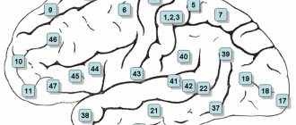
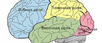
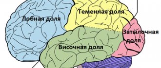
The superolateral surface has a convex shape and is adjacent to the bone structures that form the cranial vault. The flat medial surface is located opposite the similar surface of the second hemisphere. Each hemisphere consists of 5 lobes. The frontal lobe of the cortex, covering the cerebral hemispheres, is the largest part of the telencephalon. The central sulcus, anatomically transversely crossing the brain, is the border between the frontal and parietal lobes within the cerebral hemispheres.
The Sylvian fissure, also known as the lateral fissure, runs within the cerebral hemispheres perpendicular to the central one, separating the temporal lobe of the telencephalon from two segments - the frontal and parietal. The parieto-occipital sulcus is the border of the parietal segment on the posterior side. The insular lobe within the hemispheres of the brain is located deep in the Sylvian fissure. The insular lobe is visible if you lift the areas of the frontal and temporal lobes that cover it.
The Sylvian fissure, also known as the lateral fissure, which separates the temporal lobe from the brain structures of the frontal and parietal, is one of the largest. It is formed at the 14th week of intrauterine development. Small grooves dividing the lobes of the cerebral cortex into convolutions are formed at 24-38 weeks, forming a peculiar relief of the surface of the hemispheres, individual for each person.
The sulci and gyri of the 3rd order continue to develop after birth, most intensively in the 1st year of life. In the frontal lobe of the cortex within the cerebral hemispheres, a precentral groove runs parallel to the central one, separating and limiting the precentral gyrus - the center where the initiation and regulation of conscious motor activity, directed by volitional effort, occurs.
The precentral gyrus is perpendicularly crossed by the frontal sulci (superior, inferior). They divide the frontal gyri into segments. In the posterior part of the frontal gyrus, located below, there is Broca's center, which is responsible for speech function. In the parietal lobe, the postcentral sulcus runs parallel to the central one.
The postcentral gyrus, located between these grooves, is a center of sensitivity, which is responsible for the body’s reactions to pain, tactile, and temperature influences. The temporal grooves (superior, inferior) lie in the temporal lobe and are located parallel to the Sylvian fissure.
The temporal sulci are divided into segments by the temporal gyri, where in the upper part there is a center responsible for hearing function and Wernicke's center, responsible for speech function. The calcarine groove, which provides visual function, lies in the occipital lobe, where it borders on the parieto-occipital groove.
Functions
The cerebral hemispheres are covered with a cortical layer, which contains structures that support high mental functions. The higher activity of the nervous system implies a set of reflexes (conditioned, unconditioned) and mental reactions that determine adequate behavior in changing conditions under the influence of natural and social factors.
The role of the cortical sections is great in ensuring interaction with the environment. Main areas of activity: synthesis, integration, analysis. The structures of the cortex combine excitations that arise in different functional areas, differentiate and integrate stimuli coming from receptors located throughout the body.
Impulses transmitted by receptors are separated by nature and intensity, forming appropriate responses. The functions of the cortical layer of the cerebral hemispheres are differentiated and localized in different parts of the brain. Taking into account the functional direction, the following zones are distinguished:
- Motor. Located in the area of the central (precentral) gyrus, located in front. In this area, impulses are formed that cause contraction of skeletal muscles, moving the limbs, head, neck and other parts of the body. The upper segments of the motor zone interact with muscle receptors in the lower extremities. The middle segments are interconnected with the muscle receptors of the torso, the lower segments with the muscle receptors of the head.
- Sensitive. Includes visual and auditory zones, areas of skin, taste and olfactory sensitivity.
- Associative. They are located in the parietal, frontal lobes and other parts of the cortical layer. The designated areas provide connections between highly specialized processes, such as the ability to read written text, hear speech, and speak back.
The area of skin sensitivity is located in the parietal lobe in the region of the central (postcentral) gyrus, located posteriorly. Impulses coming from skin receptors arrive here. The area of visual perception lies in the occipital lobe. In the temporal gyrus, which lies above, there is a region of auditory perception.
In the limbic system (brain structures located below the telencephalon, surrounding the thalamus on both sides) of the cortex there is an area responsible for taste perception. This receives impulses from receptors located in the mucous membrane of the tongue and oral cavity.
Within the limbic system there is also a zone of olfactory perception, which receives impulses from receptors located in the nasal cavity. Speech functions are localized in the cerebral cortex - in Broca's and Wernicke's areas, which, in short, are located in the frontal and temporal lobes.
Broca's area (motor center of speech) is located in the area of the frontal gyrus, which lies below. Wernicke's area (the center of speech perception) is located in the area of the temporal gyrus, which lies above. In the parietal and occipital lobes there are areas responsible for the perception and understanding of written speech; the angular gyrus separates the designated sections from each other.
Possible diseases
Any violation of the anatomical integrity of the nerve cell does not pass without leaving a trace. However, the severity of the pathological disorder and its duration are directly influenced by the nature of the provoking factor. Thus, when cerebral blood flow deteriorates due to an atherosclerotic plaque, which leads to post-hypoxic changes in the brain, ischemic stroke is characterized by:
- local feeling of numbness,
- partial/complete loss of movement in any part of the body,
- muscle weakness.
If the injuries lead to the death of a large area of the cortex, the person completely loses one of his higher nervous functions and becomes disabled. In the case of tumor damage to subcortical structures, disorders may occur in the regulation of structures dependent on them - autonomic abnormalities, thermoregulation, endocrine disorders.
Of course, diseases of the cortical structures are immediately noticeable. Meanwhile, atrophy of white fibers can occur hidden, for example, with dyscirculatory encephalopathy. Initially, small areas of the brain are affected, which affects a person’s daily activities. Later, the process covers all areas of brain activity - for example, Alzheimer's disease, multiple sclerosis. When performing magnetic resonance imaging, single lesions can be detected in the white matter of the frontal lobes - leukoaraiosis, or their localization in the cerebellum. Then, in addition to intellectual disorders, the patient is characterized by motor disturbances. The selection of optimal treatment regimens should be carried out by a neurologist, taking into account the anatomical and functional characteristics of the gray/white matter of the brain.