Your orthopedist
Everything about the musculoskeletal system on one site: Back, Joints, Bones and more
home › Anatomy › Where is the spinal cord located and why does it need reliable protection
Why is this part of the central nervous system needed?
The article talks about why the spinal cord needs reliable protection. The anatomical structures that protect this organ are described.
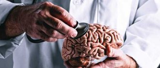
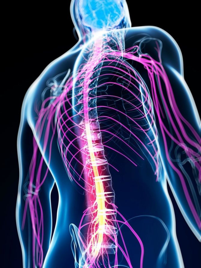
Everyone knows where the spinal cord is located - in the spine. More precisely, in the spinal canal, which reliably protects the brain from damage. However, such protection can also be violated. Then severe pathological conditions develop, sometimes extremely life-threatening.
- General information
- Location
- What kind of damage can there be?
What is the spinal cord
The spinal cord, together with the brain, is part of the central nervous system of the human body. Both of them begin to form in the early stages of embryo development. Around the 20th day of pregnancy, certain folds form on the body of the embryo, which will eventually turn into what experts call the neural tube of the fetus. It represents the rudiment of the future central nervous system. During the development of the tube, its front part will turn into the brain, and the back will serve as the basis for creating the spinal cord.
Content:
- What is the spinal cord
- How does the spinal cord control the body?
- Functions of the spinal cord
- Consequences of central nervous system injury
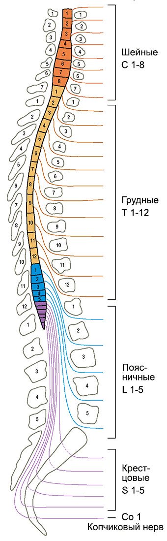
Humanity has been studying the structure and role of the spinal cord since antiquity. For example, the ancient Greek physician Galen studied the central nervous system of animals and gladiators, analyzing how different types of injuries affect the functionality of the body. Modern scientists already know for sure that the spinal cord is a cylindrical bundle of nerve fibers that is connected to the brain and runs through the spine from the neck to the lower back. Scientists also know that in the body of an adult, this organ can reach 40-45 cm in length, 1-1.5 cm in width and weigh about 35 g. It has the shape of a thick-walled tube with a narrow channel inside, somewhat flattened in the anteroposterior direction, located inside the spinal canal, which is divided into segments (they correspond to the sections of the spine):
- 7 cervical;
- 12 breast;
- 5 lumbar;
- 5 sacral;
- 3-4 coccygeal.
Interestingly, in the adult body, the spinal cord does not occupy the entire cavity of the spinal canal, but only about 2/3 of its length. Experts explain this feature by the fact that the spinal column grows faster than the spinal cord, therefore, as a person grows, this part of the central nervous system takes up less and less space in the spinal canal. That is, in the body of adults, the spinal cord in its lower part reaches only the first lumbar vertebrae.
The spinal cord is covered by three meninges (protective membranes). The first, located closest to the spinal substance (internal), is called the pia mater of the spinal cord. It consists of a network of vessels that are responsible for the blood supply to the brain. On top of it is the middle arachnoid membrane, under which contains cerebrospinal fluid - cerebrospinal fluid. By the way, when a spinal cord puncture is performed, the cerebrospinal fluid is taken for analysis, that is, a needle is used to penetrate the space between the inner and middle membranes. The uppermost (outer) shell is hard. It protects the nerve roots leading to the intervertebral foramina.
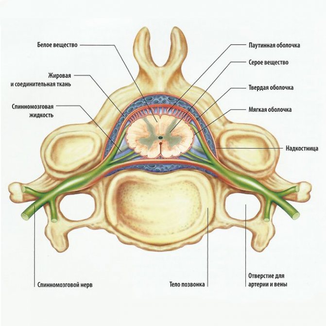
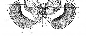
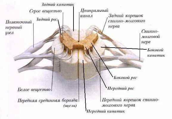
If the spinal cord is cut across, you can see that its tube has an oval, almost round shape. The upper layer of this organ consists of white matter, under which lies the gray spinal substance. In cross section, it has the shape of a butterfly, from which horns of gray matter extend forward, backward and sideways. In the central part of the spinal substance is the intermediate nucleus of the gray matter. This part is the most important. She is responsible for collecting all the information and making decisions about the activation of one or another reaction of the body.
White matter is made up of bundles of nerve fibers called axons. They allow different levels of the central nervous system to exchange impulses. It is known that each axon bundle differs in the specificity of the signals transmitted. So, some of them (ascending) transmit information to the brain, while the rest (descending) are responsible for transmitting impulses from the brain to neurons in different muscles and glands located throughout the body. White matter axons are covered with an insulating myelin sheath, which facilitates the rapid and free transmission of impulses. Myelin is whitish in color, hence the name white matter.
The gray substance of the spinal cord is formed from nerve cells and their branched processes - dendrites. This substance is responsible for the perception and processing of information. For example, if we talk about the brain, it is the gray matter in it that gives a person the ability to think. If we talk about the spinal cord, the horns of the gray matter also process various information messages from the body. Moreover, each horn has its own function.
Shells
The outside of the spinal cord is covered by three structures:
- Arachnoid medium.
- Solid outer.
- Soft inner.
The epidural space is located between the periosteum of the spinal canal and the dura mater. It is filled with venous plexuses and fatty tissue. Between the arachnoid and dura mater there is the subdural space. It is permeated with thin connective tissue crossbars. The soft membrane is separated from the arachnoid by the subarachnoid subarachnoid space. It contains liquor. Cerebrospinal fluid is formed in the choroid plexuses located in the ventricles of the brain. Renshaw cells protect the central nervous system from overexcitation.
How does the spinal cord control the body?
Each segment of the spinal cord controls the functionality of a specific part of the body. Thus, the segment of the central nervous system located along the cervical vertebrae sends impulses to the back of the head, neck and upper limbs. The thoracic segments are responsible for the functionality of the torso (chest, abdomen). The lumbar zone of the central nervous system controls the lower extremities, and the sacrococcygeal segments control the pelvis and internal organs located in this area.
The relationship between the segments of the spinal cord and the parts of the body is always strictly observed. At each “floor” of the body, nerve endings extend from the brain and are distributed throughout the area controlled by these segments.
The spinal cord is a tube that runs inside the spine, and spinal nerve processes emerge from the intervertebral spaces. Understanding the structure of this part of the central nervous system, it becomes clear how dangerous the slightest violation of the natural positions of the vertebrae is for a person. Any displacement of even one vertebra can lead to compression of the spinal nerves and disruption of interaction between the parts of the central nervous system and the part of the body for which the segment is responsible.
Many people know that spinal injuries can lead to paralysis. But this is not the only danger that can arise from a spinal cord injury. By and large, most health problems can arise due to misalignment of the vertebrae - even gastritis or ulcers. Just imagine, the vertebra is displaced, pinching the nerve ending, which is why the brain sends incorrect signals to the digestive system and gastric juice begins to be produced in excess, corroding the gastric mucosa. The result is gastritis, and then an ulcer.
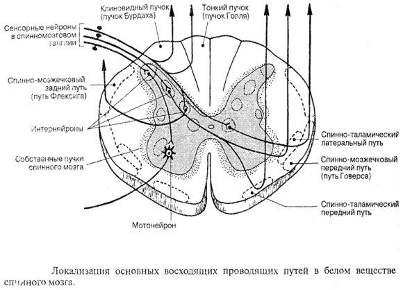
Ascending Paths
- Anterior spinothalamic. This is the afferent pathway of pressure and touch.
- Lateral spinothalamic. This is the path of temperature and pain sensitivity.
- Posterior and anterior spinocerebellar. These are afferent pathways of muscle-articular sensitivity of the cerebellar direction.
- Bundles of Burdach (wedge-shaped) and Gaulle (thin). These are afferent pathways for transmitting cortical muscle-articular sensitivity from the lower half of the torso and legs and from the upper body and arms, respectively.
Functions of the spinal cord
So, the gray matter of the spinal cord has 3 horns, which actually perform the main functions of this organ:
- analyze sensory signals;
- govern internal organs (vegetative function);
- control muscles (motor function).
The collection of sensory signals occurs separately on each “floor” of the body. That is, each segment of the spinal cord accumulates information about what is happening in the territory “controlled” by it. This information is also strictly structured. It concerns three aspects:
- pain, that is, damage to cells and tissues on the “floor”;
- skin sensitivity, that is, heat, cold, vibrations, etc.;
- muscle sensitivity, that is, about the condition of muscles, tendons and joint position.
The spinal cord receives all information through clusters of nerve cells known as the spinal ganglia (spinal ganglia). The number of these nodes exactly corresponds to the number of spinal segments - 31. The cells included in the ganglion have a special structure: 1 long dendrite and 1 axon. The dendrite collects information about what is happening on the controlled “floor”, and the axon connects to the dorsal horn of the gray spinal substance, to which it transmits the received data. In this area, the primary processing of sensory signals occurs and then the transfer of information to the intermediate nucleus of the gray spinal matter, which determines the further reaction of the body. But here it is worth mentioning another interesting point. The neurons of the intermediate nucleus receive signals not only from the horn, but also from the brain, which also retains the right to vote on what the reaction to the impulse received from the horn should be.
To make this information easier to understand, we will use an example. Let's say a person took a glass of boiling water. As a result, sensory fibers send pain information to the spinal cord. Consequently, he is ready to give the signal to throw a red-hot object. But at the same time, another signal comes from the brain that, for example, the glass contains something valuable or the glassware itself is valuable, so the vessel should be brought to the surface. These conflicting signals arrive at the intermediate nucleus of the spinal gray matter, where neurons analyze both options and either trigger or block a response. If the intermediate nucleus approves of the reaction, it transmits the corresponding signal to the anterior and lateral horns of the gray substance.
The anterior horns of the spinal gray matter contain so-called motor neurons, which cause muscle contractions. Autonomic neurons (sympathetic and parasympathetic nervous system) are concentrated in the lateral horns, which regulate the activity of internal organs.
Basic “working” elements
The functions possessed by the spinal cord center would be impossible without two substrates - white and gray. They are located directly in the canal of the brain itself, and the amount of one or another substance predominates in different areas. The bulk of the gray substrate is concentrated in the upper part of the tube and in the lumbar region. White matter predominates in the chest area, and the lower it is, the more its amount decreases and gradually comes to zero. When we take a cross section of the spinal cord, we also see that the gray matter is the middle, which looks like the letter H, and it is surrounded on all sides by a white membrane.
Consequences of central nervous system injury
Even partial damage to the spinal cord can cause disruption of the functionality of a wide variety of systems and organs.
Bones and joints
A spinal cord injury can impair the absorption of calcium and some other important minerals into bone tissue. But at the same time, these substances can accumulate in the urinary system, causing the formation of stones. Experts usually explain such processes by a decrease in human motor activity. In addition, decreased mobility can lead to stiffness in joints, including the knees, elbows, and shoulders. To prevent this, there are special sets of exercises for people with disabilities.
Urinary system

The urinary system consists of the kidneys, which filter the blood and produce urine, and the bladder, which collects and then removes it from the body. After a spinal cord injury, the kidneys continue to produce urine, but the bladder may not work as well as before. After an injury, a person may not feel when his bladder is full of fluid, or his urinary system, due to a lack of necessary impulses, may lose the ability to eliminate urine naturally. As a result, a catheter may be needed to empty the bladder. Another option: urine may, on the contrary, come out involuntarily and the person loses the ability to control this process.
Respiratory system
Even after a spinal injury, human lungs continue to function. However, the ability to inhale and exhale air is controlled by the muscles. Depending on the level of injury, a person may lose the ability to cough or take deep breaths. And they are necessary for complete straightening of the lungs and deep air circulation.
The most important muscle for breathing is the diaphragm. It is a large arcuate muscle that is located directly under the lungs. And if the diaphragm is paralyzed due to damage to the spinal cord, special equipment may be needed to maintain respiratory function.
Leather
Damage to the spinal cord can also negatively affect the skin. Skin plays a very important role for humans. It protects the body from germs and other pathogens from the outside world. The spinal cord is an important organ that helps protect the skin from damage. For example, when sitting in one position for a long time, the brain sends signals in the form of a feeling of discomfort, which encourages you to change your position and prevent damage or pressure on the skin. In the same way, the central nervous system protects against burns, cuts and other possible damage to the skin (for example, a person reflexively withdraws his hand from a hot or cutting object). With spinal cord injuries, such signals may not appear, which greatly increases the risk of damage to the skin and, consequently, the penetration of microbes into the body.
Reproductive system

After a spinal cord injury in the lumbar or sacrococcygeal segment, the functionality of the reproductive system may be affected. Men usually experience erectile dysfunction, problems with ejaculation and fertility. In women, after injury, sensitivity in the genital area may be impaired, although fertility itself may not be directly affected.
Intestines
It is well known that the digestive system is responsible for the breakdown of food consumed, the absorption of nutrients from it and the removal of metabolic products. A fairly common consequence of central nervous system injury is impaired intestinal motility. That is, the digestive system continues to absorb and digest food, but the process of eliminating excrement is disrupted. In a healthy body, when the rectum is full, an exchange of impulses occurs between the intestines and the brain. If the spinal cord is damaged, such signals may not be received. As a result, a person loses the ability to control the process of defecation.
Autonomic nervous system
The autonomic nervous system regulates the activity of internal organs, blood and lymphatic vessels, as well as endocrine and exocrine glands. Body temperature, blood pressure, digestion and other important functions depend on it. After a spinal injury, some of its functions may be impaired. Quite often (especially if the damage occurs in the thoracic segment) after an injury the body loses the ability to thermoregulate. What does it mean? If the autonomic nervous system works correctly, then the body temperature remains stable regardless of the weather conditions outside or the indoor microclimate. Due to spinal cord damage, body temperature may rise or fall according to indoor or outdoor temperatures. Disturbances in thermoregulation are most noticeable in parts of the body located below the area of injury.
What kind of damage can there be?
Despite such massive protection, cases of spinal cord damage are possible:
- in case of car accidents;
- when falling from a height;
- some infectious diseases;
- tumor processes;
- degenerative diseases of the spine.
In injuries and degenerative diseases, the cause of damage is various disturbances in the location of the vertebrae. In infectious diseases, the brain is damaged by microorganism toxins. During tumor processes, compression of the brain occurs from the outside.
The symptoms that the patient will experience will depend on the location of the injury. You can learn more about this from the video in this article.
Knowing where the spinal cord is located, experts suggest certain diseases. This knowledge also helps in carrying out some diagnostic and therapeutic procedures.
Segmentation
The sections of the spinal cord are divided into five parts, the significance of which depends not on the location, but on the section in which the exiting nerves leave the spinal canal. In total, a person can have 31-33 segments, five parts:
- cervical part – 8 segments, at its level there is more gray matter;
- chest – 12;
- lumbar – 5, the second region with a large amount of gray matter;
- sacral – 5;
- coccygeal – 1-3.
The human spinal cord is divided into several sections with segments. The number of segments is 32. Each segment controls certain innervation functions.
Spine structure
- the cervical spine has 8 segments;
- thoracic includes 12 segments;
- the subsequent sections (lumbar and sacral) consist of 5 segments;
- The coccygeal region has only 2 segments.
The diameter of the spinal cord varies from place to place. In two sections there is thickening of the spinal cord, which occurs in the first couple of years of a child’s development due to the increased load on them. The cervical enlargement of the spinal cord and the lumbar enlargement are responsible for motor activity and limb function.
Structure of a spinal cord segment
The structure of the spinal cord segment is such that it contains pairs of roots connected to other organs through nerves. The roots leave the spinal canal and form nerves that branch to various tissues and organs. The anterior and posterior roots of the spinal cord are conductors of information.
Sensitivity is provided by the dorsal roots, which transmit information from the stimulus through the activation of receptors and carry it to the dorsal roots. At the junction of the anterior and posterior roots is the spinal ganglion, a collection of neurons located under a protective sheath.
The roots go into the holes between the vertebrae, so some of them run level, and the other part is inclined. For example, in the cervical region such roots are located horizontally, and in the thoracic region - along an oblique line. The remaining sections force the roots to bend almost vertically downwards. Some of the roots from these sections are so close to each other that in the photo the resulting bundle is called the cauda equina in the spinal cord.
Each segment is responsible for its own zone of innervation. The innervation zone includes internal organs, bones, muscles and skin. Knowing the zone of innervation, one can clearly determine which segment of the nerve is responsible for it and suggest the pathology of this area. The segmental principle, which shows where the cause of the pathology is located, helps both in the diagnosis and treatment of diseases.
Reflexes
The functional diversity of neurons in spinal structures creates conditions for reflex activity, both with the participation of its own elements and central ones - the cortex and subcortical centers.
Each such reflex arc necessarily contains the following links:
- afferent – perceives impulses;
- intercalary – connects sensory and motor neurons;
- efferent - transmits the impulse to the immediate performer.
Among the motor reflexes, attention should be paid to myotatic ones, which are aimed at stretching/contracting muscles, as well as skin ones - plantar and abdominal. With their help, every person can perform voluntary and involuntary movements.
In addition to motor reflexes, there are other reflexes. Thus, autonomic ones are usually divided into sympathetic and parasympathetic. Their common task is to control and ensure the constancy of the internal environment of the body to maintain human life.
Conclusion
Thus, the loss of certain functions, for example, leg movements, makes it possible to determine which part was damaged. Injuries to this organ are among the most serious, and the damage is often irreparable. The main thing is to monitor the health of your spine and not overload it unless absolutely necessary.
The organ is located in the spinal canal, and its length is no more than 45 cm, which is less than the length of the spine itself. This is due to the fact that the brain grows only until the age of five, and the spine, as a rule, until the end of puberty.
Features of gray matter
This substrate consists mainly of nerve fibers, cells and processes. Initially, it seems that the gray matter is the most central part of the brain, but in fact it functions as just another shell, so to speak. In the very center there is a very narrow cavity, which widens slightly only in the area of the cervical vertebrae (at this stage the diameter is less than 1 mm). This cavity is the very channel through which the spinal cord transmits all the necessary information to the brain.