How does our brain work? How many neurons are there and what are the functions of the neocortex? Modern scientists are scrupulously studying the features of our brain and discovering more and more interesting details.
Thanks to the development of higher nerve centers, a person determines himself and his place in society, consciously controls his behavior and is capable of adapting to a new environment. All of these benefits are related to the functions of the cerebral hemispheres, which we will look at.
Features of the human brain
The human brain weighs approximately 1 kg 200 grams - these are average figures. It consists of 5 main parts: the telencephalon, diencephalon, midbrain, hindbrain and medulla oblongata.
Large grooves (depressions) separate the 4 main parts of the cerebral hemispheres: the frontal lobe from the parietal lobe; and the parietal one - from the occipital; The temporal lobe is adjacent to the other three. The last, fifth lobe is the insula, which is located deep in the lateral fossa. The harmonious interaction of all neurons ensures the growth and development of our individuality, our character and abilities.
One can single out a separate function of the cerebral hemispheres - continuous development. The human brain is developing all the time. Everything that an individual reads, sees, perceives, he literally absorbs into himself. New information is especially important for children under 2 years old, at this time their neurons are actively building connections for the future.
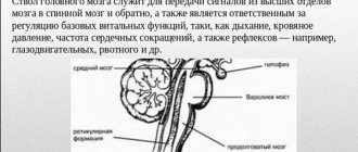
Inhale and exhale
Excited inspiratory neurons reach the intercostal muscles and cause them to contract, and the muscles of the diaphragm also begin to contract. An inhalation occurs, which delivers another portion of oxygen to the body. When you inhale, the lungs expand, and the receptors located in the pulmonary lobes come into motion. They, in turn, send impulses to the medulla oblongata. The respiratory center receives impulses and turns them into a brake for inspiratory neurons, which lose activity. The expiratory neurons of the respiratory center begin to be excited. They cause the group of muscles responsible for contracting the chest to react, and thus exhalation occurs.
Large hemispheres. Structure and functions
There are 14 to 17 billion neurons in the cortex; and there are many times more connections between cells. Neurons are connected by synapses. And various neurotransmitters help activate connections - chemicals that activate a nearby synapse.
The hemispheres of the brain have a special structure. Thanks to folds consisting of grooves and convolutions, the area of the cortex increases significantly. According to some data, the total area of the cortex of the average person is 2200 square meters. cm.
Beneath the cortex is the subcortex, or white matter of the brain. The hemispheres are connected to each other by the corpus callosum. And even deeper are the ventricles of the brain - spaces filled with cerebrospinal fluid.
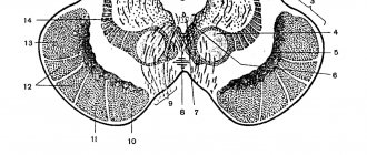
The cortex consists of layers of nerve cells, which alternate with layers of their branches - axons. There are 6 layers in total:
- molecular layer;
- external granular;
- external pyramidal - contains mainly pyramidal neurons;
- internal granular;
- internal pyramidal;
- layer of spindle neurons.
Fusiform neurons gradually pass into the white matter of the brain. Conscious actions occur in the cortex and speech is formed. In the lower deep parts under the cortex there are centers of unconscious reflexes and control of internal organs and organ systems.
Emotions and breathing
In addition to inspiratory and expiratory neurons, other factors also influence the breathing process. Since the respiratory center is located in one of the parts of the brain, it is influenced by many associated factors. Breathing may increase due to physical exertion, emotional experiences, feelings of fear or danger. The activity of the respiratory center also depends on the hormonal state of the body. But in any case, metabolic processes in the human body are regulated by enriching the blood with oxygen.
Brain zones
To understand the functions of the cerebral hemispheres, you must first understand their structure. The hemispheres are divided conditionally into several centers in which certain mental and physiological processes take place. These centers are not separate structures. All neurons of all networks constantly interact with each other. This is confirmed by many researchers.
But it is still possible to identify some areas in the gray matter of the brain that are more specialized for individual tasks.
Neurophysiologists distinguish the following brain areas:
- Occipital zone.
- Temporal - responsible for the sense of smell and taste. These two feelings are strongly interconnected.
- Visual zone. Here the signals coming from the eyes are deciphered.
- The parietal is the so-called zone of musculocutaneous sensitivity.
- The frontal lobe is a person’s conscious behavior, his attitudes and work activity. The back of the frontal lobe is the motor center.
The functions of the cerebral hemispheres, as we see, are distributed across zones. Some areas have multiple functions. For example, the hands are connected in the cerebral hemispheres with two zones - motor and sensitive.
And if any of these areas are damaged during a traumatic brain injury, the function of this area will suffer or completely disappear. It is possible to restore the lost function if another part of the brain - the one where the neurons associated with the damaged tissues were located - can take on all the work of the lost center.
Scisne?
One of the most important functions our brain performs is movement control.
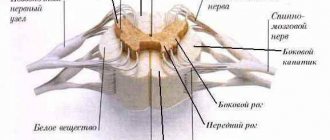
There are many motor centers of the brain. If you add them all together, you get about the same amount as all the sensory systems combined. That is, our movement is not something similar to vision or hearing. This is all together: vision, hearing, skin sensitivity, smell, and taste... If you sum it all up, you get a volume approximately equal to the total volume of, say, the cerebellum, basal ganglia and other structures that control movements. The movements themselves are muscle contractions. And if we start with the simplest thing, then we need to say a few words about motor neurons. Motor neurons are special nerve cells that work with striated muscle cells and trigger their contractions by releasing acetylcholine. And the lion's share of motor neurons is located in the gray matter of the spinal cord, in its anterior horns, and the axons are directed to the muscles of our limbs, our torso. In addition, many motor neurons are located in the brain, and through the cranial nerves they reach the muscles of our head, facial muscles, extraocular muscles, tongue muscles, and so on. The axon of a motor neuron usually branches at the end, and eventually one motor neuron controls several muscle cells. A collection of muscle cells that are controlled by a single motor neuron is called a motor unit. Our muscles are organized in such a way that the motor unit can have different sizes. And the finer the movement, the finer the muscle control must be, the smaller the motor unit.
The most subtle movements are performed by our oculomotor muscles. There, a motor unit consists of only 5–10 muscle cells, that is, one motor neuron works with a very small number of contractile units. If we are talking, for example, about fingers or facial muscles, one motor neuron controls several dozen muscle cells. Biceps, triceps, limb muscles - there is one motor neuron, the commander for several hundred muscle cells. The grossest movements are performed by the muscles of our torso, back muscles, abdominal muscles - there one motor neuron commands thousands of muscle cells. And accordingly, the back muscles cannot make any subtle movements.
If you look at who controls the motor neurons, it turns out that there are many centers both inside the spinal cord and inside the brain that send commands directly to these cells and to those neurons in the gray matter of the spinal cord that surround the motor neurons. And many different types of movements use the same motor neuron. That is, the specificity of the organization of our motor systems is such that the same muscle contraction can be included in a variety of movements. Generally speaking, the movements we make fall into four broad types. These are reflex movements, locomotor movements, voluntary movements and automated movements.
Reflex movements (from the word “reflex”) are a reaction to some stimulus. They arise as a response to some sensory stimulation. The brain has at its disposal chains of nerve cells that fire if the very first neuron - a sensory neuron - produces an electrical impulse. These reflex arcs can be innate, or we can learn to implement this or that reflex, but in any case, the triggering factor is the sensory signal. There is a signal - there is a reflex, there is no signal - we do nothing. Everyone knows examples of basic innate motor reflexes (say, the reflex of withdrawing from the source of pain or, for example, the knee reflex). Here it is also important to realize the biological meaning of these reflexes, because if the reflexes are innate, then it means that evolution for some reason shaped them. And let’s say, in the case of the reflex of withdrawing from the source of pain, the meaning is obvious: we avoid damage to our skin surface or some deeper structures.
And in the case of, for example, the knee-jerk reflex, it is not very clear why this is necessary. The knee reflex is a situation where a light but sharp blow occurs to the quadriceps tendon, that is, under the kneecap, and in response to this blow, the quadriceps muscle contracts, the knee straightens, and your leg moves forward. As a reflex it looks funny, but it’s not very clear why it exists. In fact, reflexes like the knee jerk - they are called myotatic - are needed primarily to maintain posture. Myotatic reflexes occur when some additional load is applied to the muscle. And they represent, as it were, stubbornness at the muscular level. Imagine that you are standing in the middle of a subway car and suddenly this car starts to slow down. The moment braking begins, the load on your legs begins to be redistributed. And on the leg on the side where you started to fall, the tendons are stretched more strongly, its muscles are stretched more strongly, and in response to this stretch, arcs similar to the arcs of the knee reflex are activated, the muscle tenses more, and as a result, the body as a whole tries to maintain the pose.
Another well-known example of a reflex is the reaction of a baby grasping a finger. If you touch his palm, he grabs your finger. This is an ancient reflex that allowed monkey babies to cling to their mother's fur. In fact, baby monkeys are doing this even now. Women have not been covered with such thick hair for a long time, but the reflex remains. This is like a physiological rudiment that shows where our species came from. Of course, everyone also knows such reflexes as yawning, hiccupping, and coughing. Again, each of these has its own biological meaning.
Let's say, when we cough, we clear our airways from some foreign objects. And hiccups are needed in order to expel, for example, excess air from the stomach. Interestingly, these reflex arcs tend to have a long evolutionary history. The same hiccup is a reflex that, apparently, we inherited from amphibians. That is, at the moment when a tadpole, which previously breathed with gills, grows lungs, it needs a similar reflex, so the tadpole simultaneously breathes with both lungs and gills. And at that moment, when he basks in the sun and dives into the water, he needs to close his lungs and switch to breathing with his gills. What exactly is hiccupping? You begin to inhale, and then suddenly close your glottis, and the sound that is heard when you hiccup is a sign that our larynx is closing. This is what the tadpole needs to prevent water from getting into the developing lungs. And then the neural circuits are transferred to other vertebrates and begin to be used, for example, to ensure that the digestive system functions correctly and well.
Yawning is still a rather mysterious reflex because there are several explanations for why we yawn. There is an idea that we yawn to cool the brain because there is intense ventilation of the various cavities inside the head. There is an idea that blood flow increases at this point. There is an idea that when yawning, stimulation of facial muscles gives an activation flow to the brain and from this we seem to awaken. Finally, yawning in monkeys is also used as a type of communication: when yawning, the dominant male shows his fangs and generally says that “I’m not afraid of you at all, I’m bored in your presence.” All these factors apparently coexist and explain the presence of the yawn reflex in our behavior.
The second type of movement is called locomotion. Locomotion is movement in space. That is, this category includes movements associated with rhythmic flexion and extension of the limbs, which allow swimming, walking, and running. And these are separate programs, largely innately given, but not reflexes, because in the case of locomotion, the motor program and movements mainly circulate along a certain closed circuit. Let's say we have four limbs, and for us this circuit includes the centers of the two forelimbs and the two hind limbs. And accordingly, already inside each limb there are flexor and extensor muscles, for example: biceps, triceps or pectoral muscles, ribbon muscles of the shoulder. These muscles must alternately work rhythmically, and therefore rhythm within each limb is necessary to organize locomotion.
There are special neural circuits that provide this rhythm. But a separate limb is only the first level. Further, in order for us to walk or run effectively, we need to coordinate the movements of all four limbs. The simplest way of locomotion, or, as they say, gait, is a step. If we look at how we walk, we see that, as a rule, the step begins with one of the hind limbs. In a person, therefore, with one of the legs, for example, with the right leg. Then the hand on the same side comes into motion, then the diagonal leg, and then the hand on the same side. And then the excitation inside the spinal cord moves in a figure eight. And this contour of the figure eight is given innately. Accordingly, this is not a reflex, but an evolutionarily determined program that allows us to alternately drag each limb forward and move in space.
Naturally, we inherited this program from our four-legged ancestors, and that is why we wave our arms when we walk. Once upon a time this movement of the forelimbs had a very specific physiological meaning, but now it is again something like a rudiment. There is no particular sense in this movement of the hands, but it is easier for the brain to wave your hands than not to wave, because if you do not wave, then, accordingly, you will have to spend separate nervous forces on it. But a step is only the first speed of our body; there is also a second and third. The second speed is called trot. Then one front and one hind limb works diagonally at the same time. We run with this gait or, for example, a soldier walks at a marching pace. At this moment, two limbs are already activated simultaneously, so the movements are faster.
The third speed is a gait called gallop. It is uncharacteristic for humans, because, running on two legs, it is difficult for us to switch to this gallop. Kangaroos do this well. When galloping, two front and two hind limbs are simultaneously activated alternately, and this is accompanied by a very powerful extension of the back. As a result, the gallop turns out to be a very fast, very powerful gait. It's a bit of a shame that we lost the ability to gallop like that, because if we had continued to use all four limbs to run, our speed would have been 60-70 km/h. When trotting, we cannot develop such speed. And so, imagine, I put on a helmet, went out onto the highway - and went to work, and maybe no cars would have been needed... But, unfortunately, we stood on our own two feet. We use the front ones to work with tools, and, accordingly, the third speed is not available to us. Although the children, of course, try to gallop. By the way, cetaceans (whales, dolphins), when they swim, also move at a gallop. That is, when a fish swims, it bends in a vertical plane, and this is an ancient version of locomotion using only the body. And in cetaceans, the function of the hind limbs is already performed by a huge, powerful caudal fin.
The third type of movements are voluntary movements that are generated by the cerebral cortex. And inside the cerebral cortex there is a frontal lobe, which is responsible for selecting and launching a behavioral program. The selection of a behavioral program is carried out by the anterior part of the frontal lobe, the so-called prefrontal cortex, and then, when the program has already been selected, it is transmitted to the motor cortex, to the zones that Brodman once designated as numbers 4 and 6. And the main role, of course, is played by the fourth zone - the motor cortex, where the muscle map of our surface is located. And accordingly, the nerve cells that are located there directly send their axons to the motor neurons of the spinal cord. Especially fast transmission is characteristic of fine motor skills of the fingers.
Voluntary movements are good because they are new movements that can be realized in new conditions. But voluntary movement takes up too many resources from the cerebral cortex, and we sometimes overly concentrate on these movements. Therefore, evolution came up with a fourth type of movement, which is formed when voluntary movements are repeated. When voluntary movements are repeated, the cerebellum and basal ganglia remember the parameters of these movements and first begin to help the cerebral cortex, and then replace it. These are automated movements, the fastest and most precise movements we make.
Vyacheslav Dubynin, Doctor of Biological Sciences, Professor of the Department of Human and Animal Physiology, Faculty of Biology, Moscow State University, specialist in the field of brain physiology.
PostScience
Functions of the cortex
So, what are the functions of the cerebral cortex? The cerebral cortex is responsible for conditioned reflexes formed during the accumulation of experience. Also, all higher mental processes take place in the cortex. The areas of memory, speech, and thinking are concentrated here. It is a more recent biological structure compared to the ancient central brain and is poorly understood. But it is known that our personality and character traits, the ability to assimilate and analyze information are inherent in the cortex.
Associative areas play a large role in the formation of skills and habits. We can say, exaggerating the information, that the most basic function of the cerebral cortex is associative. After all, personality is formed on the basis of these mechanisms.
3 associated areas:
- parieto-occipital-temporal;
- prefrontal associative;
- limbic.
The joint work of these centers ensures a comprehensive analysis of information coming from outside. Without these higher centers, a person would not be able to carry out work purposefully.
LiveInternetLiveInternet
According to some researchers, many types of frontal lobe pathology may be associated with self-awareness, that is, the ability to be aware of oneself and one's relationships with others and the environment. Observations of children have shown that "meta-consciousness" - awareness of what you are aware of - is determined by the development of areas of the frontal lobe in relatively late infancy. Therefore, it is the frontal lobes that appear to be the site of fusion of zones. determining our stable personality.
These vexing questions about the source of consciousness and personality can be answered in terms of connections between different parts of the brain, rather than in terms of one isolated location. But if we still have the “engine” of the entire intellectual apparatus - those specific connections that distinguish, for example, you from me. - then we can say with a certain degree of confidence that it is located in the frontal lobe of the cerebral cortex. Your right front lobe of the brain registers negative emotions: how can you benefit from this fact? Sometimes it seems to us that our emotional self - fears, pleasures, outbursts of anger - is something diametrically opposed to the cold calculation, planning and logic that are necessary for setting and solving problems. This idea is only partly true. And that's why.
Emotions are transmitted to the frontal lobes
The frontal lobes are a kind of crossroads of the emotional centers of the brain. Negative emotions - disgust, fear and anger - are registered in the right frontal lobe, and joy - in the left. An electroencephalogram (EEG) shows increased activity in these particular areas when a person is deliberately provoked to produce certain emotional reactions, for example, by showing a dog eating its own vomit.
Emotions and Reason
The frontal lobes are also responsible for executive functions such as goal setting, conscious self-regulation, and planning. People with damage to the left frontal lobe may have difficulty planning simple sequences of actions or even carrying out conscious actions, and may become lethargic, apathetic, and depressed. Consistent behavior - movement towards a real or imaginary goal - requires not only planning a strategy for achieving this goal, but first of all the desire to act.
Reduced left frontal lobe activity correlates with depression
Some people experience decreased activity in the left frontal lobe. Such people are often considered introverted and shy. Their left frontal lobe also responds poorly to positive stimuli, such as happy endings in movies. People with unusually high activity in the right frontal lobe have increased anxiety and a tendency to fear. Depression and sadness are considered negative emotions along with fear, anger and disgust.
But even with this in mind, depression is better understood as low activity in the left frontal lobe, that is, the “happiness” zone. This condition is not necessarily associated with increased activity in the “negative” right frontal lobe, such as anger. Does this mean that one half of me is an energetic and purposeful person, and the other half is an insecure coward?
Since the right hemisphere of the brain controls the left half of the body, and the left hemisphere controls the right, it would be logical to assume that joy is reflected more strongly on the right side of the face, and disgust, anger and fear - on the left. It is likely that this is indeed the case. (There is an opinion that the “secret” of Lisa’s smile lies precisely in the fact that the left half of her face is smiling, and not the right. Thus, the “negative” part of the face expresses joy, while the “positive” remains neutral, which leads to an overall somewhat mysterious expression.) Some psychologists argue that the right hemisphere of the brain is dominant for all emotions. This assumption is probably true for the perception of emotions on the faces of other people, but not for the sensation of emotions themselves.
Although the left hemisphere dominates the expression of positive emotions, the right hemisphere is most likely involved in recognizing and transmitting signals of both negative and positive emotions. In addition, the role of the right hemisphere in the perception of positive facial expressions is much more significant, despite the dominance of the left hemisphere in the formation of positive emotions.
The left hemisphere plays a role in both the transmission of positive emotions and their formation. A joyful facial expression is recognized better when it falls in the right half of the visual field (left hemisphere). In addition, when watching films, a certain pattern emerged: what happens in the right half of the visual field is more often perceived as pleasant than that. what is shown in the left half (right hemisphere).
Physical activity
The most important function of the cerebral hemispheres is physical activity. In the anterior sections of the precentral gyrus there is a center where the projection areas of the feet and legs are localized. In the middle part of this gyrus there are cells that work with signals from the upper limbs, and the deepest part of the precentral gyrus is responsible for the work of the facial muscles.
The coordinated work of the receptors of the nerve pathways and these brain centers provides us with walking, working with our hands and other motor activities. Moreover, all this is controlled automatically. An athlete no longer thinks about how to bend his leg while running. It is enough just to give the start signal consciously.
Evolutionary development
The modern school biology course covers topics from simple to complex. First we talk about cells, protozoa, bacteria, plants, fungi. Later there is a transition to animals and humans. To some extent, this reflects the hypothetical course of evolution. Looking at the structure of, for example, worms, it is easy to notice that it is much simpler than that of humans or higher animals. But these organisms have something important - a nerve ganglion that performs the functions of the brain.
The nervous system is generally extremely complex. It includes not only the brain and spinal cord, but also numerous processes consisting of special cells, as well as all the sensory organs. Thanks to this system, human life is possible in the form it exists. And, of course, the main organ in it is still the brain, which even in itself has a rather complex structure.
Memory and speech
The medial temporal zone and hippocampus play a role in memory formation. However, they are not the place where accumulated information is stored. These are more like service areas. It is believed that a person remembers everything he once saw or heard. The main problem lies in the ability to reproduce information and recode it into words.
The speech area is the border of the temporal and parietal zones. Moreover, in humans, two zones are distinguished: Wernicke’s center, which is responsible for speech perception, and Broca’s center, which is responsible for pronunciation itself.
Formation and functions of the brain convolutions
It has been revealed that the main sections of the contents of the cranium begin to form from the mother's womb. And each of them is responsible for a separate side of the human personality. Thus, the function of the temporal gyri is associated with the perception of written and spoken speech.
Wernicke's center is located here, damage to which leads to the fact that a person ceases to understand what is being said to him. At the same time, you can still pronounce and write down words. The disease is called sensory aphasia.
In the area of the inferior pubic gyrus there is a formation responsible for the reproduction of words, which is called Broca's speech center. If an MRI reveals damage to this brain region, the patient experiences motor aphasia. This means full understanding of what is happening, but the inability to express your thoughts and feelings in words.
This happens when there is a disruption in the blood supply to the cerebral artery.
Damage to all departments responsible for speech can cause complete aphasia, in which a person may lose contact with the outside world due to the inability to communicate with others.
The anterior central gyrus is functionally different from the others. As part of the pyramidal system, it is responsible for performing conscious movements. The functioning of the posterior central eminence is inextricably linked with human senses. Thanks to her work, people feel heat, cold, pain or touch.
The angular gyrus is located in the parietal lobe of the brain. Its meaning is associated with visual recognition of the resulting images. It also contains processes that allow sounds to be deciphered. The cingulate gyrus, located above the corpus callosum, is a component of the limbic system.
It is responsible for emotions and control of aggressive behavior.
Memory is of particular importance in human life. She plays an important role in her own education and in the education of new generations. And storing memories would be impossible without the hippocampal gyrus.
Doctors who study neuropathology note that damage to one of the brain regions is more common than disease of the entire organ. In the latter case, the patient is diagnosed with atrophy, in which a large number of irregularities are smoothed out. This disease is closely associated with serious intellectual, psychological and mental disorders.
Useful to know: Midbrain: structure, functions, development
to contents ^
What is the best way to remember information?
One of the functions of the cerebral hemispheres, as we now understand, is remembering and reproducing encoded information in words. If you keep the same words in your thoughts and constantly repeat them, the information will remain only in the speech zone and will disappear in a few days.
To remember information more deeply, it is necessary to use imaginative thinking, associating each abstract concept with bright objects.
In deep memory, we retain only those aspects of reality that are associated with vivid impressions and strong, lasting emotions. And our emotions are “based” deep in the white matter - in the amygdala. The functions of the cerebral hemispheres are associated with purely conscious intentions to remember.
Stress and depression impair the brain's ability to remember things. Starting to study material in a restless or irritable state is simply useless.
Structure
The main organ of the nervous system consists of three parts:
- two hemispheres;
- trunk;
- cerebellum.
It also has five departments:
- final, constituting 80% of the mass;
- intermediate;
- rear;
- average;
- oblong.
Each section consists of a specific set of cells (white and gray matter).
White matter is presented in the form of nerve fibers, which can be of three types:
- association – connect cortical areas in one hemisphere;
- commissural - connects the two hemispheres;
- projection – connect the cortex with the underlying formations.
Gray matter consists of neuron nuclei, their functions include the transmission of information.
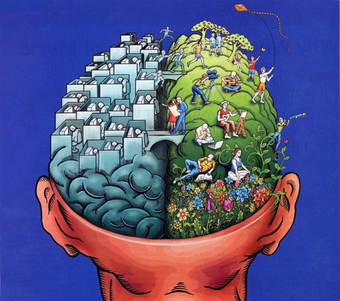
Rice. 2. Lobes of the cerebral cortex.
The following table will help you understand in more detail the structure and functions of the brain:
Conclusion
What can be said about the functions of the cerebral hemispheres? All brain centers are closely interconnected. When talking about specific areas, scientists mean a cluster of neurons that are more involved than other interconnected networks in a particular mental process.
The formation of memory, the ability to speak and think in words is the most complex mental process. This takes a lot of energy, and many nerve cells are occupied by speech.
The cerebral cortex is connected directly with conscious processes, and the subcortex is connected with the unconscious, deep parts of the personality, which Freud called “It”.
Breathing and Metabolism
Respiration provides the body with metabolic gas exchange, which involves two chemical compounds: oxygen (O2) and carbon dioxide (CO2). When there is an excess of carbon dioxide in the blood, the central nervous system sends an impulse that activates breathing, while the flow of oxygen increases. Conversely, if the body is oversaturated with oxygen, the respiratory function is inhibited, the number of chest contractions decreases, and oxygen begins to enter the blood in minimal quantities. Thus, the body maintains a balance of gas exchange.