Any strong blow to the head area can injure the brain, including those cases where the skull remains intact. Despite the fact that the brain is enclosed in soft shells and “floats” in the cerebrospinal fluid, it is not 100% protected from inertial impacts on the inner surface of the skull. If the skull is fractured, the brain can be damaged by bone fragments.
Any general practitioner, when meeting for the first time and taking a medical history, will definitely ask whether his new patient has a history of traumatic brain injuries. Brain damage can affect a person’s emotional and mental state, the functioning of his internal organs and vital systems for years.
Understanding Trauma
The human brain is responsible for all the functions performed by the rest of the body.
Even minor brain injuries make it difficult or impossible to perform these functions. Those. loss of ability to work and acquisition of disability. A fairly large part of head injuries is cerebral contusion, when, as a result of the impact of significant force on the cranium, the brain matter and its structure, as well as intracranial blood vessels, are damaged.
This can happen as a result of a domestic or work injury, an accident or a deliberate criminal attack, from being hit in the head with a heavy object or hitting the head on a hard surface.
- light,
- average and
- heavy.
The symptoms observed in the victim, the method of treatment and rehabilitation depend on how severe the brain injury was.
A brain contusion is treated exclusively inpatiently; this cannot be done at home, because... dangerous to the life of the victim. Depending on the circumstances, he must be immediately hospitalized in the intensive care, neurosurgical or trauma department of the hospital.
Traumatic brain injury is damage to the bones of the skull or soft tissue. The latter, for example, include the meninges, nerves, blood vessels and others. Head injuries are divided into several groups. Let's look at some of them in more detail.
Types of brain injuries and their signs
According to the Research Institute named after. N.V. Sklifosovsky, in Russia the main causes of brain injuries are falling from a height (usually while drunk) and injuries received during criminal actions. In total, these two factors alone account for about 65% of cases. Another 20% are road traffic accidents and falls from heights. This statistic differs from the global one, in which road traffic accidents account for half of brain injuries. Globally, 200 out of every 10,000 people suffer a brain injury every year, and these numbers are trending upward.
Brain concussion . It occurs after a minor traumatic impact on the head and represents reversible functional changes in the brain. Occurs in almost 70% of victims with traumatic brain injuries. A concussion is characterized (but not required) by a short-term loss of consciousness - from 1 to 15 minutes. Having returned to consciousness, the patient often does not remember the circumstances of what happened. In this case, he may be bothered by headache, nausea, less often vomiting, dizziness, weakness, pain when moving the eyeballs. These symptoms subside spontaneously after 5–8 days. Although a concussion is considered a mild traumatic brain injury, about half of victims have various residual effects that can reduce the ability to work. In case of a concussion, an examination by a neurosurgeon or neurologist is required, who will determine the need for a CT or MRI of the brain, or electroencephalography. As a rule, hospitalization is not required for a concussion; outpatient treatment under the supervision of a neurologist is sufficient.
Compression of the brain . Occurs due to hematomas in the cranial cavity and a decrease in intracranial space. It is dangerous because due to the inevitable infringement of the brain stem, the vital functions of breathing and circulation are disrupted. Hematomas that cause compression must be removed urgently.
Brain contusion . Damage to the brain substance due to a blow to the head, often with hemorrhage. May be mild, moderate or severe. With mild bruises, neurological symptoms last 2–3 weeks and go away on their own. Moderate severity is characterized by mental disturbances and transient disorders of vital functions. With severe bruises, the patient may remain unconscious for several weeks. Brain contusions, their degree and condition during treatment are diagnosed using computed tomography. Treatment is medicinal: neuroprotectors, antioxidants, vascular and sedative drugs, B vitamins, and antibiotics are prescribed. Bed rest is indicated.
Axonal damage . Axons are long, cylindrical extensions of nerve cells that can be damaged by a blow to the head. Axonal injuries are multiple axonal breaks accompanied by microscopic hemorrhages in the brain. This type of brain injury leads to the cessation of cortical activity and the patient falling into a coma, which can last for years until the brain starts working again. Treatment consists of maintaining vital functions and preventing infectious diseases.
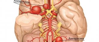
Intracranial hemorrhage . A blow to the head can cause destruction of the wall of one of the blood vessels, which leads to local hemorrhage into the cranial cavity. Intracranial pressure instantly increases, causing damage to brain tissue. Symptoms of intracranial hemorrhage are severe headache, depression of consciousness, seizures, vomiting. There is no single treatment tactic for such cases; depending on the individual picture, medical and surgical methods are combined to remove and resolve the hematoma.
Consequences of head injuries
Various consequences of a brain injury can manifest themselves during its treatment, during the rehabilitation period (up to six months) and the long-term period (usually up to two years, but possibly longer). First of all, these are mental and autonomic dysfunctions that can complicate the patient’s entire future life: changes in sensitivity, speech, vision, hearing, mobility, memory and sleep disorders, confusion. The development of post-traumatic forms of epilepsy, Parkinson's disease, and brain atrophy is possible. The more severe the injury, the more negative consequences it brings with it. Much depends not only on proper treatment, but also on the rehabilitation period, when the patient gradually returns to normal life and it is possible to track the onset of post-traumatic diseases in time in order to begin their treatment.
History knows cases where brain injuries led to the emergence of new talents in the victim - for example, increased ability to study foreign languages or exact sciences, fine arts or music. This is called acquired savant syndrome (acquired savantism). Often these abilities are based on old memories - for example, the patient may have studied Chinese for some time at school, forgotten it completely, but spoke it again after an injury and continued learning with better success.
First aid for head injuries
Anyone can find themselves in a situation where there is a person nearby with a head injury. Knowing the rules for providing first aid, you can alleviate his condition and even save his life.
- Signs of a serious traumatic brain injury include blood or clear fluid (CSF) leaking from the nose or ear and bruising around the eyes. Symptoms may not appear immediately, but several hours after the injury, so if there is a strong blow to the head, you must call an ambulance immediately.
- If the victim has lost consciousness, breathing and pulse should be checked. If they are absent, you will need to perform artificial respiration and cardiac massage. If there is a pulse and breathing, the person is placed on his side before the ambulance arrives, so that possible vomiting or a sunken tongue will prevent him from suffocating. You cannot sit him down or lift him to his feet.
- In case of a closed injury, ice or a cold wet towel should be applied to the site of impact to stop tissue swelling and reduce pain. If there is a bleeding wound, you should lubricate the skin around it with iodine or brilliant green, cover the wound with gauze and carefully bandage your head.
- It is strictly forbidden to touch or remove fragments of bones, metal or other foreign bodies protruding from the wound, so as not to increase bleeding, damage the tissue even more, or cause infection. In this case, a gauze roll is first placed around the wound, and then a bandage is made.
- The victim can only be transported to the hospital in a supine position.
The hospital conducts an examination, determines the severity of the patient’s condition, and prescribes diagnostic procedures. For open wounds with bone fragments or other foreign bodies, the patient requires urgent surgery.
Main symptoms of head injury
The symptoms of a head injury fit into 3 main syndromes:
- General cerebral, associated with a nonspecific brain reaction to injury.
- Local, depending on the immediate location of the brain injury (the most dangerous are injuries affecting the medulla oblongata, since it contains the centers for regulating breathing and cardiac activity).
- Meningeal, caused by irritation of the meninges.
General cerebral symptoms occur with a bruise of any severity. Their presence and connection with the traumatic factor allow the doctor to make a preliminary diagnosis.
These symptoms include:
- diffuse pain in the head;
- nausea causing vomiting;
- dizziness;
- decreased attention;
- weakening of memory up to its loss for some events.
It is indicated by:
- Strong headache;
- tension in the muscles of the neck and back;
- repeated vomiting, after which there is no relief, etc.
Local (focal) symptoms allow for topical diagnosis, i.e. guess in which lobe of the brain the pathological focus is located.
Thus, when the back of the head is bruised, visual functions are affected. This is due to the fact that the peripheral nerve pathway from the eyeballs ends in the occipital lobe and switches to the central one.
Therefore, a person may experience temporary blindness, double vision and other ophthalmological signs.
They should be differentiated from similar symptoms, but associated with direct injury to the eye, which leads to retinal detachment. A patient with a contusion of the back of the head requires additional consultation with an ophthalmologist.
Focal symptoms of contusion of the frontal lobes also have a characteristic picture:
- the unconscious state is replaced by mental and motor excitement;
- confusion;
- aggression;
- euphoria and incorrect assessment of one’s condition;
- reduction of criticism, etc.
Forecasting consequences
In practice, this type of injury never goes away without a trace.
A brain contusion of the second severity can provoke the formation of water on the brain - hydrocephalus. Also, such an injury can later remind itself of itself with attacks of headaches, vegetative-vascular distance, and impaired coordination of movements.
With severe severity of this injury, according to statistics, up to 50% of patients die. Survivors experience severe mental, motor and speech abnormalities, as well as epileptic seizures. Thus, suffering a serious brain injury can cause disability.
With the first degree of severity, the prognosis is almost always favorable. In this case, the main thing is to maintain bed rest. Patients with moderate severity in most cases manage to restore their ability to work and resume their social position without any consequences.
Severe head injury
- light,
- average and
- heavy.
In this case, intracerebral hematomas are noted in both frontal lobes in the form of limited blood accumulations due to various injuries with rupture of blood vessels. In this case, a cavity is formed that contains coagulated or liquid blood.
A severe injury is characterized by prolonged loss of consciousness (up to several weeks). There is often pronounced motor agitation.
Disorders of vital functions in the body are also noted. However, in comparison with the moderate degree, in the severe case they are more pronounced.
For example, there is a disorder of respiratory function with impaired patency and rhythm. The patient experiences hyperthermia and dominance of primary brainstem neurological symptoms.
In particular, swallowing disorders, floating eye movements, ptosis or mydriasis, gaze paresis, decerebrate rigidity, nystagmus, increased or depressed reflexes of the mucous membranes, skin, tendons, etc. are detected.
Neurological symptoms in the initial period (in the first hours or days) prevail over focal hemispheric manifestations. The patient may experience paresis of the limbs, subcortical disorders of muscle tone, etc.
In some cases, focal or generalized epileptic seizures are likely. Regression of focal manifestations occurs quite slowly.
Why is such a head injury dangerous? The consequences can be quite serious. Severe residual effects are often observed, mainly in the mental and motor spheres.
Damage classification
In medical practice, there are three main degrees of severity of bruise:
- The first one is easy.
- Average.
- Severe degree (concussion).
The manifestation of symptoms characteristic of a given injury directly depends on the severity of the injury. The most severe condition and consequences are observed in patients with the third degree, in which even a fracture of the base of the skull is possible.
Severe head injury
Head contusions are conventionally classified into 3 degrees, determining the severity of a person’s condition and its further prognosis.
Light damage is characterized by the following criteria:
- Loss of consciousness lasting no more than a few minutes;
- Its rapid restoration without auxiliary methods;
- General cerebral symptoms prevail over focal ones;
- Involuntary movements made by the eyeballs;
- Sometimes sensitivity and motor activity may decrease on the opposite side of the body relative to the side of the brain injury (this symptom is more typical for a moderate injury, but can also occur with a mild injury);
- Regression of clinical symptoms and morphological changes takes 2-3 weeks. Almost no residual changes are observed.
Moderate brain contusion is accompanied by a pronounced impairment of the general condition.
Its signs are:
- Longer loss of consciousness – up to 2-4 hours;
- Consciousness is stunned for several hours, up to a maximum of 24 hours;
- General cerebral symptoms are moderately expressed;
- There are manifestations of meningeal syndrome;
- Focal symptoms are loss of speech, perverted sensitivity, inability to move the limbs of the right or left side normally, increased breathing, and others.
A severe head injury (severe) poses a serious threat to life.
It may be accompanied by a coma that persists for several days. These patients have disturbances in the functioning of the respiratory and cardiovascular systems, which require medication and hardware correction. Otherwise, death occurs.
Other signs of a severe injury include:
- Loss of memory for events that preceded the injury;
- Visual impairment;
- Motor restlessness;
- Increased mental excitability, etc.
Concussion: severity levels for skull injury – Website about
29.12.2019
Quite serious head injuries that require emergency hospitalization and long-term treatment are traumatic brain injuries (TBI), which damage the tissues, bones of the skull, and the membranes of the brain.
There are open (TBI), with a violation of the integrity of soft tissues and the tendon helmet covering the cranial vault, and closed (CLT) injuries, for example, concussion, bruise, compression of the brain with the absence of violations of the integrity of tissues and tendon stretching of the skull.
According to statistics, fatal injuries account for 2/3 of TBI cases.
Causes and consequences of injury
Damage to the skull can occur due to:
- Motor vehicle accident.
- Impact (beating, domestic, sports injury).
- Accidental/deliberate fall from a hill. You can get fatally injured by falling even from a small height, not exceeding your own height.
When physical damage occurs to the bones of the cranial vault, brain structures, nerve roots and blood vessels, brain function is disrupted. Even the slightest changes occurring in neurons significantly affect the function of the damaged area.
With a strong impact, the non-branching processes of neurons are twisted and stretched. The entire force of the damaging impulse falls on the cerebral hemispheres and medulla oblongata. The difference in pressure between intracerebral structures leads to the formation of microhemorrhages associated with the rupture of the smallest capillaries.
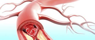
Damage to intracranial blood vessels is considered one of the most severe and dangerous head injuries.
As a result, hemorrhage occurs in neighboring areas of the brain, which causes acute attacks of headaches, nausea, and episodes of vomiting. The consequences of such conditions can be the most tragic.
Often, patients with TBI, even after long rehabilitation, experience limb paresis and cognitive impairment.
Classification of CBI
By type of skull injury they are divided into:
- Isolated.
- Combined (with damage to other organs).
- Combined (when the body is affected by other negative factors).
Depending on the type of damage to brain tissue, there are:
- Brain compression . There is a growing and non-growing type, it occurs due to large formations that significantly reduce the intracranial space. When the bones of the cranial vault are fractured, the brain is compressed by fragments, fragments of bone tissue and other foreign bodies, which is called non-increasing compression. The growing type includes any hematomas: intrathecal, intraventricular, epidural. In such patients, along with the formed hematomas, clinical manifestations of primary damage to brain structures are noted almost immediately after injury.
- Brain contusion . Brain structures are injured, and a necrotic focus of nerve fibers is formed.
- Concussion . Occurs when exposed to a small traumatic impulse. Most victims with TBI face this problem. The pathology is characterized by short-term fainting after a blow. Often, patients complain of a sensation preceding vomiting, and less often, of its episodes: dizziness, lethargy, pain when rotating the eyeballs.
The most harmless, but no less serious, requiring therapeutic measures, closed craniocerebral injury is a concussion with stretching of nerve endings without the occurrence of vascular disorders and the manifestation of pronounced changes in the structure of the brain.
Severity of concussion
Concussions are divided both by area of impact and severity:
- Easy . Characterized by the absence of loss of consciousness or its temporary loss for several minutes. The victim may have headache, vomiting, pale (or red) face, and sweating. Increased blood pressure is common in elderly patients. With hypotension it can become very low. Similar symptoms are observed within 15 minutes and disappear without consequences. If a person is under the influence of alcohol/drugs at the time of injury, unpleasant symptoms will persist longer.
- Average . Characterized by loss of consciousness for up to five minutes, dizziness, pain in the head, nausea. A pronounced sign of this condition is disturbance, confusion, and tinnitus. When examining the victim, the presence of small bruises in the eye area is revealed. Photo and sound sensitivity and loss of concentration are also recorded. Instrumental diagnostics of brain injury (computer, magnetic resonance imaging, electroencephalography) does not reveal abnormalities in brain tissue and other pathologies inherent in cerebral head injury.
- Severe degree . Significant hematomas around the eyes, fainting lasting more than five minutes, intense cephalalgia, vomiting, memory impairment and other neurological signs are noted. In this case, the functioning of the cardiac and pulmonary systems suffers, which requires immediate medical intervention.
Mild and moderate concussions threaten health no less than a severe form of closed skull injury. Therefore, the patient needs appropriate therapy and supervision by specialists.
The consequences of a concussion without proper treatment can occur at any time and lead to:
- Depressive state.
- Sudden mood swings.
- Brain swelling.
- Loss of visual acuity and hearing.
- Memory impairment.
- Neurosis.
- Migraine attacks.
- Meningioma, brain cancer.
How can I help you
It doesn’t matter what kind of concussion the person received (mild, moderate, severe). The main thing is to immediately call doctors and provide the most competent first aid for a traumatic brain injury, the basics of which everyone needs to know.
You need to do the following:
- Examine the victim for open wounds. If there are any, treat them and bandage them.
- Apply something cold to the bruise.
- Do not give water, food or medicine.
- Try to limit the patient's movements.
- If he is unconscious, first aid for a concussion is to turn the body on the right side and bend the left arm/leg at the elbow/knee.
- Provide oxygen flow.
- Place a small elevation (jacket, pillow) under your head.
- When vomiting, turn your head or lower it to prevent choking on vomit.
You cannot revive a person by shaking, tugging, sitting, or lifting him in case of injury in the absence/presence of skull fractures and other injuries. It is prohibited to hit the victim on the cheeks or head. It is not advisable to try to move or transport it without an initial examination by a specialist.
Diagnostic and therapeutic measures
Even mild TBI is fraught with functional disorders of the nervous system (up to epilepsy), impaired cerebral circulation and liquor dynamics. After hospitalization, the diagnosis can be confirmed using modern research methods (ultrasound, MRI of head and neck vessels, radiography) and examination by specialized specialists: a neurologist, an ophthalmologist, a traumatologist.
The main treatment for a concussion is based on ensuring the patient's rest (bed rest with limited use of books, tablets and other gadgets). Further therapy and length of hospitalization depend on the degree of brain damage, the general condition of the patient and the severity of clinical signs:
- Caffeine bromide mixture helps normalize sleep.
- Painkillers can help reduce the severity of headaches.
- Metabolism in nerve tissues can be improved by administering a 40% glucose solution, nootropic drugs, and vitamins B and C.
- Liquor circulation improves Eufillin, Trenatal.
During the rehabilitation period after a concussion (2-3 months), you should not drink alcohol, move to a place with different climatic conditions, or stay in open sunlight.
It is important to provide a recovering person with easy work. As home remedies, experts recommend using soothing herbs that normalize the functioning of the nervous system: motherwort, peppermint, lemon balm, mistletoe. Fried and salty foods are excluded from the diet.
30.11.2018
(1
Source:
This is serious! Concussion of the 2nd and 3rd degree of severity
A concussion is damage to brain function due to trauma that leaves no physical damage to the brain itself. This occurs because the brain collides with the inner shell of the skull at the time of injury, and the processes of nerve cells are stretched.
A concussion is the mildest of all types of traumatic brain injuries. It is not accompanied by a violation of the structure of the brain. But its cells do not perform their functions well. This is the most common type of traumatic brain injury.
Severity
According to severity they are divided into:
- 1 mild degree;
- 2 medium degree;
- 3 severe degree.
A mild degree is assigned when memory loss does not occur and there has been no loss of consciousness.
Symptoms of a concussion (lethargy, headache, nausea) remain for about 15 minutes.
Average degree. The victim does not lose consciousness. As a result of the injury, the following occurred: memory loss, symptoms accompanying the concussion last for several hours:
- slowness in action;
- pulse disturbance;
- blanching and redness of the skin;
- nausea;
- vomit;
- headache.
Severe degree. Loss of consciousness occurs and can last up to 6 hours. With a severe concussion, the symptoms vary.
Causes of concussion 2 and 3 degrees
The main cause of a severe concussion is trauma. However, this is not always a head blow, since the concussion does not damage the integrity of the skull bones. Injury can occur:
- in an accident;
- when the car brakes sharply;
- when performing sports activities;
- when falling on ice (without hitting the head);
- when hit on the head.
Symptoms of bruise
Common symptoms of a concussion include:
- lethargy;
- headache;
- dizziness;
- noise in ears;
- slow speech;
- nausea or vomiting;
- problems with coordination;
- double vision;
- fear of light and sounds;
- memory losses;
- pain when moving the eyes.
At a moderate degree it occurs:
- short-term memory loss;
- prostration;
- headache;
- dizziness;
- nausea;
- vomit;
- weakening of reflexes;
- decreased heart rate;
- constriction of the pupils.
For severe forms:
- brain contusions with intracranial hematomas are detected;
- the main symptom is loss of consciousness;
- difficulty swallowing as a result of fluid entering the respiratory tract;
- severe pallor;
- slow pulse;
- exhaustion;
- lack of reflexes;
- small pupils;
- disappearance of reaction to light;
- shallow breathing.
The main symptoms that allow you to distinguish between the mild and severe condition of a trauma victim
- With a grade 1 concussion, the main symptoms will disappear within 20 minutes and normalcy will return.
- With a grade 2 concussion, disorientation lasts more than 20 minutes.
- With a 3rd degree concussion, the victim loses consciousness and does not remember what happened.
It takes from a couple of hours to five days to restore memory.
Why is it important to correctly determine the severity of the injury caused?
A very important factor in the treatment of severe or moderate brain contusion is time.
Source: https://fiz-disp.ru/stroenie/sotryasenie-golovnogo-mozga-stepeni-tyazhesti-pri-travme-cherepa.html
Contusion of soft tissues of the head
A bruise of the soft tissues of the head, which is not accompanied by brain damage, does not pose a serious danger to humans.
This is a fairly common condition that can be caused by a blow to the head with a blunt object, without breaking the integrity of the skin. It most often occurs in athletes, but can also occur in everyday life.
A lump on the head with such a bruise is the leading symptom. She appears in the place where the blow was struck. When it is felt, it is painful. There may be minor abrasions on the skin, but there is no epithelial defect as such.
Cones are the result of 2 mutually determining processes:
- Hemorrhages in tissue due to mechanical rupture of blood vessels;
- Swelling due to the release of plasma into surrounding tissues.
Usually, no specific treatment is required for a head injury. Immediately after an injury, it is recommended to apply ice to the injured area. This will lead to vasospasm and reduce hemorrhage.
Subsequently, warming physiotherapeutic procedures (UHF, electrophoresis) are recommended to accelerate resorption. If the head hematoma after a bruise is massive, then surgical treatment may be required, consisting of two stages:
- Opening the hematoma (an incision is made on the skin under anesthesia);
- Treatment of the hemorrhage cavity and drainage (introduction of special tubes through which the contents will flow out and, if necessary, the introduction of antiseptics).
In some cases, soft tissue hematomas can fester (and this does not depend on their size). The risk of developing this complication increases in patients with diabetes.
When hemorrhage suppurates, it is opened and antibacterial therapy is prescribed. This approach will prevent the transition of purulent inflammation of soft tissues to the brain.
Physiotherapeutic treatment
To improve blood circulation in the brain tissue, the following are prescribed:
- medicinal electrophoresis with vasodilators;
- galvanization of the brain.
To increase the metabolism of nervous tissue, it is recommended:
- transcerebral UHF therapy;
- medicinal electrophoresis with drugs that improve metabolism;
- laser treatment;
- air baths.
In order to reduce high cerebrospinal fluid pressure, low-intensity decimeter therapy and therapeutic sodium chloride baths are prescribed.
To improve the rheological properties of blood, laser irradiation is performed.
Moderate bruise
This head injury is characterized by a longer loss of consciousness (up to several hours). The patient has severe amnesia.
The following signs of head injury are also observed: severe headache, repeated vomiting, mental disorders. Transient disturbances in vital functions are likely.
In particular, tachycardia or bradycardia, increased blood pressure, tachypnea (shallow rapid breathing without disruption of rhythm and patency), low-grade fever (body temperature rises to 37-37.
9 degrees). Stem and meningeal symptoms, dissociation of tendon reflexes and muscle tone, and bilateral pathological manifestations are common.
The focal symptoms are quite clear. Its character is determined by the localization of the injury.
Oculomotor and pupillary disorders, speech disorders, sensitivity disorders, paresis of the limbs and others are detected. These symptoms usually gradually subside over three to five weeks.
However, in some cases, the described clinical picture persists for quite a long time. With a moderate injury, fractures in the bones of the base and vault of the skull and extensive subarachnoid hemorrhage are often found.
On CT, focal changes are detected in the form of small high-density inclusions or a homogeneous moderate increase in density. This corresponds to minor hemorrhages in the area of the bruise or hemorrhagic penetration of brain tissue without gross destruction.
Brain contusion: consequences and rehabilitation
Brain contusion (brain contusion) is a type of brain damage that occurs as a result of traumatic brain injury (TBI).
Depending on the type and severity of the injury, pathological changes during a bruise can be varied: from single to multiple, affecting vital structures. Manifestations of brain tissue contusion are detected in 10% of victims.
This pathological condition, depending on the nature of the damage and clinical manifestations, can be mild, severe or moderate.
Minor bruise GM
The duration of loss of consciousness after a head injury characterizes the severity of brain damage.
Due to the impact of a traumatic factor, the patient loses consciousness. This condition usually lasts for several minutes.
After regaining consciousness, complaints of dizziness, repeated vomiting, nausea and headache appear. Characterized by amnesia and mild neurological symptoms (meningeal symptoms, clonic nystagmus, slight anisocoria, etc.).
Breathing and body temperature do not change significantly; blood pressure and heart rate may increase. Within 3 weeks the patient recovers and the symptoms disappear.
Moderate bruise GM
The clinical picture is characterized by a loss of consciousness for a longer period (up to several hours). The patient experiences repeated vomiting, intense headache, more severe amnesia and mental disorders.
An increase in blood pressure and body temperature, increased respiration, pulse, and meningeal signs are detected. Focal neurological symptoms appear, the manifestations of which depend on the location of the injury. These may be speech disorders, motor disorders (paresis), oculomotor disorders, etc.
The condition improves within 3-5 weeks, focal symptoms may persist longer. During examination, damage to the skull bones and subarachnoid hemorrhage are often diagnosed. The latter develops as a result of rupture of the vessels of the pia mater, and sometimes rupture of the cerebral sinuses.
Its manifestations can occur acutely (severe headache, agitation, delirium, disorientation, back pain and radicular symptoms) or increase gradually.
Severe bruise GM
After a traumatic injury, consciousness is turned off for an even longer period, which can last for days (and sometimes even weeks).
Patients develop motor agitation and various neurological manifestations: impaired swallowing, paresis, paralysis, inhibition of tendon reflexes, changes in muscle tone, convulsions, multiple nystagmus, gaze paresis, pathological reflexes, etc. Examination reveals massive subarachnoid hemorrhage and skull fractures.
This condition is accompanied by high temperature, increased blood pressure, and disturbances in the frequency and rhythm of breathing. General cerebral and focal symptoms slowly undergo reverse development and do not completely disappear.
Long-term consequences of bruise
- Post-traumatic encephalopathy.
- Episyndrome.
- Mental disorders.
- Residual neurological symptoms (motor, sensory, speech disorders, etc.).
Diagnostics
To recognize the severity of damage and its nature in traumatic brain injury, an integrated approach is needed. Dynamic observation plays an important role, since the patient’s condition can change quickly.
When making a diagnosis, the fact of injury, duration of loss of consciousness, clinical manifestations, data from a neurological examination and additional research are taken into account.
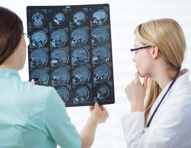
To obtain complete information about the state of the brain, the following examination methods are used:
- computer and magnetic resonance imaging (detects areas of contusion, hemorrhage, allows you to assess their size and character, as well as the condition of the ventricles of the brain, etc.);
- radiography of the skull (detects cracks and fractures of bone tissue);
- echoencephalography (determines the displacement of brain structures);
- lumbar puncture and examination of the cerebrospinal fluid (allows to recognize subarachnoid hemorrhage and intracranial hypertension, cannot be performed if there is a threat of wedging of the brain stem into the foramen magnum).
Treatment
After an injury, patients receive first aid at the scene of the accident by an emergency medical team. If the patient is unconscious, he is turned on his side or face down.
First aid measures are aimed at preventing aspiration of vomit and clearing the airways, stopping bleeding.
Such patients are required to be hospitalized in a hospital.
The nature and scope of treatment is determined by the condition and age of the victim, the severity of edema of brain tissue, liquor hypertension, impaired cerebral hemodynamics, etc.
All patients with a bruise of brain tissue are advised to rest, bed rest for a period of 7 days to 2 weeks, constant medical supervision is required. Drug therapy includes the prescription of the following medications:
- analgesics (ibuprofen, analgin, ketorol);
- antiemetic drugs (metoclopramide, domperidone);
- sedatives (phenazepam, Relanium, adaptol);
- with severe agitation - haloperidol, sodium hydroxybutyrate;
- diuretics (furosemide, diacarb, manitol);
- antihistamines (tavegil, suprastin);
- hemostatic agents for hemorrhage (dicinone, etamsylate);
- Medicines that improve blood circulation in brain tissue (sermion, vinpocetine);
- metabolic agents (piracetam, cerebrolysin);
- nootropic drugs (zncephabol, nootropil);
- B vitamins (milgamma, neirovitan).
To sanitize the cerebrospinal fluid and reduce its pressure, a therapeutic lumbar puncture is used.
Severe bruises of the brain require resuscitation and intensive care.
Surgical treatment is indicated for large areas of tissue crushing and the absence of effect from conservative treatment.
Measures for caring for patients with bruises of the brain consist of the prevention of bedsores, pneumonia, and passive exercises to prevent contractures.
Patients who have suffered a brain injury are subject to long-term follow-up.
During the recovery period, they are recommended to take courses of vascular therapy, exercise therapy, physiotherapeutic and sanatorium-resort treatment.
The latter can be prescribed several months after the injury in the absence of pronounced motor and mental disorders. In the presence of gross residual defects, the issue of the patient’s ability to work is resolved.
Physiotherapeutic treatment
To improve blood circulation in the brain tissue, the following are prescribed:
To increase the metabolism of nervous tissue, it is recommended:
- transcerebral UHF therapy;
- medicinal electrophoresis with drugs that improve metabolism;
- laser treatment;
- air baths.
In order to reduce high cerebrospinal fluid pressure, low-intensity decimeter therapy and therapeutic sodium chloride baths are prescribed.
To improve the rheological properties of blood, laser irradiation is performed.
First aid at home and when to go to hospital
First aid for a head injury - its quality and timeliness - determine the effectiveness of further treatment. Therefore, you need to know how to provide it correctly.
The priority activities are:
- Turn the injured person's head to the side to prevent possible vomit from entering the respiratory tract;
- Removal of all removable dentures and removal of foreign bodies from the mouth;
- If consciousness is preserved, then the person must lie down - standing or sitting is prohibited;
- Fixation of the cervical spine using any means that are at hand.
It should be remembered that if you receive any head injury, you should always consult a doctor, because... In some patients, bruises may occur with minimal symptoms at the beginning, but then lead to serious consequences.
Causes and mechanism of development of the condition
Brain contusion accounts for about a quarter of all closed head injuries. This consequence can occur with any aggressive mechanical impact on the head, as a result of which a zone of increased pressure is formed. This leads to the primary destruction of brain tissue and the blood vessels passing through it.
A zone of low pressure appears on the side of the skull opposite the impact, which also causes extensive damage to nerve cells.
The situation is aggravated by the displacement of the central nervous system, which disrupts the transmission of impulses from the cerebral cortex to its deep structures. Inhibition of the reticular formation causes loss of consciousness. An additional negative factor is the appearance of pinpoint hemorrhages in the gray matter. They can be microscopic or extensive, which in the latter case leads to the formation of a hematoma. If it is not eliminated, then after a few hours or days it will begin to put pressure on the brain and its membranes.
The main cause of brain contusion is head trauma. It can be a consequence of a traffic accident, an industrial accident, carelessness at home, a fight, or an attack. The risk group for this condition includes overly active children, athletes and extreme sports enthusiasts, and people with epilepsy.
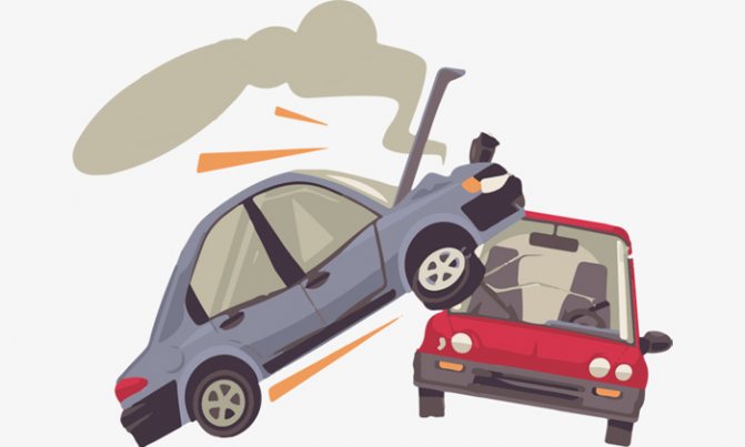
Brain damage can be caused by a traffic accident.
Diagnosis and treatment
Diagnosis of patients with suspected head contusion is carried out comprehensively:
- X-ray (to exclude fractures and identify local lesions in the brain);
- Spinal puncture (increased number of red blood cells is determined);
- Computed tomography (with its help you can identify not only the site of the injury, but also the therapeutic reserve zone - edema and ischemia).
The principles of treatment for brain contusion are determined by the nature and stage of pathological changes. Depending on this, primary and secondary damage to nervous tissue is distinguished.
Primary are those that are directly caused by the impact of a traumatic factor. These injuries are represented by a variety of conditions:
- Violation of the structure of nerve cells and glia (surrounding nervous tissue);
- Breaks in connections between nerve cells;
- Vascular thrombosis;
- Rupture of the vessel wall;
- Increased permeability of cell membranes and energy starvation (the number of ATP molecules decreases), accompanied by cell death.
There is a zone of increased sensitivity around the immediate pathological focus. These are living nerve cells, but easily vulnerable when exposed to any pathological factor (lack of glucose or oxygen).
It is this zone that represents the therapeutic reserve, i.e. with proper treatment, these cells will replace the dead ones, and there will be no loss of the function for which the bruised lesion was responsible.
Secondary damage develops as a result of the inflammatory process, which is always present during injury. Depending on the intensity of inflammation, nerve tissue cells can be either restored or damaged. Treatment should be aimed at creating conditions for recovery.
Treatment of a head contusion can be conservative or surgical. The latter type of assistance is required in 10-15% of cases for patients diagnosed with brain contusion.
Indications for surgical treatment are:
- Hematoma, the internal diameter of which exceeds 4 cm;
- Significant displacement (more than 5 mm) of brain structures, with the exception of the hemispheres;
- Severe intracranial hypertension, which cannot be eliminated by pharmacological methods.
Conservative treatment includes:
- Diuretics to reduce the severity of cerebral edema;
- Oxygen therapy (if necessary, tracheal intubation is performed);
- Infusion therapy and maintaining blood pressure at an adequate level;
- Anticonvulsants;
- Antihypoxants that reduce the severity of ischemic changes, increase the resistance of nervous tissue to oxygen starvation and promote its recovery.
The bruise is dangerous because as a result of damage to the tissue and structure of the brain and intracranial vessels, within 2-3 days the patient may develop cerebral edema, a hematoma, an ischemic stroke, etc.
Preference is given to conservative treatment, all medications are aimed at combating cerebral edema and intracranial hypertension.
In severe cases, surgical intervention is performed, i.e. craniotomy and removal of damaged brain matter or swelling.
After discharge from hospital treatment, the patient undergoes a long rehabilitation journey, and not all of his neurological and mental symptoms disappear over time; many remain forever.
Diagnostics
To recognize the severity of damage and its nature in traumatic brain injury, an integrated approach is needed. Dynamic observation plays an important role, since the patient’s condition can change quickly. When making a diagnosis, the fact of injury, duration of loss of consciousness, clinical manifestations, data from a neurological examination and additional research are taken into account. To obtain complete information about the state of the brain, the following examination methods are used:
- computer and magnetic resonance imaging (detects areas of contusion, hemorrhage, allows you to assess their size and character, as well as the condition of the ventricles of the brain, etc.);
- radiography of the skull (detects cracks and fractures of bone tissue);
- echoencephalography (determines the displacement of brain structures);
- lumbar puncture and examination of the cerebrospinal fluid (allows to recognize subarachnoid hemorrhage and intracranial hypertension, cannot be performed if there is a threat of wedging of the brain stem into the foramen magnum).
Consequences of a bruise
The consequences of a head injury are varied and depend on the severity of the condition. In mild cases, symptoms usually quickly regress without leaving a trace. With severe bruises, there is a high probability of certain complications:
- Apallic syndrome - a person is conscious, but indifferent to his surroundings, unable to fixate objects and people, reacts only to painful stimuli (state of waking coma);
- Paresis – loss of the ability to move muscles;
- Brain cysts;
- Abscess is the formation of a purulent cavity in the brain;
- Persistent intracranial hypertension;
- Chronic headache is a condition where the head hurts after a bruise for 6 months or more;
- Meningitis is an inflammatory lesion of the meninges;
- Secondary epilepsy.
The success of treatment will depend on the timeliness of seeking help and the severity of the lesion.
Consequences of TBI
Depending on the severity of the traumatic brain injury, the consequences range from mild to extremely severe. Neurologists like to repeat that the human brain is a very plastic organ, sometimes capable of recovering even from critical injuries. At the same time, making predictions about the speed and completeness of recovery even after moderate injuries is not an easy task, because the consequences of injuries can make themselves felt several weeks, months and even years after the incident, especially when it comes to TBI in children.
The most common consequences of skull and brain injuries are:
- partial or complete paralysis of the limbs on one or both sides;
- loss of sensation in the legs, arms, or skin of the torso;
- chronic headaches;
- loss of vision, hearing, ability to speak;
- lack of ability to breathe independently, swallow;
- loss or weakening of control over the function of the pelvic organs (the patient is not able to control the processes of urination and defecation);
- development of post-traumatic epilepsy (convulsive seizures, in some cases accompanied by loss of consciousness);
- tremor (shaking) in the limbs;
- partial memory loss, deterioration of attention, changes in character and other consequences associated with higher nervous activity.
It is important to understand that a particular patient will not necessarily experience all of the listed consequences of TBI: it depends on which parts of the brain were affected and how severe the injury was. Some symptoms - such as paralysis of the limbs or breathing problems - appear only in the early post-traumatic period, and then disappear safely. Others, such as excruciating headaches, can make themselves felt many years after a seemingly minor injury (this is often observed in professional athletes).
Features of recovery of patients with traumatic brain injuries
Unlike strokes, which in many patients develop according to a similar scenario, TBI implies a significant variety of options for the development of events. Depending on the mechanism of injury and its combination with other injuries to the body, the patient’s condition and prognosis are very different. In some cases, immediately after the incident, people fall into a coma for several days or weeks, which can anticipate the death of the patient or act as an “energy-saving” mode for restoring damaged brain functions.
And yet, doctors note that in most cases, the patient’s condition after a TBI improves over time: and by the speed of positive changes that occur, it is possible to assume how good the prognosis will be. Therefore, it is so important to think about rehabilitation measures even before the patient is discharged from the neurological hospital, literally from the first days of treatment. Working with a psychologist, early physical activity, and even physical therapy can significantly increase the chances of the victim returning to normal life without long-term impairment. On the contrary, seeking rehabilitation help too late does not always work: after several months after a TBI, it will be very difficult to reverse some pathological changes.
Thus, every patient who has experienced traumatic brain injury requires a multidisciplinary approach. For example, severely ill patients with disorders of stem functions - breathing and swallowing - will need the help of rehabilitation specialists and neuropsychologists. The loss of speech functions puts work with a speech therapist in first place in priority. In cases of mental changes, chronic headaches and problems falling asleep, a neuropsychologist and occupational therapist will play the main role in rehabilitation.
FAQ
- Can you feel dizzy after a head injury?
Depending on the severity of the bruise and its massiveness, dizziness may persist for several months. If it is very intense, the doctor may prescribe specific medications that will help eliminate this unpleasant symptom.
Over time, with a slight injury, dizziness goes away on its own.
- What to do if you bruise the back of your head?
In this case, immediately after injury you must:
- apply ice or a towel soaked in cold water to the injured area;
- take a horizontal position and turn your head to the side;
- call an ambulance or go to the hospital yourself (when transporting in a car, it is recommended to lower the seat as much as possible).
(Visited 28,522 times, 4 visits today)
CT indicators
In case of severe trauma, in a third of cases, focal lesions in the brain are observed in the form of heterogeneous areas of increased density. In this case, an alternation of zones is observed.
Areas with increased and decreased density are identified. In the most severe course of the condition, the destruction of the brain substance is directed deeper and can reach the ventricular system and subcortical nuclei.
Observations of dynamics show a gradual decrease in the volume of compacted areas, their merging and transformation into a more homogeneous mass. This occurs 8 or 10 days after the incident.
Regression of the volumetric effect of the pathological substrate occurs more slowly, which indicates the presence of unresolved clots and crushed tissue at the site of the contusion. At this point, they become equally dense relative to the surrounding edematous medulla.
Disappears after 30-40 days. The volumetric effect indicates the resorption of the substrate and the formation of areas of atrophy or cystic cavities instead.
Therapeutic measures for bruises
Regardless of the extent of the damage, the patient should receive medical care. If there is a head injury, the victim must be transported to the hospital as soon as possible.
To make an accurate diagnosis, radiography and CT are indicated. The patient needs bed rest.
Its duration in mild cases is 7-10 days. with average – up to 14 days.
In case of severe TBI, resuscitation measures must be taken. They begin in the prehospital period and continue in hospital settings.
To normalize breathing, it is necessary to ensure free passage in the upper respiratory tract - free them from mucus, blood, and vomit. An air duct is inserted and a tracheostomy is performed (dissection of the tracheal tissue and installation of a cannula or the formation of a permanent opening - a stoma).
Inhalation using an oxygen-air mixture is also used. If necessary, mechanical ventilation is used.
Treatment
After an injury, patients receive first aid at the scene of the accident by an emergency medical team. If the patient is unconscious, he is turned on his side or face down. First aid measures are aimed at preventing aspiration of vomit and clearing the airways, stopping bleeding. Such patients are required to be hospitalized in a hospital.
The nature and scope of treatment is determined by the condition and age of the victim, the severity of edema of brain tissue, liquor hypertension, impaired cerebral hemodynamics, etc.
All patients with a bruise of brain tissue are advised to rest, bed rest for a period of 7 days to 2 weeks, constant medical supervision is required. Drug therapy includes the prescription of the following medications:
- analgesics (ibuprofen, analgin, ketorol);
- antiemetic drugs (metoclopramide, domperidone);
- sedatives (phenazepam, Relanium, adaptol);
- with severe agitation - haloperidol, sodium hydroxybutyrate;
- diuretics (furosemide, diacarb, manitol);
- antihistamines (tavegil, suprastin);
- hemostatic agents for hemorrhage (dicinone, etamsylate);
- Medicines that improve blood circulation in brain tissue (sermion, vinpocetine);
- metabolic agents (piracetam, cerebrolysin);
- nootropic drugs (zncephabol, nootropil);
- B vitamins (milgamma, neirovitan).
To sanitize the cerebrospinal fluid and reduce its pressure, a therapeutic lumbar puncture is used.
Severe bruises of the brain require resuscitation and intensive care.
Surgical treatment is indicated for large areas of tissue crushing and the absence of effect from conservative treatment.