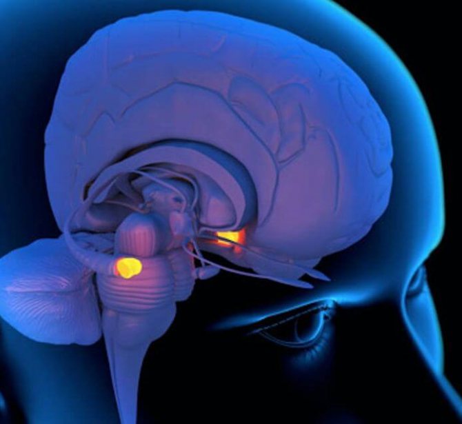
The pineal gland is an important element of the endocrine system. The formation of an elliptoid shape in the brain, despite its small size, produces several important hormones and neurotransmitters, among them serotonin.
When the pineal gland is damaged, sleep and wakefulness patterns are disrupted, insomnia develops, and the condition of blood vessels worsens. Melatonin deficiency negatively affects the endocrine system. If you experience frequent headaches, insomnia, general weakness, or decreased visual acuity, you need to visit specialized specialists and undergo diagnostic tests to rule out severe damage to the pineal gland.
[contents]
Functions of the pineal gland
For many years, British medical scientists have made attempts to understand what the pineal gland of the brain is responsible for. But even today, the potential and role of this gland have not been fully studied.
Affecting all processes of the body, the pineal gland is involved in stabilizing metabolic functions and the functioning of the central nervous system.
At the very core of the human brain there is a gland - the pituitary gland, which affects growth, metabolic functions of the body, and development. If the pituitary gland works at full strength, a person’s skeleton is more actively formed and secondary sexual characteristics (menstruation, erection, active development of the genital organs, etc.) are progressively developed. To balance the processes, the pineal gland inhibits the growth of the pituitary gland until the onset of puberty.
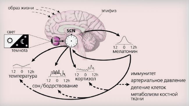
In addition, the pineal body takes part in the organization of stable biological rhythms that determine sleep or wakefulness, rest or a strong “burst of strength” - physical or emotional uplift. Performs the following functions:
- The pineal gland maintains circadian rhythms and sleep patterns in the right way.
- Slows down sexual development in adolescents.
- Suppresses the release of excess growth hormone until puberty.
- Inhibits the growth of tumors.
- Increases immunity.
Particular activity of the pineal gland is observed in children. As you grow older, the mass of the gland decreases and its functionality weakens.
Genesis, anatomical structure and histology
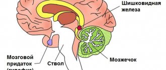
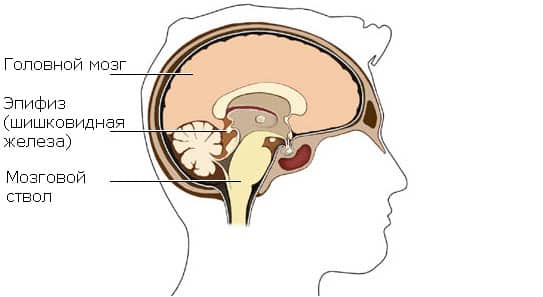
The pineal gland is part of the diencephalon, which, in turn, is located between the midbrain and the cerebral hemispheres. The normal dimensions are small, about 1 cm wide and 1.5 cm long, with a weight of only 0.15-0.2 g (in women the pineal gland is usually larger than in men).
The pineal shape of the gland is due to the developed capillary network of this organ. In addition to blood vessels, nerve fibers of the sympathetic system pass through the pineal gland.
The pineal gland appears in the human embryo already in the second month of development; with age, its size increases, it penetrates into the region of the midbrain and is fixed there between the superior visual hillocks of the quadrigeminal midbrain.
The location of the pineal gland in the center of the brain gives it special significance; some scientists even consider it the superior appendage of the brain, similar to how another important endocrine gland, the pituitary gland, is considered the inferior cerebral appendage. The pinkish-gray color of the pineal gland is due to its good blood supply.
On the outside, the pineal body of the epiphysis is covered with dense connective tissue. The growth of the pineal gland stops at the onset of puberty, and as the body ages, its reverse development is observed.
The pineal gland is a small organ of a grayish-red hue, which belongs to the interstitial
brain
. In the human body, it is formed, starting in utero from the fifth week, from the caudal end of the roof of the interstitial cord in the form of epithelium, which lines all structures of the brain and spinal cord.
Over time, during the development of the pineal gland, its walls thicken, the lumen disappears, and particles of mesoderm are introduced into it parallel to the blood vessels.
The pineal gland is located deep beneath the hemispheres of most of the brain. The upper part, which is tilted back and is directly located in the recesses between the parts
brain
. The base of the epiphysis is directed forward and, due to a small stalk, is attached to the lateral ventricle.
Along it from the gland on both sides there are two white bundles that are adjacent to the thalamus. Form 3 of the ventricle has a slight protrusion into the base of the epiphysis, which represents the residual part of the primary lumen of the epiphysis embryo.
The shape of the pineal gland in humans constantly changes throughout life. In infancy up to a year, it has a rounded body, and in a mature person it has a flattened shape from top to bottom with the dimensions of the lateral surfaces within 5 mm.
The end and upper part of the organ is covered by the posterior part of the choroidal membrane of the lateral stomach, and on the lower back side by the pia mater. From the connective tissue that surrounds the pineal gland. In the inner part there are layers that divide the multi-structured fabric into several parts of different shapes and sizes.
The structure of the totality of the main elements of the pineal gland:
- The main pineal cells are enriched with lipoid and pigment inclusions.
- Protoplasms of cells having eosinophilic granulation and punctation.
- At the moment, it has been proven that there are nerve cells in the structure of the pineal gland.
Structure of the pineal gland:
- Pinealocyte
- Neurons
- Perivascular cellular formations of the immune system that protect the body by consuming phagocytosis.
- Leydig cells
- Peptidergic neuron-like structures.
The enrichment of the glands with blood and its movement occurs with the help of two main arteries: the main and internal carotid. On the upper part of the quadrigeminal area, an arterial network is formed around the pineal gland, allowing up to a dozen branches to penetrate into the thickness of the organ through the connective tissue. The abundant blood supply to the gland indicates its active functionality.
It is worth noting that the main growth and development of the pineal gland occurs during puberty. Over time, the amount of connective tissue in the pineal gland increases and the number of parenchyma cells decreases significantly.
Accordingly, the characteristic features of embryogenesis emphasize the importance of the penial gland. And considering this organ to be rudimentary, especially during puberty, is a significant mistake.
The influence of pineal gland hormones on the human body
The most important function of the pineal gland in the brain is to produce the hormones melatonin, serotonin, histamine and norepinephrine. The healthy functioning of the organ in childhood ensures the child’s intellectual development and his ability to learn.
The effect of hormones on the body as a whole:
- Supports stress resistance of the central nervous system;
- Control blood pressure;
- Regulate sleep starting from embryonic development;
- Supports carbohydrate and fat metabolism in the body.
Malfunction of the pineal gland affects the biological rhythms of the body: insomnia, shallow sleep and feeling tired in the morning.
Causes of pineal gland dysfunction:
- Cystic formation in the epiphysis of the brain;
- Degeneration of cellular composition with age-related changes in the body;
- Amyloidosis;
- Accumulation of calcifications;
- Neoplasm in the pineal gland;
- Blockage of a vessel providing blood supply to an organ by a thrombus;
- Stroke;
- Inflammation and lack of blood supply (with intoxication, infectious diseases, hemorrhagic diathesis);
- Hypertension.
Each hormone produced by the pineal gland is responsible for certain conditions of the human body.
Melatonin
The pineal gland secretes melatonin into the blood, which is produced from serotonin and is involved in the regulation of circadian rhythms. The substance has virtually no effect on the depth of sleep. The pineal gland is the main source of melatonin in the human body (produces approximately 80% of this substance), in addition, melatonin is produced in the gastrointestinal tract (in particular, in the cells of the appendix) and a number of other organs.
Melatonin synthesis is blocked by bright light, the effect of which on the gland occurs through nerve pathways included in the photoneuroendocrine system. Melatonin is characterized by daily fluctuations in concentration. Its maximum level is observed between 00:00 and 05:00. Less melatonin is produced in summer and in winter. A change in the rhythm of production occurs when moving to other time zones.
With a normal daily routine (with sleep at night), a person produces 70% of the total daily production of melatonin at night. With insufficient sleep duration, its synthesis at night decreases and approaches daytime levels.
The functions of melatonin include:
- regulation of blood pressure;
- decreased mental and physical activity;
- slower growth and sexual development in children;
- increased antibody production;
- slowing down aging;
- participation in the regulation of the functions of the thyroid gland and thymus;
- inhibition of the production of gonadotropins, as well as other hormones of the anterior pituitary gland;
- decrease in the concentration of glucose and cholesterol in the blood;
- improving learning ability;
- antioxidant effect.
Melatonin is one of the few hormones that have nuclear and membrane receptors. Its transport is carried out through serum albumin, after being released from which melatonin binds to receptors located on the membrane of target cells and penetrates into the cell nucleus, where it produces its effect. Hydrolyzed in the liver and excreted in the urine.
With age, due to a decrease in the activity of the pineal gland, the level of melatonin decreases, which leads to the appearance of age-related sleep disorders (shallow sleep, insomnia). Also, a violation of melatonin secretion can occur against the background of stress, prolonged exposure to a computer monitor.
Violation of melatonin synthesis causes the following conditions:
- weekend insomnia;
- insomnia due to a shift work schedule;
- delayed sleep phase syndrome;
- jetlag (time zone change syndrome);
- daytime sleepiness;
- nightmares;
- deterioration of skin and hair condition;
- depressive states.
Serotonin
Serotonin is one of the main neurotransmitters, it is also known as the “happy hormone” or “good mood hormone.” Sunlight is required to produce serotonin. Norepinephrine inhibits its release.
Serotonin is involved in the regulation of vascular tone, as well as (together with dopamine) in the regulation of the hormonal function of the pituitary gland by the hypothalamus.
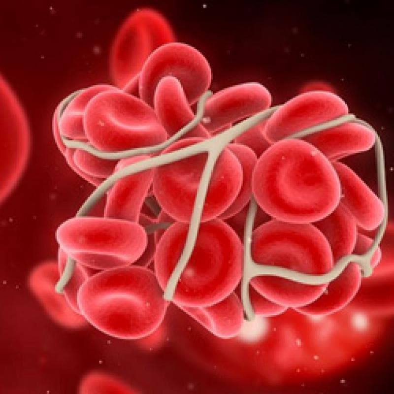
Plays an important role in blood clotting processes:
- increases the functional activity of platelets;
- increases the tendency of platelets to form blood clots;
- stimulates the liver's production of blood clotting factors.
A large amount of serotonin is also produced in the intestine and is involved in the regulation of secretion and motility of the digestive tract, in particular, it increases secretory activity and peristalsis.
Serotonin is found in large quantities in the uterus and a number of other organs and tissues, and is involved in the coordination of labor. In men, serotonin, when its level in the blood increases, can delay the onset of ejaculation.
Serotonin takes part in the development of allergic and inflammatory reactions:
- increases the permeability of blood vessels;
- enhances the migration of leukocytes to the site of inflammation;
- increases the number of eosinophils in the blood.
Winter lack of ultraviolet radiation, necessary for the synthesis of serotonin, can lead to the development of seasonal depression. A decrease in the level of the hormone in the blood can lead to increased sensitivity to pain and the development of migraines.
The pineal gland produces the “happiness hormone” – serotonin.
Excess serotonin causes serotonin syndrome. This can occur with the concomitant use of antidepressants that belong to the classes of selective serotonin reuptake inhibitors and monoamine oxidase inhibitors, as well as narcotics. In this case, a person experiences mental disorders, neuromuscular disorders, signs of autonomic dysfunction, and death is possible.
The amino acid tryptophan, from which serotonin is produced, is found in dairy products, soy, dark chocolate, plums, tomatoes, and dried fruits.
Adrenoglomerulotropin
Adrenoglomerulotropin is a hormone of the pineal gland, which is formed as a result of the biotransformation of melatonin. Among the main target cells of this substance are the cells of the zona glomerulosa of the adrenal cortex.
Adrenoglomerulotropin stimulates the synthesis of aldosterone, which is the main mineralocorticoid in humans (not in all vertebrates).
The psychoactive effects of adrenoglomerulotropin on humans have not been fully studied.
Dimethyltryptamine
Dimethyltryptamine is an endogenous psychedelic substance produced in small amounts by the pineal gland during REM sleep. This substance in certain concentrations can cause visual and auditory hallucinations, impaired perception of time and surrounding space, and an altered state of consciousness. Based on the results obtained by performers in separate studies, it can be assumed that dimethyltryptamine is used by the human body to create the visual effects of natural dreams.
Dimethyltryptamine is synthesized from the proteinogenic amino acid L-tryptophan; its structure resembles serotonin.
Norepinephrine
The hormone is a biogenic amine and is synthesized from tyrosine (an amino acid), which the body receives from food. The process of norepinephrine synthesis occurs in the pineal gland of the brain and adrenal glands.
The hormone improves the functioning of brain structures and promotes the absorption of glucose in muscle fibers.
Norepinephrine begins its work in stressful situations, increasing heart rate and increasing blood pressure, which is why it is called the “stress hormone.”
Functions and hormones
Neuropathologists and neurosurgeons who study the pineal gland of the brain have long studied what it is and why it is needed.
The main function of the pineal gland is the regulation of circadian (otherwise known as cycadal) rhythms, metabolism, and other endocrine functions. It secretes vital hormones melatonin, serotonin and some others. They are most actively released at night, in the dark, when a person has been sleeping for several hours - this is from midnight until dawn. Artificial lighting deceives the pineal gland, blocking the release of hormones. Therefore, absolute darkness is necessary for night sleep.
Melatonin hormone
Melatonin is produced in the blood only at night; light blocks its release. With the onset of dawn, another hormone begins to be produced. During the day, there is significantly less melatonin in the body; the pineal gland does not produce iron at this time.
A supply of melatonin can be obtained from food, although in quantities that are completely insufficient for health.
Melatonin is also called the “hormone of youth”, which prevents early aging of the body. If there is lighting, it is practically not produced. This is why it is not recommended to sleep with light on. During the polar day, doctors recommend tightly curtaining the windows in the bedrooms.
Melatonin is also directly related to sexual function; it is also responsible for the cyclicity of menstrual function in women.
The lack of this hormone leads to psychosomatic diseases, metabolic disorders, and sleep disorders. Because of this, general health worsens, insomnia occurs, and women may experience ovarian dysfunction. Its high level gives healthy longevity and good physical fitness, vision and memory.
Now a synthetic melatonin has been created, which is similar in structure to natural one. It is prescribed to people with severe insomnia.
Hormone serotonin
This hormone of the pineal gland regulates the psycho-emotional state of a person, relieves stress, and regulates smooth muscle tension. Helps cope with various states of anxiety, obsessive thoughts, aggressiveness, and so on. Its popular name is “happiness hormone”. Unlike melatonin, it is produced during the daytime.
Additional positive emotions also promote the production of serotonin.
Excess light interferes with the conversion of serotonin to melatonin.
With a lack of this hormone, depression and various phobias develop. Alcoholism and drug addiction can also be caused by a lack of this hormone. More complex mental illnesses are also possible.
Harmonizes the rhythm of wakefulness and sleep
To maintain life, the body needs to regulate the rhythm of rest and wakefulness. At night, it reduces nervous tension and produces substances that have a hypnotic effect on the body. If at this time you prevent this blockage and take substances that, on the contrary, invigorate, you can disrupt this function. Which will subsequently require drug treatment.
Suppression of sexual desire
Pineal gland hormones suppress sexual desire in childhood. In children and adolescents, the epithalamus is larger relative to the size of the brain than in adults, and more melatonin is produced per unit of weight. Accordingly, attraction does not arise at this age. With the onset of puberty, the suppression gradually ceases.
Inhibits the development of tumors
Pineal gland hormones prevent the uncontrolled production of growth hormone. This helps the body recognize pathological cancer cells in time and prevents them from developing. We have the pineal gland to thank for this obstacle.
Helps to navigate in space
Thanks to the hormones of the pineal gland, a person can determine his location.
It also stimulates the immune system, and in general all metabolic processes of the body. With age, the pineal gland begins to decrease in size and atrophy. This inevitably leads to the occurrence of oncology.
What does the pineal gland produce?
Functioning as a gland, the pineal gland supplies hormones to the human body:
Melatonin . It is the most important and indispensable regulator of the circadian rhythm and, produced at the beginning of twilight, helps to fall asleep. Entering directly into the blood, the hormone notifies all cells of the body about the onset of night. Almost all tissues and organs contain receptors that are sensitive to melatonin, but they work depending on many factors present in a particular person. That is why the effect of the hormone on the body may vary.
This:
- Full healthy sleep.
- Calm nervous system and stress resistance.
- Stabilization of blood pressure.
- Decreased blood glucose levels.
- Reducing cholesterol levels.
- Strengthening the body's protective properties.
- Ensuring sufficient potassium in the body.
Among other things, being involved in the formation of the state of sleep, this hormone enhances the effect of sedatives, and in some cases, it is used in preparations as a medicine for minor disturbances in night sleep.
Among representatives of the younger generation, the functionality of melatonin has the highest indicators: the hormone increases the ability to learn and improves the body's protective functions, strengthens vascular walls.
If there is too bright lighting in the evening and at night, a person begins to experience sleep disorders and diseases of the nervous system, depression, and depression. To avoid such complications or at least reduce their occurrence, preference should be given to natural changes in lighting.
- Serotonin (“happiness hormone”) is produced in daylight. Improves a person’s psychological and emotional state, facilitates motor activity and has other functions. It accumulates in the gland, and at night is converted into the most important hormone of the pineal gland - melatonin.
- Histamine. Supports vital functions of the body.
- Norepinephrine. It affects the metabolic functions of the body and regulates blood pressure.
- Peptide hormones. Stimulate the functioning of the sex glands, normalize reproductive functions in women and regulate the duration of the menstrual cycle. They also affect the immune system, metabolic functions of the body and the condition of blood vessels.
Structure
This small gland has a very complex structure; it is all surrounded by blood vessels. About 200 ml of blood passes through it per minute.
This small organ, located deep in the brain, is involved in all metabolic processes that occur in the body.
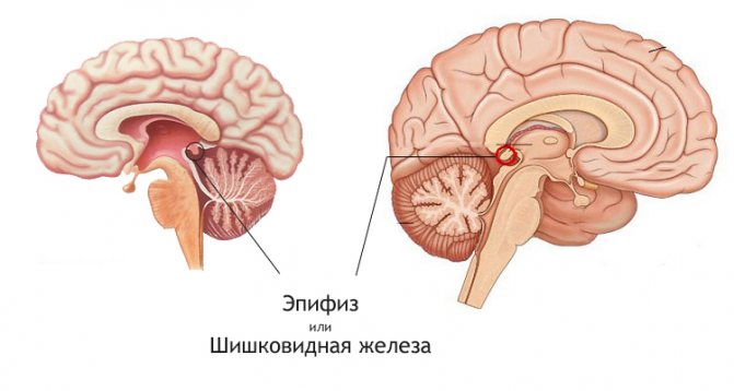
It is all covered with connective tissue, and the pineal gland itself, or pineal gland, is divided into hemispheres. They are also divided into small divisions. It is these divisions that create the resemblance to a pine or fir cone.
Hyperfunction and hypofunction of the pineal gland
If for some reason the pineal gland malfunctions, a person begins to experience serious disorders in the functionality of the entire body.
Pelizzi syndrome begins to develop, which most often occurs in boys and young men aged 5-20 years. The syndrome is characterized by the following signs: accelerated growth of the skeleton and muscles, changes in voice, mental disorders, and strong attraction to the opposite sex.
The most pronounced signs of pathology include the early development and formation of the genital organs and secondary signs - testes, as in adult men; pubic hair in boys; menstruation and even the possibility of early pregnancy in girls.
Where is the pineal gland located? Location of the gland
The superior cerebral appendage is located at the bottom of the third cerebral ventricle. In the oblong depression between the upper colliculi of the quadrigeminal, where the epiphysis is located, leads come from the pineal gland that attach it to the visual tuberosities.
Thus, one side of the epiphysis is located in the region of the midbrain, and the other is adjacent to the third ventricle. In simple terms, the pineal gland is a type of gland located between the hemispheres of the brain. The volume of the gland in an adult is from 25 to 430 mg, while the mass of the pineal gland depends on many factors: the person’s age, his state of health, gender, environmental conditions and climate (the average is 0.2 grams).
What is the pineal gland of the brain
Between the hemispheres of the brain there is a formation similar to a gray-red pine cone - the pineal gland. The gland is located behind the third ventricle of the brain next to the canal connecting its cavity with the fourth ventricle. Anatomically, this region is superior to the thalamus, and is therefore called epithalamic. The pineal gland belongs to the endocrine glands, although given its functions it is closer to the diffuse endocrine system, the cells of which are found throughout the body.
The hormones produced by this organ inhibit many endocrine glands - the pituitary gland, the thyroid gland and the reproductive glands. The main feature is the change in the functional activity of the pineal gland depending on the degree of illumination. This happens because light impulses from the eyes are sent along nerve fibers to the epiphyseal cells.
The pineal gland cannot perceive objects, but does respond to light. Thanks to this feature, esotericists attribute to it the functions of the third eye. Previously, it was considered the container of the human soul.
The pineal gland has the ability to respond to the earth's magnetic field. This feature is a kind of built-in compass that allows you to navigate in space. In general, the pineal gland is capable of changing hormonal levels and the body's reactions to changes in the external environment, helping a person adapt to new conditions.
We recommend reading the article about pituitary adenoma. From it you will learn about the causes of pituitary adenoma, symptoms of a brain tumor in men and women, classification of the disease, as well as the diagnosis and treatment of pituitary adenoma.
What it is
The pineal body is a formation up to 1 cm in size, one of the structures of the diencephalon. Small lobules and cords of the epiphysis consist of light and dark cells.
Location - brain, area - above the quadrigeminal tuberosities, color of the formation - grayish-red. The cells of the important gland are associated with the receptive area of the eyes. The pineal gland is most active when it gets dark.
Vessels in the pia mater deliver oxygen and nutrients to the pineal gland. Nerve fibers also approach the elliptoid formation.
The formation of an important endocrine gland occurs already in the fifth week after conception. The pineal gland's own hormones are active during intrauterine development.
The pineal gland is a small but important structure in the brain. This element is less known to people far from medicine, but the role of the endocrine gland, shaped like a pine cone, cannot be underestimated. When the functioning of the pineal gland is disrupted, pathological changes occur in the body, circadian rhythms are disrupted, hormonal and metabolic pathologies arise, problems with sleep and the development of the reproductive system.
What is an ovarian teratoma and how to get rid of the formation depending on its shape? We have the answer!
Leptin hormone is elevated: what does this mean and how to bring the levels of an important substance back to normal? Read the answer at this address.
Functions of the Pineal Gland
The main task of the pineal gland is to synchronize the work of the organs of the endocrine system with the degree of illumination. The second important point is the effect on the cyclic activity of serotonin.
Other important functions of the pineal gland:
- inhibits the activity of important parts of the brain (hypothalamus and pituitary gland) at night,
- sufficient production of pineal gland hormones provides a hypnotic effect,
- suppresses excessive nervous stimulation,
- maintains the tone of veins, capillaries and arteries,
- prevents premature sexual development in children with the help of physiological regulators,
- normalizes circadian rhythms, maintains optimal duration of wakefulness and night sleep,
- pineal gland hormones influence the production of aldosterone and other regulators.
The role of the hormone melatonin
Obesity, insomnia, depression, blood pressure surges, and non-insulin-dependent diabetes mellitus often develop against the background of improper functioning of the pineal gland. Failures in the production of melatonin are one of the factors causing pathological changes.
The neurotransmitter serotonin transforms into melatonin. Transformations have different speeds at different times of the day. The main percentage of the important substance accumulates at night (up to 75%), during the day the production of melatonin is no more than a quarter of the total amount. In winter, when it gets dark earlier, melatonin production increases.
But human living conditions are not the same as several centuries ago. The presence of artificial lighting makes it possible to extend daylight hours by several hours at any time. The less time spent sleeping at night, the lower the production of melatonin.
A decrease in the amount of an important hormone also occurs when working on night shifts, daily shifts, getting up late, staying awake after 11 pm or later. The more actively melatonin production decreases, the higher the risk of pathologies associated with impaired functioning of the pineal gland.
↑ Diseases associated with impaired sexual differentiation
Among the diseases associated with impaired sexual differentiation, in which pathological changes in the organ of vision are observed, we will consider the following: Shereshevsky-Turner syndrome, trisomy X, Klinefelter syndrome.
Diseases associated with impaired sexual differentiation are based on chromosomal abnormalities
.
Shereshevsky-Turner syndrome
caused by monosomy on chromosome X (karyotype XO). With this anomaly, newborns experience lymphatic swelling of the dorsal surfaces of the feet, legs, and neck. The swelling goes away after a few months; folds of skin remain on the neck, running from the occipital bone to the shoulders; these folds give a sphinx-like expression to the face. Other characteristic signs of Shereshevsky-Turner syndrome are short stature, infantility, hypoplasia of the female genital organs, and low-set ears. Qualitative skeletal abnormalities are noted (diffuse osteoporosis, shortening of the metacarpal bones, high hard palate, etc.) A low hairline on the back of the neck, age spots, and telangiectasia are often observed. With this syndrome, there may be congenital heart defects, large vessels, kidney malformations (absence of one kidney, anomalies of the pelvis and ureters, horseshoe kidney). Mental development is normal.
Of the eye syndromes with Shereshevsky-Turner syndrome,
epicanthus, drooping upper eyelid (Fig. 70),

Rice. 70.
Congenital bilateral ptosis. Epicanthus.
color anomaly, strabismus, exophthalmos, cataract, choroidal coloboma (Fig. 71),
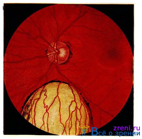
Rice. 71.
Coloboma of the choroid.
lack of pigment in the retina, glaucoma and some other changes.
The domestic literature provides a description of eye symptoms
in 29 people with this disease [Kaplan A.I. et al., 1969]. One patient had facial asymmetry with the location of the eye sockets at different levels and a wide nasal bridge, 10 had epicanthus, 12 had ptosis of the upper eyelid, 3 had a Mongoloid eye shape, 10 had strabismus, 4 had nystagmus, and 8 had pigment deposition on the lens capsule. One patient each had a coloboma of the iris and choroid, decoloration of the optic nerve head, a decrease in visual acuity to 0.06 and a narrowing of the boundaries of the visual field by 15-20°. 5 people had a color anomaly (protanomaly and deuteranomaly). The above changes in the organ of vision occurred either in isolation or in various combinations.
Klinefelter's syndrome
. There are chromatin-positive and chromatin-negative Klinefelter syndromes. In chromatin-positive Klinefelter syndrome, the sex chromosome set is XXV. Carriers of this syndrome are characterized by tall stature, a eunuchoid physique, often with gynecomastia, infertility. Quite often, mental retardation is noted with this syndrome.
Various changes in the organ of vision have been described
, color anomaly, hypertelorism, epicanthus, ptosis, ametropia, strabismus, coloboma of the iris, choroid, congenital cataract, pigmentary degeneration of the retina, optic nerve atrophy are found in this syndrome. A.I. Kaplan and co-authors, in a study of 17 patients with Klinefelter syndrome, revealed the following changes: 3 had epicanthus, 2 had a mongoloid eye shape, 3 had nystagmus, 1 had an irregularly shaped pupil, 4 had unilateral nystagmus, 4 had clouding on the lens capsule, 6 had a decrease in visual acuity, 2 had a narrowing of the visual field by 15°. These changes occurred either in isolation or in various combinations. No color anomalies were noted in any case.
Trisomy X
.
In this disease, the chromosome set (karyotype) contains three X chromosomes. Patients have
normal growth and sexual development may be normal. The offspring of women with trisomy X are healthy. Along with this, with normal sexual development and regular menstruation, it is noted that the latter disappear after a few years and secondary amenorrhea occurs. Mental development with trisomy X may not be impaired, but with this anomaly the incidence of mental defects is increased. A contributing factor in the occurrence of trisomy X in newborns is the advanced age of the mother. In total, a little more than 50 cases of trisomy X have been described, in which pathological changes in the organ of vision were observed. These include bilateral optic atrophy, chorioretinitis, and unilateral corneal opacification. Due to the small number of observations, it is difficult to express an opinion on the specificity of these changes.
Diagnosis and treatment of pineal gland diseases
A study of the pineal gland, if a pathological focus in the organ is suspected, is carried out using computed tomography of the brain, X-ray or magnetic resonance imaging.

The projection of the epiphysis in the absence of pathologies is visualized only along the midline. Cysts of the pineal gland of the brain or tumor neoplasms displace the epiphysis in the opposite direction in relation to the pathological focus.
If malignant formations in the epiphysis of the brain are suspected, a biopsy of the pineal gland is performed, followed by a histological examination of the obtained material.
Based on the data obtained, the neurologist makes a diagnosis and prescribes the necessary therapy. Drug treatment is aimed at relieving symptoms and inhibiting the growth of cystic formation:
- Diuretics (for fluid accumulation in the brain tissue);
- Anticonvulsants (for epileptic seizures);
- Venotonics (restore cerebral blood circulation and strengthen the vascular wall);
- Adaptive drugs (to restore sleep patterns);
- Painkillers (relieve headaches).
Cystic transformation of the pineal gland (pathological change in tissue) caused by echinococci, in the absence of positive dynamics in treatment with anthelmintic drugs, is an indication for surgical intervention.
With the rapid growth of cystic formation in the pineal gland and the deterioration of the general condition, instrumental methods of intervention are resorted to:
- Shunting (fluid from the pathological focus is drained into the abdominal cavity through a catheter);
- Endoscopy (drainage is installed into the cystic formation to remove fluid and empty the pathological focus);
- Trepanation (opening the skull and complete excision of the cyst or removal along with the epiphysis).
Instrumental intervention is contraindicated during pregnancy and old age, as well as in patients with a low platelet count in the blood.
Complications due to untimely treatment:
- Paralysis;
- Paresis;
- Decreased vision up to complete loss;
- Mental retardation;
- Dementia (dementia);
- Loss of auditory perception.
Detection of a small tumor in the pineal gland of the brain and the absence of pathological symptoms does not require therapy. The growth of microcysts is possible due to hormonal changes in the body; women planning pregnancy need to consult a specialized specialist.
Calcification of heart valves
Calcification is (as mentioned above) the formation of accumulations of undissolved calcifications in various organs or tissues in which such salts should not normally be contained.
The cause of pineal calcification can be congenital pathologies, various infections and metabolic disorders. Physiological calcification of the pineal gland is most often (40%) found in patients under 20 years of age. In this case, compact neoplasms with a diameter of up to 1 cm are formed in the organ.
In cases where calcifications are large in size, it is worth studying them in detail, as they can become the basis for malignant neoplasms. Dystrophic (pathological) calcification in the pineal gland occurs as a result of trauma, chemotherapy, ischemia, and so on, and is characterized by the deposition of cholesterol and lime in neoplasms.
Calcification of the pineal gland is accompanied by dysfunction of the latter, which can provoke the development of cancer, multiple sclerosis and schizophrenia due to blockade of melatonin synthesis. Filling of the pineal gland (calcification) with calcifications increases the likelihood of developing nervous exhaustion, anxiety, depression and gastrointestinal pathologies.
Ligament calcification is a fairly common occurrence associated with age-related changes in the body, injury and inflammation. Ligamentous calcification is often asymptomatic and discovered incidentally during X-ray examinations.
Such involutive processes in cartilage and ligaments during calcification of joints are accompanied by a loss of shock-absorbing properties, plasticity and elasticity in the joints.
Most often, tendon calcification develops in the spine (cervical/lumbar spondylosis deformans), due to tears in the area where the annulus fibrosus and longitudinal vertebral ligament attach to the edge of the vertebra, as a result of which the intervertebral disc is displaced, tearing the ligament away from the vertebra. This is where calcifications/ossifications develop.
In addition, similar processes are often found in the spinal-costal joints (ribs 9-10), hip and phalangeal joints (Eberden and Bouchard nodes), being a local demonstration of the aging of the body.
- Calcification of the aortic valve. The cause of this disease is rheumatic valvulitis, leading to degenerative changes in tissues. In this case, the valve flaps are deformed and soldered together. At the same time, calcifications form on them, which block the aortic ostium. In some cases, the process extends to the interventricular septum, the mitral valve leaflet and the wall of the ventricle (left). As a result, aortic insufficiency develops.
- Calcification of the mitral valve. It is quite difficult to diagnose such a pathology, due to the fact that its symptoms are similar to the clinical picture of cardiosclerosis, hypertension and rheumatism. More often this disease is detected in elderly patients.
- Calcification of the aorta. Develops in patients over 60 years of age. The clinical picture of the disease depends on the level of vessel damage.
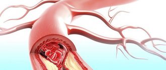
- Calcification of cerebral vessels. Calcification is in this case a synonym for atherosclerosis. Due to the accumulation of lipids on the walls, insufficiency of blood circulation in the brain occurs, which is fraught with the development of strokes, dementia, and so on.
- Calcification of the coronary arteries. In this case, cholesterol and fats settle on the walls of these vessels, that is, atherosclerotic plaques are formed, which leads to a loss of elasticity and a change in the shape of the vessel, as a result of which the blood supply to the myocardium is disrupted, and in the case of complete blockage of the lumen, tissue necrosis.
Organ pathologies, their symptoms and treatment
In addition to disruption of daylight hours, other negative factors affect the functioning of the endocrine gland:
- congenital abnormalities of brain development,
- severe neuroendocrine pathologies,
- damage to brain cells.
Hypoplasia of the pineal gland is indicated by the early onset of puberty. Doctors rarely diagnose congenital malformations of the pineal gland.
Functional disorders can be eliminated quite easily; you just need to review your daily routine and treat background pathologies. For the proper production of melatonin and other important substances in the pineal gland, you need to include foods of various categories in your diet. Balancing nutrition is a prerequisite for the normal functioning of the endocrine system.
On a note! Pineal gland diseases occur in men and women. The more provoking factors, the higher the risk of damage to the pineal gland. Lack of attention to the signs of pathological changes can cause severe damage to the nervous system, ischemic stroke, and hypertensive crisis. If problems with the pineal gland occur in children, then against the background of active growth of the tumor, the functioning of the adrenal cortex is disrupted, which provokes a shift in the timing of puberty to an earlier period.
Pineal tumors
Volumetric neoplasms in the tissues of the pineal gland are benign and malignant. Often the cause of development is the influence of ionizing or X-ray radiation.
While the tumor is small, the patient does not suspect the existence of a pathological focus in the pineal gland. If the neoplasm grows, reaches 3 cm, and cell malignancy actively occurs, then negative symptoms appear. Patients complain of headaches that cannot be controlled, and blurred vision.
After examination with a tomograph, the doctor confirms or denies the suspicion of tumor development. Treatment is strictly surgical. If tissue histology shows the presence of atypical cells, then the patient, under the guidance of an oncologist, receives chemotherapy or undergoes a course of radiation to suppress the growth of cancerous structures.
Hemorrhage into the pineal gland tissue
A dangerous process develops in adults against the background of vascular atherosclerosis. In children, a serious condition rarely occurs. Sometimes a stroke occurs due to an aneurysm (congenital abnormality).
Against the background of hemorrhage in the brain, various parts of the nervous system are often affected. An MRI is prescribed to confirm the diagnosis. A neurologist, neurosurgeon, cardiologist, and other specialists are involved in treatment.
Parasitic diseases
Most often, this type of pathological process is detected in residents of regions where livestock farming is developed. In most cases, echinococci penetrate the pineal body. The size of parasites is from 2 to 8 mm. Waste products poison the body, the movement of dangerous tapeworms disrupts the functioning of the pineal gland and provokes the development of tumor-like formations.
With echinococcosis, cysts are prone to active growth. The cavity with a dense capsule must be removed: non-surgical methods and medications do not have any effect. A cyst in the pineal gland is diagnosed using immunological tests and tomography. To eliminate the pathological process, you need the help of a qualified infectious disease specialist and neurosurgeon,
↑ Obesity
Obesity
is a pathological condition in which there is an accumulation of fat in places of its physiological deposits.
Of all the classifications, the one proposed by V.G. Baranov should be considered the most successful. According to this classification, primary and secondary obesity
. Primary obesity is an independent disease, the origin of which is still unknown. Obesity, which developed as a result of a number of pathological conditions (Itsenko-Cushing's disease and syndrome, adiposogenital dystrophy, trauma or inflammatory process in the hypothalamus, etc.), V. G. Baranov proposes to call secondary.
To assess the degree of obesity, the Broca index is used as an indicator of ideal weight (body weight in kilograms is equal to height in centimeters minus 100). This indicator is adjusted due to age, constitution, and the state of skeletal muscles (±10% of ideal body weight).
There are three degrees of obesity
: first - body weight exceeds ideal weight by 10-29%, second - body weight is increased by 50-99% and third - increased by 100% or more.
Obese patients
for excess body weight, shortness of breath, pain in the heart, drowsiness, increased sweating, headache, dizziness, increased appetite, tendency to constipation.
For obesity
In the initial period, fat is deposited evenly, and later mainly on the abdomen, chest, neck, back and pelvic area. Hyperhidrosis is noted, and skin diseases (eczema, furunculosis, pyoderma) often occur.
Obese patients are much more likely than usual to experience cardiovascular disorders
. Atherosclerosis often occurs due to lipid metabolism disorders, bradycardia, arterial hypertension, and coronary heart disease. It is also possible to develop circulatory failure, accompanied by edema, cyanosis, and shortness of breath. In some cases, respiratory failure is noted due to the high position of the diaphragm.
In obese patients
pyelitis, urethritis, and cystitis develop more often than usual; The reproductive system also suffers (in women - menstrual irregularities, infertility, etc., in men - weakened libido, impotence).
From the nervous system
Various changes are observed: drowsiness, insomnia, headaches, myalgia, neuralgia, neuritis.
Eye symptoms
with obesity, they are expressed in a more frequent occurrence of eyelid diseases - recurrent styes, skin eczema, as well as changes in the fundus of the eye caused by atherosclerosis and arterial hypertension. Angiosclerosis of the retina develops, and arteriosclerotic retinopathy and hypertensive retinopathy may also develop.
—
Article from the book: Pathology of the organ of vision in common diseases | Komarov F.I., Nesterov A.P., Margolis M.G., Brovkina A.F.
Preventive recommendations
Some types of pineal diseases are difficult to prevent: the exact causes and mechanisms of failures in the structure and functionality of the organ are not known. Neurologists advise adults to remember their daily routine and, unless absolutely necessary, not to go to bed later than 10 p.m. in order to prevent disruption of natural rhythms.
Night shifts cause serious damage to health. All systems, including the endocrine system, experience the negative impact of working at a time when the body should be resting. Melatonin deficiency provokes insomnia, irritability, and increased fatigue. The best option is to avoid such a work schedule. If there is no other opportunity for professional activity, then after the shift you need to rest fully, and not give up sleep for six to seven hours. Disruption of the circadian rhythm in combination with short sleep duration provokes the development of hypertension, nervous disorders, anxiety, general weakness, hormonal imbalance and metabolic processes. The likelihood of heart attacks and strokes also increases.
Other preventive measures:
- during pregnancy, stop working in hazardous work, give up alcoholic beverages and smoking, avoid infection during influenza epidemics,
- to reduce the risk of hemorrhage in the tissue of the pineal gland and prevent ischemic stroke, you need to contact a cardiologist in time, treat arterial hypertension and atherosclerosis,
- It is important to eat properly so that the parts of the brain receive a sufficient amount of oxygen and substances, without which the stable functioning of an important organ is disrupted. Hypoxia plus thrombosis, atherosclerosis, excess bad cholesterol in blood vessels is a dangerous combination, against the background of which the risk of severe damage to brain cells sharply increases,
- Regularly receive foods that contain a substance from which melatonin is transformed in the body. The following types of food contain the valuable amino acid tryptophan: fish, meat, nuts, legumes, dried dates, mushrooms. For vigor and good mood, you need to consume dairy products, especially yogurt and cottage cheese,
- X-rays of the skull and neck area should be carried out strictly for medical reasons: excessive penetration of radiation can cause tumors of the pineal gland.
When pineal gland diseases are detected, you need to adjust your daily routine and diet, and give up bad habits. If cysts, tumors, or parasitic infections are detected, surgical treatment is prescribed. You need to understand that disruption of the frequency of sleep and rest can provoke a deficiency of melatonin, an important hormone that regulates circadian rhythms.
How does age affect hormones?
In the human body, the pineal gland begins to develop around the sixth week after conception. In a newborn, the pineal gland weighs 7 mg, then its weight begins to increase and reaches a peak at approximately five years. Then comes the phase of reverse development - involution. In adulthood, the weight of the organ averages 150 mg.
The process of age-related changes in the pineal gland affects not only mass and size, but also structural changes. Thus, in the pineal gland there are more dark stromal cells and fewer pineal cells, which synthesize hormones. The result is a decrease in endocrine activity. But the pineal gland does not completely give up its position: until old age, individual cells produce a valuable secretion. They are necessary not only to regulate sleep, but also, thanks to their antioxidant effects, slow down the aging process.
Pineal gland hormones stimulate immune cells, normalize fat-carbohydrate metabolism, restore reproductive function, and also prevent the appearance of tumors.
Since the pineal gland changes over the years more functionally than organically, it is possible to restore the activity of this organ with the help of pharmaceuticals and the elimination of unfavorable conditions for the functioning of the gland.
To establish normal hormone synthesis, you should exclude:
- night wakefulness and daytime sleep;
- excess artificial light;
- long work at monitors.
Maintaining a natural sleep/wake schedule is extremely important for health and youth.
If there are problems with the supply of melatonin, pharmaceutical products containing this hormone of synthetic or animal origin are used. They may be needed, for example, by residents of the Far North or people forced to work in shifts. And for the treatment of chronic diseases, a protein extract of the pineal gland, created from animal cells, with serious antioxidant and immunostimulating effects is used.