Magnetic resonance imaging is a diagnostic method based on the phenomenon of vibration of hydrogen atoms in the induction field created by the device during the study. Due to its high information content and safety, MRI is one of the best medical imaging technologies.
Preparing the patient for an MR scan
Using a magnetic tomography scan, various organs are studied, including the brain, spinal cord, and spine. Directly or indirectly assess the functionality of the central nervous, circulatory, endocrine and other systems. MRI can be performed on patients of any age without restrictions on the number and frequency of diagnostic sessions.
For maximum accuracy of results, there are few corresponding characteristics of the tomographic apparatus. Proper preparation for research is necessary. One of the frequently asked questions to doctors is: “Do they undress during an MRI?” Let's talk about this in our article.
Do I need to undress during an MRI?
MRI examination – magnetic resonance imaging – is a modern method for detecting diseases of internal organs and the spine, based on the action of a magnetic field.
Layer-by-layer scanning with a three-dimensional image displayed on a computer monitor allows you to determine the cause and course of the disease and choose the most favorable treatment method. All life on Earth has its own magnetic field and, when exposed to a magnet of greater power, responds by changing the normal behavior of atomic nuclei. Since a person is mostly composed of liquid, a response to the influence of hydrogen nuclei is recorded.
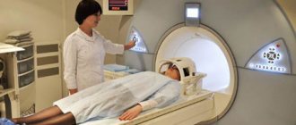
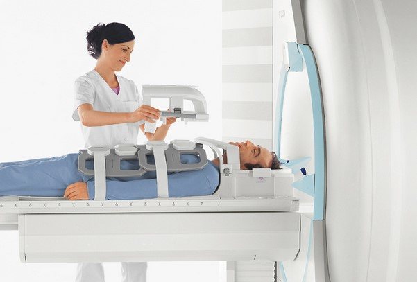
How an MRI examination is performed: the patient takes a position lying on his back on the retractable table of the device
How is the examination carried out?
The MRI machine is a large hollow cylinder into which the patient is placed lying down. The entire procedure lasts from 30 minutes to a couple of hours and depends on the complexity of the particular case.
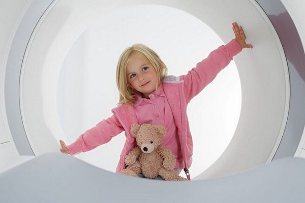
In this case, there is no harmful effect on the body. The only inconvenience is being in a confined space, which is not suitable for those suffering from claustrophobia.
There is an open MRI device, but, unfortunately, not every medical institution is equipped with it.
How an MRI scan is done:
- the patient takes a position lying on his back on the retractable table of the device;
- a correct diagnostic result is possible only if you remain motionless during the examination, this is especially true when scanning the spine. Even a slight change in posture causes blurring of the picture on the monitor. The person’s arms and legs are secured with soft belts and special bolsters. For young children, MRI diagnostics are performed under general anesthesia. If a person is nervous, a sedative can be taken orally or intravenously;
A person’s arms or legs are secured with soft straps and special bolsters.
How to dress for a magnetic tomography procedure
Magnetic tomography is performed more often to confirm the diagnosis, after a comprehensive examination of the body by other methods.
Before the MRI procedure, you must remove all items of clothing and jewelry that contain metal. You should dress in a disposable robe or other things without metal rivets, hooks, buttons, etc.
It is necessary to warn the doctor in advance about existing dentures, dental fillings, implants with metal components that can change a person’s magnetic field and distort diagnostic results.
What contraindications are taken into account when an MRI is performed?
Contraindications for the procedure:
Before the MRI procedure, you must remove all items of clothing and jewelry that contain metal, dress in a disposable robe, or other items without metal objects.
- tattoos on the body containing metal elements in the dye;
- artificial devices that improve the functioning of internal organs: cardiac implants, blood vessels, hearing aids;
- intrauterine devices, cosmetic devices implanted into the body;
- early pregnancy;
- recent surgery on the heart, brain or blood vessels;
- nervous diseases, epilepsy;
- patient weight over 120 kg;
- the presence in the body, and especially in the eyes, of metal pieces that entered the body as a result of injury.
Consequences of failure to comply with safety requirements
You should notify your doctor about all such cases in order to avoid harmful effects on the organs of a high-power magnetic field:
- burn at the tattoo site;
- corneal damage;
- malfunction of implants;
- The effect of a magnetic field on the fetus has not been fully studied.
If a person suffers from kidney or liver failure, it is necessary to take a biochemical test of blood and urine so that the doctor can fully assess the condition of the patient’s internal organs if a dye is injected. An allergy test is also performed.
How to do an MRI with an axial load: after tomography of the spine in a horizontal view, the table, together with the patient and the magnet, is raised to a vertical position
Tomography of the spine and spinal cord
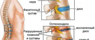
Unlike common diagnostic methods, MRI allows you to identify diseases of the spine at the initial stage, assess the condition of the skeletal system and the soft tissues located around it, tendons, lymph nodes, blood vessels, cartilage tissue of the joints, and interdisk space. The study makes it possible to identify incipient foci of inflammation, tumors in the formation stage, and the penetration of metastases of malignant neoplasms into the spinal cord and surrounding tissues.
When is an MRI of the spine prescribed?
MRI helps to evaluate spinal diseases:
- Osteochondrosis and its complications in the form of hernia formation.
- Changes in the spinal column after injuries and tumors.
- Curvature of the spine: kyphosis, scoliosis, lordosis.
- Bacterial, fungal, viral damage to the spinal substance, multiple sclerosis.
- Infringement of the nerve endings of the spine.
- Changes in vascular circulation.
- Congenital pathologies.
- The appearance of pain in the spine of unknown origin.
Vertical diagnostic method
How to do an MRI with axial load:
- after tomography of the spine in a horizontal view, the table, together with the patient and the magnet, rises to a vertical position;
- gravity begins to act on the spinal column - the vertebrae shift under the load of the human body;
- disorders in the form of a hernia become more pronounced;
- The level of instability of the organ is determined if it is required to securely fix it. The information is used by neurosurgeons for traumatic lesions of the skeletal system.
The results of the MRI study are assessed by a neurologist. Images are prepared within 1-2 hours after the examination. The patient awaits the result; if necessary, another series of images is taken. Since the procedure does not have a harmful effect on the body, there is no need to worry about health complications after the diagnosis.
So that's what you are, brain! Painless and non-threatening examination. Answers to questions that interested me before the MRI. Without long cool stories about life, doctors and the weather outside the window
A neurologist from a paid clinic sent me for an MRI of the brain, so you won’t read any life hacks in my review on how to get the procedure for free, other than contacting a neurologist at a state clinic or using a VHI policy, whoever has one.
I tried to answer the questions that interested me before the study, and described my experience without artistic details and unnecessary long stories. Just something that might be useful for someone who hasn't had an MRI yet and doesn't know how it will go.
Background.
I visited a neurologist with complaints of headaches in the forehead and left temple and found out that I needed to do an MRI of the brain. On the same day, I looked for nearby medical centers where they do the study. The clinic where I went does not have a tomograph, otherwise I would have undergone the procedure there.
It turned out that in St. Petersburg MRIs are done literally 24/7. You can sign up for night time, and the price will be two times lower. I have already had urgent fluorography done at a similar center. You come by appointment at the appointed time, take a photo, and after 10-15 minutes you receive a photo and a transcript. It's the same with MRI. I signed up for the daytime, so the price is regular, without discounts.
Cost of the study.
The procedure cost 2500 rubles.
You have to pay an additional 350 rubles for the photo.
Where did you do it?.
St. Petersburg, Entuziastov Ave., 33, “NDC-MRI”. They have branches all over the city. A small MRI center - everything is neat, polite, fast.
How to prepare for an MRI exam.
No way for MRI of the brain. There is no need to do anything special. I was told about this when I signed up, but I, of course, decided to read the reviews. I learned everything I wanted and didn’t want about panic, fears and a noisy tomograph.
I won’t say that I was afraid - the procedure is non-contact, but someone else’s experience still sets me up in a certain way.
You start to think, what if I get scared, panic, start itching, sneezing, my legs go numb, I urgently need to go to the toilet, and so on.
During the recording, I was asked if I worked in metal production (as far as I understand, this is due to the possible presence of metal shavings in the body), if I had a pacemaker and braces. If the answer to even one question is “yes”, the research cannot be done.
Before the procedure, they ask me to fill out a questionnaire, where they also ask if I have a fear of closed spaces, if I was in the war, if I developed PTSD (post-traumatic stress disorder) there.
That is, the entire preparatory stage is connected with finding out whether it will become scary and whether there is a threat to the tomograph.
Which tomograph to choose for MRI.
You can choose any one, it all depends on the capabilities of the clinic and the patient’s weight. I did it in an open-type tomograph. It is not as intimidating as the closed one, but I read that the open one is less informative in terms of image quality. I don't know, I'm not a doctor.
Unlike a classic device, an open-type tomograph does not have a closed scanning chamber. During the procedure, the person is placed in a special room in which magnets are located above and below. The space around the table on which the patient lies remains free.
Do I need to undress for an MRI?.
One of the main questions that interested me was whether to undress? It seems clear that they will examine the head, but it doesn’t matter, especially since they wrote different things everywhere. It turned out there was no need to undress.
As for clothes, I only needed to remove my bra, since it has metal wires, and my shoes (well, that’s understandable). No one asks you to take off a sweater or other outerwear for an MRI of the brain. Panties, tights, skirts, trousers, T-shirts, etc. remain on you.
As for jewelry and hairpins, you need to remove everything: watches, rings, chains, etc.
In the clinic where I did the research, there are special changing rooms - you don’t need to take off and put on your underwear in front of the doctor (for those who are shy).
How is an MRI scan performed?.
A large orthopedic pillow is placed under the knees so that the legs lie comfortably, a blanket is covered on top, and something like a plastic box is placed above the head. Here I closed my eyes so as not to see how I would “enter” the belly of the tomograph.
During the examination, especially when the device is rattling (more on this below), you should not move, blink, scratch, or sneeze - the pictures will be blurry.
To contact the doctor, I was given a special clicker with a big red button. They told me to press it if I suddenly started to panic. Why panic? I almost fell asleep.
Tomograph noise.
Here everyone has very different impressions. For some, it’s a hell of a noise and requires earplugs, but for others, it doesn’t matter. I'm from the latter. Yes, the tomograph really makes different and quite loud sounds: it squeaks, rumbles, imitates the sounds of a drill working somewhere several floors away. But I personally didn’t hear anything disgusting or unbearable. Moreover, after 10 minutes I relaxed and almost dozed off.
How long does an MRI scan take?.
I was in the device for exactly half an hour. The photo is printed immediately. We had to wait about half an hour for the pictures to be deciphered. In general, the entire study, including hibernation in the tomograph and waiting for decoding, takes about an hour, no more. After the diagnosis, there were no unpleasant consequences. She got up and went. There was no headache, no dizziness, no nerves, everything was calm.
Bottom line.
After the examination, you receive a photo and a transcript that the doctor needs. They gave me a huge picture - a brain in four projections. What I read about, I saw practically alive. So that's what you are, brain!
And yes, there won’t be a photo of the brain, because I’ve never posted photos of the insides in reviews and I don’t think it’s useful or understandable for anyone other than doctors, and they’re unlikely to be interested in looking at other people’s brains even on a review site.
In general, there is no need to be afraid of the examination - it is absolutely painless, comfortable, fast and relatively inexpensive. The worst thing about undergoing an MRI is waiting for the results. Everything else depends on personal impressionability, and plus I’m an adult woman without claustrophobia, so my experience is extremely positive.
How is an MRI done?
MRI examination – magnetic resonance imaging – is a modern method for detecting diseases of internal organs and the spine, based on the action of a magnetic field. Layer-by-layer scanning with a three-dimensional image displayed on a computer monitor allows you to determine the cause and course of the disease and choose the most favorable treatment method.
All life on Earth has its own magnetic field and, when exposed to a magnet of greater power, responds by changing the normal behavior of atomic nuclei. Since a person is mostly composed of liquid, a response to the influence of hydrogen nuclei is recorded.
How an MRI examination is performed: the patient takes a position lying on his back on the retractable table of the device
Tomographs are divided into 3 types, differing in the power of the consumed magnetic field. The most in demand are devices with a voltage of 1.5-3 Tesla, which allow obtaining clear contrast images of the organ being diagnosed.
What causes secondary neuropathy?
The following causes of secondary neuritis are identified:
- infection entering the human body (as a rule, a person has previously suffered diseases such as measles or herpes);
- facial injuries;
- hypertension and atherosclerosis caused by disturbances in the functioning of blood vessels located in the spine;
- malignant tumors, the focus of which is at the base of the brain;
- consequences of an anesthetic injection given by a dentist.
To determine the factor that could provoke the appearance of neuritis, the doctor prescribes an MRI procedure for facial nerve neuropathy.
Magnetic resonance imaging (MRI) of the brain - review
A neurologist from a paid clinic sent me for an MRI of the brain, so you won’t read any life hacks in my review on how to get the procedure for free, other than contacting a neurologist at a state clinic or using a VHI policy, whoever has one.
I tried to answer the questions that interested me before the study, and described my experience without artistic details and unnecessary long stories. Just something that might be useful for someone who hasn't had an MRI yet and doesn't know how it will go.
Background.
I visited a neurologist with complaints of headaches in the forehead and left temple and found out that I needed to do an MRI of the brain. On the same day, I looked for nearby medical centers where they do the study. The clinic where I went does not have a tomograph, otherwise I would have undergone the procedure there.
It turned out that in St. Petersburg MRIs are done literally 24/7. You can sign up for night time, and the price will be two times lower. I have already had urgent fluorography done at a similar center. You come by appointment at the appointed time, “take a photo,” and after 10-15 minutes you receive a photo and a transcript. It's the same with MRI. I signed up for the daytime, so the price is regular, without discounts.
Cost of the study.
The procedure cost 2500 rubles.
You have to pay an additional 350 rubles for the photo.
Where did you do it?.
St. Petersburg, Entuziastov Ave., 33, “NDC-MRI”. They have branches all over the city. A small MRI center - everything is neat, polite, fast.
How to prepare for an MRI exam.
No way for MRI of the brain. There is no need to do anything special. I was told about this when I signed up, but I, of course, decided to read the reviews. I learned everything I wanted and didn’t want about panic, fears and a noisy tomograph.
I won’t say that I was afraid - the procedure is non-contact, but someone else’s experience still sets me up in a certain way.
You start to think, what if I get scared, panic, start itching, sneezing, my legs go numb, I urgently need to go to the toilet, and so on.
During the recording, I was asked if I worked in metal production (as far as I understand, this is due to the possible presence of metal shavings in the body), if I had a pacemaker and braces. If the answer to even one question is “yes,” the study cannot be done.
Before the procedure, they ask me to fill out a questionnaire, where they also ask if I have a fear of closed spaces, if I was in the war, if I developed PTSD (post-traumatic stress disorder) there.
That is, the entire preparatory stage is connected with finding out whether it will become scary and whether there is a threat to the tomograph.
Which tomograph to choose for MRI.
You can choose any one, it all depends on the capabilities of the clinic and the patient’s weight. I did it in an open-type tomograph. It is not as intimidating as the closed one, but I read that the open one is less informative in terms of image quality. I don't know, I'm not a doctor.
Unlike a classic device, an open-type tomograph does not have a closed scanning chamber. During the procedure, the person is placed in a special room in which magnets are located above and below. The space around the table on which the patient lies remains free.
Do I need to undress for an MRI?.
One of the main questions that interested me was whether to undress? It seems clear that they will examine the head, but it doesn’t matter, especially since they wrote different things everywhere. It turned out there was no need to undress.
As for clothes, I only needed to remove my bra, since it has metal wires, and my shoes (well, that’s understandable). No one asks you to take off a sweater or other outerwear for an MRI of the brain. Panties, tights, skirts, trousers, T-shirts, etc. remain on you.
As for jewelry and hairpins, you need to remove everything: watches, rings, chains, etc.
In the clinic where I did the research, there are special changing rooms - you don’t need to take off and put on your underwear in front of the doctor (for those who are shy).
How is an MRI scan performed?.
A large orthopedic pillow is placed under the knees so that the legs lie comfortably, a blanket is covered on top, and something like a plastic box is placed above the head. Here I closed my eyes so as not to see how I would “enter” the belly of the tomograph.
During the examination, especially when the device is rattling (more on this below), you should not move, blink, scratch, or sneeze - the pictures will be blurry.
To contact the doctor, I was given a special clicker with a big red button. They told me to press it if I suddenly started to panic. Why panic? I almost fell asleep.
Tomograph noise.
Here everyone has very different impressions. For some, it’s a hell of a noise and requires earplugs, but for others, it doesn’t matter. I'm from the latter. Yes, the tomograph really makes different and quite loud sounds: it squeaks, rumbles, imitates the sounds of a drill working somewhere several floors away. But I personally didn’t hear anything disgusting or unbearable. Moreover, after 10 minutes I relaxed and almost dozed off.
How long does an MRI scan take?.
I was in the device for exactly half an hour. The photo is printed immediately. We had to wait about half an hour for the pictures to be deciphered. In general, the entire study, including hibernation in the tomograph and waiting for decoding, takes about an hour, no more. After the diagnosis, there were no unpleasant consequences. She got up and went. There was no headache, no dizziness, no nerves, everything was calm.
Bottom line.
After the examination, you receive a photo and a transcript that the doctor needs. They gave me a huge picture - a brain in four projections. What I read about, I saw practically alive. So that's what you are, brain!
And yes, there won’t be a photo of the brain, because I’ve never posted photos of the insides in reviews and I don’t think it’s useful or understandable for anyone other than doctors, and they’re unlikely to be interested in looking at other people’s brains even on a review site.
In general, there is no need to be afraid of the examination - it is absolutely painless, comfortable, fast and relatively inexpensive. The worst thing about undergoing an MRI is waiting for the results. Everything else depends on personal impressionability, and plus I’m an adult woman without claustrophobia, so my experience is extremely positive.
Source: https://irecommend.ru/content/tak-vot-ty-kakoi-mozg-bezboleznennoe-i-nestrashnoe-issledovanie-otvety-na-voprosy-kotorye-in
What is optic neuritis
- Accounts for about 50% of cases of bilateral visual impairment.
- Prevalence 3:100,000
- First manifestation of demyelinating disease in 12-30% of cases
- Optic neuritis - acute inflammation of the optic nerve (II pair of cranial nerves)
- Autoimmune disease (eg, systemic lupus erythematosus, disseminated encephalomyelitis)
- Infectious or viral etiology (eg, cytomegalovirus infection, rubella, mumps, toxoplasmosis)
- Radiation damage (with exposure of 10 Gy or more).
Optic neuritis, depending on the location of the damage, can be divided into:
- Papillitis is a lesion of the intraocular area.
- Retrobulbar optic neuritis is inflammation of a portion of the nerve outside the eyeball. It can occur both in a structured form and in a chronic one.
- Neuroretinitis is papillitis and inflammation in the nerve fiber layer.
The whole truth about MRI and computed tomography
Can bones be seen on an MRI? Which is safer - MRI or computed tomography (CT)?
Why can MRI be replaced by CT, but CT cannot be replaced by MRI?
Answers to these and many other questions are given by Anton Ivanovich Chugaev, chief physician of the MRI 24 network of diagnostic centers.
Radiologist Anton Ivanovich Chugaev.
What is MRI?
Magnetic resonance imaging is a modern method for diagnosing diseases using nuclear magnetic resonance. The method is based on measuring the electromagnetic response of hydrogen atoms.
It sounds complicated, but in reality everything is much simpler. MRI shows the signal from tissues that contain hydrogen atoms. The more hydrogen atoms in tissue, the better they are visible on MR images.
And since water contains the most hydrogen atoms, the more water there is in the tissue, the brighter the signal on the MRI image. The less water, the weaker the signal.
What does magnetic resonance imaging show? What can be seen on an MRI and what cannot?
MRI photograph of the knee joint.
Tissues and media that contain water are best seen on MRI. Therefore, MRI shows the brain very well, because the brain is 75% water.
In addition to the brain, the following are very clearly visible on MRI:
- Tumors and cysts;
- Healthy and damaged muscles, ligaments and any other soft tissues;
- Ligaments, menisci of joints;
- Vertebral bodies and joints.
It is very convenient to use MRI to study bone pathology. The hard bone itself is not visible, but the adjacent bones of the joints, soaked in bone marrow, are clearly visible. Thanks to this, the doctor immediately notices bone problems.
Limitations of MRI
1. It is very difficult, and sometimes impossible, to distinguish between hard bodies such as skull bones and foreign bodies on MRI.
2. Lungs are not visible on MRI images. There is too much air in the lungs for this, and the MRI does not see air.
Who should not have an MRI?
MRI cannot be performed on patients whose body contains objects with iron parts: for example, implants, hearing aids, pacemakers, neurostimulators, ferromagnetic clips and other inclusions. This is a direct contraindication to MRI.
Before the examination, a patient with an implant* or prosthesis that does not contain ferromagnetic or electronic components must bring the doctor a special extract or conclusion from the attending physician.
The statement should describe what the implant is made of, and the doctor’s report may say whether an MRI can be done with this implant.
Under no circumstances should MRI be performed on patients who have electronic pacemakers or pacemakers. People with pacemakers should not even come close to the device.
*Cardiac bypass surgery is not a direct contraindication to MRI. Your doctor will usually allow magnetic resonance testing 1 to 3 months after bypass surgery.
How is an MRI performed? There is an opinion that during the procedure you need to lie motionless for a long time in a large apparatus. What happens if you move?
It is recommended to do MRI examinations in specialized centers using modern equipment.
To get good MRI images, exposure time is needed. This means that you will have to lie still in the device for some time. If you move, the outlines of the tissues will be blurred.
This implies another limitation. MRI cannot be done on people who are not fully conscious, who are in shock (for example, after being shot in the abdomen or after a stroke), or those who cannot obey commands or lie still.
Patients with severe pain will not be able to lie still and hold their breath at the doctor's command. When a person moves, the result is a fuzzy, blurry image that is useless to the doctor.
Patients who cannot lie still are referred for a computed tomography (CT) scan or an MRI under general anesthesia.
Are MRIs done for children?
Yes, MRIs are done for children. But children have an innate fear of closed spaces, which kids cannot consciously control. Therefore, young children are often given anesthesia before the procedure.
If the child is older, a parent may be nearby. If dad or mom holds the child's hand and calms him down, there is a chance that the child will not move, and a good result will be obtained.
In what cases is it necessary to do an MRI?
If the attending physician has referred you for the test, an MRI must be done. For example, magnetic resonance imaging is often ordered after surgery to remove a brain tumor.
Which doctor can refer you for an MRI? Can I come for an MRI on my own?
Any doctor can refer you for an MRI.
There is also nothing wrong with going to an MRI yourself. Many diagnostic clinics even have a comprehensive study that allows you to examine most body systems.
After the MRI, you will be given a report. But to understand what is written in the conclusion, you will have to consult your doctor.
You mentioned computed tomography. How does MRI differ from CT?
The MRI machine (left) looks very similar to the CT machine (right). But the operating principles of the devices are different.
The main difference is that MRI is not an x-ray method. This means that the person who came for an MRI does not receive a dose of radiation.
MRI is not harmful. If you schedule an MRI for yourself, nothing bad will happen. MRI can be done even for pregnant women, starting from the second trimester of pregnancy.
CT is a modern x-ray diagnostic method. Accordingly, the person who comes for this procedure receives a dose of radiation.
It is often impossible to do a CT scan, and neither is it possible to prescribe it for yourself. Only a doctor can refer you for a CT scan.
Advantages of CT over MRI
1. CT is less demanding on movements. Even a person in an inadequate and shock state can undergo a computed tomography scan.
2. On CT, foreign objects and hard tissues are clearly visible, which are not visible on MRI. For example, CT can accurately diagnose skull fractures.
3. Lungs are visible on CT scan.
4. CT scanning can be performed on people who have metal objects in their bodies. This is especially important if you need to urgently evaluate a person who has been involved in a car accident or anyone else who has received resuscitation measures.
Such patients are given subclavian catheters and oxygen equipment is connected. If you refuse all this during an MRI examination, the victim may die.
Are there any analogues for MRI? Can it be replaced by an x-ray examination?
CT machine from the diagnostic center MRI 24.
Although MRI and computed tomography are similar at first glance, they are very different diagnostic methods that do not replace, but complement each other.
As for the technical quality of the image, MRI images, in my opinion, are still better than CT images. But at the same time, CT is suitable for those people who cannot undergo MRI.
Therefore, when public hospitals have a choice of which device (CT or MRI) to purchase first, they always start with a CT machine.
Where is it better to do an MRI - in a large hospital or in a specialized center?
It is better to do an MRI where there is a good machine and a good specialist. Usually the best equipment and specialists are available in specialized centers.
For example, in clinics dealing with a specific pathology and having their own diagnostic department, or in specialized centers dealing with MRI and CT diagnostics.
Moreover, the examination can be done in any hospital where there is a modern MRI machine. A good modern MRI machine should have a power of at least 1.5 Tesla (Tesla).
Symptoms indicating the presence of neuritis
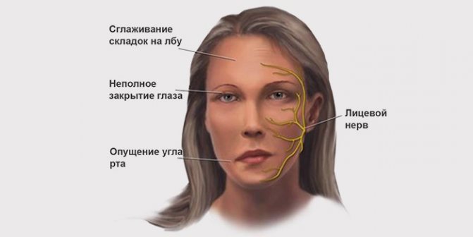
- In the early stages of the disease, the primary sign of neuritis is pain in the back of the ear and an asymmetrical appearance of the face. In this case, the corner of the mouth is lowered, and the nasolabial fold takes on a smooth outline.
- The affected part of the face twists to the opposite healthy side.
- Inability to close the eyelids, while on the affected side of the face the eyelid is wide open and a strip of white sclera is visible.
- Watery eyes, dry eye syndrome.
- Increased salivation.
- Increased hearing on the part of the affected nerve.
- Loss of taste.
A doctor examining a patient asks to perform a few simple steps to confirm the diagnosis.
- smile;
- bare your teeth;
- make lips into a tube;
- frown and then raise your eyebrows.
If you have neuritis of the facial nerve, it is impossible to perform the above actions. If deviations from the typical clinical picture are observed, the doctor prescribes an MRI to identify the absence or presence of a secondary nature of the disease, which manifests itself in the presence of a tumor, brain abscess or encephalitis.