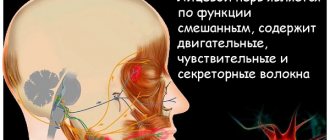
The thalamus, also called the thalamus opticum, is located next to the third ventricle of the brain. The ventricles, in turn, are cavities in which cerebrospinal fluid (CSF) circulates. It is part of the diencephalon (diencephalon). In the vast majority of people, the thalamus is divided into two parts, connected by gray matter. Around this formation is bordered by the internal capsule, which separates it from the basal ganglia. This capsule consists of nerve fibers that ensure the interaction of the cerebral cortex with underlying structures.
Core kernels
The structure of this formation is quite complex, which is explained by the wide range of functions performed by the thalamus. The main component of the thalamus is the nucleus, formed from the gray matter of the brain, that is, the bodies of nerve cells. In total, there are about 120 nuclei in the thalamus. Depending on where they are located, kernels are classified into the following groups:
- Front.
- Lateral. The posterior part of this group, in turn, is divided into the cushion, medial and lateral geniculate bodies.
- Medial.
Depending on their functions, kernels are classified into the following groups:
- specific;
- associative;
- nonspecific.
Specific kernels
This group of nuclei of the thalamus has a number of distinctive features that unite them. First, they receive impulses from long nerve pathways that transmit information from somatosensory, visual and auditory receptors to the cerebral cortex. Through these nuclei, the impulse is transmitted further to the corresponding areas of the cortex: somatosensory, auditory and visual. In addition, information from them enters the premotor and motor areas of the cortex.
Also, specific nuclei receive feedback from the cortex. Experiments have proven that when a section of the cortex corresponding to a specific nucleus is removed, this nucleus is also destroyed. And when certain nuclei are stimulated, the nerve cells of the corresponding cortex are activated.
This group receives information from the cortex, reticular formation, and brain stem. It is precisely because of the presence of these connections that the cerebral cortex has the ability, among all incoming information, to select the most important at the moment.
In addition, the structure of the thalamus includes nuclei that receive information from the red and basal nuclei, the limbic system, and the dentate nucleus (located in the cerebellum). Next, the signal goes to the motor areas of the cortex.
Thalamus
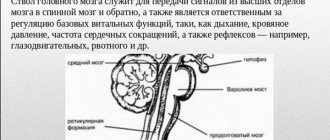
The thalamus performs several important physiological functions. It is responsible for transmitting sensory and motor information from the sense organs (except information from the olfactory organs) to the corresponding areas of the cerebral cortex of mammals or the cerebral cloak of lower chordates. The thalamus plays an important role in regulating the level of consciousness, processes of sleep and wakefulness, and concentration.
The thalamus is one of the main products of the embryonic development of the embryonic diencephalon. This fact was first established by the founder of embryology, Swedish anatomist Wilhelm Gies, in 1893.
Previously, the thalamus was considered a brain structure characteristic only of chordates. Even earlier, its existence was recognized only in vertebrates. Scientists believed that the thalamus was fundamentally absent in invertebrates, even the most highly organized ones, such as arthropods. However, in 2013, a structure homologous to the thalamus in the chordate brain was discovered in the central ganglion or brain of arthropods - the so-called “lateral accessory lobes” (LAL). These structures showed similarities both in embryonic development and gene expression patterns, as well as in anatomical location in the brain. Similarities were also found in their physiological functions (collecting information and transmitting it from various sensory pathways to more anteriorly located parts of the brain or central nerve ganglion). Thus, the thalamus may be an evolutionarily very ancient brain structure. The rudiments or precursors of the thalamus probably arose in the common ancestor of chordates and arthropods about 550–600 million years ago.
Associative kernels
A peculiarity of this group of nuclei is that they receive already processed signals from other parts of the thalamus.
Thanks to their work, it is possible to carry out integrative processes in which generalized signals are formed. They are then transmitted to the associative areas of the cerebral cortex (frontal, parietal and temporal lobes). It is thanks to the presence of this area of the cortex and associative nuclei that processes such as recognition of objects, coordination of speech with motor activity, understanding of the three-dimensionality of space and awareness of oneself in this space are possible.
Nonspecific nuclei
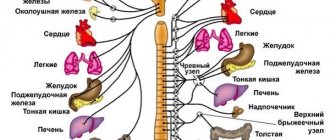
These nuclei consist of small nerve cells that receive information from neurons of other thalamic nuclei, limbic system, basal ganglia, hypothalamus, and brain stem. Through the ascending pathways, the nuclei receive signals from pain and temperature receptors, and through the reticular formation - from almost all other structures of the central nervous system.
Location
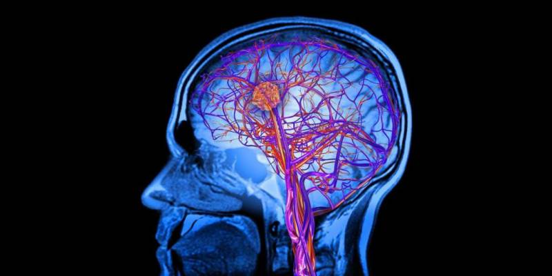
The thalamus is part of the cerebral hemispheres of the forebrain. It is located lateral to the lateral ventricles - cavities of the brain that are part of the cerebrospinal fluid (CSF) circulation system. Below it is the hypothalamus, from which the visual hillocks are separated by a groove.
Above and slightly outside the thalamus are the basal ganglia. These formations are necessary for precise, coordinated movements. These structures are separated from each other by the internal capsule - a bundle of white matter of the forebrain, through which pathways pass from the periphery to the center.
The right and left parts of the thalamus are connected to each other by interthalamic gray matter. It is present in 70% of people.
Main functions
The thalamus is a key formation in the transmission of nerve impulses to the cerebral cortex. If the cortex is damaged, it is thanks to the work of the thalamus that partial restoration of functions such as touch, sensation of pain and temperature is possible.
Another important function of the thalamus is the integration of motor and sensory activities. This is possible due to the receipt of information into the thalamus from both the motor and sensory centers of the nervous system.
In addition, the thalamus is essential for attention and consciousness. He also takes part in the formation of behavioral reactions.
Thanks to its connection with the hypothalamus, which will be discussed later in the article, the functions of the thalamus also cover memorization and emotional behavior.
Normal physiology: cerebellum, reticular formation, thalamus, hypothalamus, limbic system
When creating this page, we used a lecture on a relevant topic compiled by the Department of Normal Physiology of Bashkortostan State Medical University
Navigation:
Cerebellum
Has 3 pairs of legs . One of the most important structures of the brain, taking part in ensuring the accuracy of targeted movements and regulating the coordinated action of antagonist muscles.
The cerebellum is a corrective and controlling structure. By itself it does not cause any movement. If affected , movements will be inaccurate.
Thanks to the activity of the cerebellum, namely its hemispheres, all voluntary and involuntary movements of the upper and lower extremities become smooth.
Functions of the cerebellum:
- They coordinate and regulate voluntary and involuntary movements.
- Participates in maintaining balance and posture (anti-gravity function).
- Takes part in maintaining muscle tone.
- Performing precise, targeted movements.
These functions are performed by 3 zones: vermis, intermediate and lateral zones
1) Vermis (medial zone) - responsible for the correlation of the simplest trunk movements (balance, posture, maintaining muscle tone).
Receives impulses from:
- proprioceptors along the spinocerebellar tracts,
- vestibular nuclei of the trunk.
Directs impulses to:
- reticular formation (RF),
- Deiters nucleus.
The cerebellar vermis receives impulses about the position of the head and torso in space. It receives impulses about the position of the head in space from the vestibule of the cochlea.
When a person moves, impulses are transmitted from the receptors of the semicircular canals. This allows you to maintain balance regardless of body position and movement.
When a worm is damaged, gross static disturbances occur, that is, is lost .
In the most severe cases, the patient cannot sit or stand even with his legs spread wide apart and leans back and forth.
Violations:
- Astasia is the inability to stand without support.
- Ataxia is a disorder of balance and stability when walking, a “drunk gait.”
- Nystagmus - “shifting eyes”.
2) Intermediate zone (cork-shaped and spherical nuclei) - corrects more complex trunk movements.
Receives impulses from:
- proprioceptors along the spinal cerebellar tracts,
- vestibular nuclei of the trunk,
- cortex through the pontine nuclei.
Directs impulses to the red nucleus .
Violations:
- tremor (shaking) of the limbs,
- violation of locomotor tests (finger-nose test).
3) Lateral zone (dentate nuclei) - the youngest area of the cerebellum (corrects the most complex fast and precise movements).
Violations:
- Adiadochokinesis is a violation of supination - pronation of outstretched arms.
- Dysarthria - speech in syllables.
The consequences of removal of the cerebellum and loss of its functions were characterized by the Italian physiologist Luciani (1907) as a triad :
- Muscular asthenia or hyposthenia is a decrease in muscle performance.
- Atony is a decrease in muscle tension at rest.
- Astasia is a decrease in the stability of voluntary movements.
- Ataxia (as a result of 1, 2 and 3).
Thus, the cerebellum, having received information about the upcoming movements, adjusts the program of this movement and at the same time prepares the muscle tone for the implementation of this movement by the spinal cord. Also involved in the regulation of autonomic functions.
Reticular formation (RF)
The reticular formation is a nonspecific section of the central nervous system, a network of neurons in the thickness of the gray matter of the medulla oblongata, pons, midbrain and diencephalon, associated with all structures of the central nervous system.
Features of neurons:
- long dendrites and short, well-branched axons,
- a huge number of synapses (up to 25 thousand) between one RF neurons and neighboring ones,
- polysensory, i.e. the ability of one neuron to perceive afferent impulses from various receptors,
- high excitability and lability,
- sensitivity to certain substances (carbon dioxide, adrenaline, etc.).
RF neurons are in a state of constant tone due to:
- High sensitivity and humoral factors (neurons of the vasomotor center, sensitive to carbon dioxide in the blood, hydrogen ions).
- Collaterals from ascending pathways.
There is also an internal organization of the RF - the axons of reticular neurons have a large number of collaterals and synaptic structures.
Functions of RF neurons:
- Coordinating influence - brain stem reflexes are combined into complex motor acts.
- Complex forms of motor behavior are carried out: chewing, swallowing, singing, friendly eye movements, etc.
- Regulation of muscle tone by influencing motor neurons.
- The RF and medulla oblongata cause inhibition of neurons in the cerebral cortex.
- Midbrain RF increases cortical activity (wake-up effect).
- Regulates the activity of breathing, cardiovascular and other centers of the trunk.
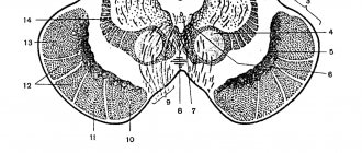
Thalamus (visual thalamus)
- large symmetrical formation,
- has the shape of an egg
- occupies 4/5 of the diencephalon,
- contains about 120 nuclei (a cluster of neuron bodies).
3 groups of cores:
- anterior - projects neuron axons into the cingulate gyrus,
- lateral (largest) - in the parietal, temporal and occipital cortex,
- medial - to any part of the cerebral hemispheres.
Classification by functional features:
- specific,
- nonspecific,
- associative.
Specific nuclei of the thalamus:
Function: rapid transmission of information from afferent systems to local areas of the cortex.
The lateral nuclei provide the transmission of impulses to the somatosensory areas of the 3-4 layers of the cerebral cortex (CBC) to the posterior central gyrus from the receptors of superficial and deep sensitivity of the limbs, trunk and head.
Dysfunction of specific nuclei of the thalamus leads to loss of specific types of sensitivity.
The nuclei of the thalamus themselves have a somatotropic localization (like the cortex).
They receive signals from receptors of the skin, eyes, ear, muscular system, as well as from interoceptors n. vagus and splanchnic nerves and hypothalamus.
Nonspecific nuclei of the thalamus form diffuse connections with all layers of the cortex.
The nonspecific nuclei of the thalamus receive impulses from:
- RF barrel,
- hypothalamus,
- limbic system,
- basal ganglia,
- specific nuclei of the thalamus.
Dysfunction of nonspecific nuclei leads to disruption of sleep onset.
Associative nuclei - do not receive direct projections from the periphery; they form connections with other nuclei of the thalamus and CBP.
Function: integrative activity of the KBP.
Functions of the thalamus:
- regulation of the functional states of the body,
- processing of all signals coming from the periphery to higher-lying centers.
The thalamus is called the “gate of consciousness”: the implementation of primary processing of information with the formation of primitive sensations.
Hypothalamus
This is the structure of the diencephalon that organizes the emotional, behavioral and homeostatic reactions of the body.
50 pairs of nuclei are isolated. Functionally divided into front, middle and rear.
Features of the hypothalamus:
- It is richly vascularized and has a permeable blood-brain barrier (BBB) to various substances.
- The nuclei of the hypothalamus are highly sensitive to shifts in the constants of the internal environment of the body: hormones, glucose, ions, etc.
- Neurons secrete peptides, neurotransmitters and other substances.
Connections of the hypothalamic nuclei:
- together,
- with the above and underlying structures of the central nervous system.
Functions:
- Impact on the vegetative functions of the body through humoral and nervous pathways .
- The hypothalamus is located centers:
- homeostasis,
- thermoregulation,
- hunger-satiation,
- thirst,
- sexual behavior,
- fear,
- rage,
- sleep-wake.
- Regulation of the pituitary gland:
- Neurons of the hypothalamus produce releasing factors (liberins) and inhibitory factors (statins) , which regulate the activity of the anterior pituitary gland (adenohypophysis).
- The hypothalamus secretes vasopressin (antidiuretic hormone - ADH) and oxytocin, which enter the neurohypophysis.
Basal ganglia. Striopallidar system
-Improvement of movements in their gradual economization and automation is ensured by the basal ganglia.
This system activates the necessary components of movement and inhibits unnecessary ones.
Basal ganglia:
- caudate nucleus,
- shell,
- pale ball,
- fence.
The globus pallidus, together with the substantia nigra, red nucleus, and subthalamic nucleus, constitute the pallidal system .
Extrapyramidal system:
- caudate nucleus,
- shell,
- pale ball,
- subthalamic nucleus
- black substance.
Information processed in the basal ganglia enters the nuclei of the anterior thalamus. It is then combined with information coming from the cerebellum. The impulses then go to the frontal motor cortex. From there to the neurons of the spinal cord.
Thus, the basal ganglia are integrative centers for organizing established types of motor activity of the body associated with learning.
Violations of the extrapyramidal system manifest themselves in the form of changes in motor function, muscle tone, autonomic functions, and emotional disorders.
Limbic system
The limbic system (LS) is a nonspecific part of the brain that determines the emotional and motivational component of human behavior.
This is a complex department of the central nervous system and consists of:
- olfactory brain,
- amygdala complex of the basal ganglia,
- fornix, hippocampus, mastoid bodies,
- transparent partition,
- orbital cortex.
LS Features:
- there are simple two-way connections between its components,
- complex paths forming many closed circles.
Thanks to drugs, the launch of vegetative, somatic and behavioral reactions is ensured, ensuring the adaptation of organisms to the external environment and maintaining homeostasis.
LS functions:
- emotional coloring of behavior, motivations and autonomic functions,
- formation of memory, learning, ability to remember new impressions,
- participation in the work of the VNS,
- determination of circadian rhythms: sleep-wake, hunger-satiety, etc.
12 0
Source: https://medfsh.ru/teoriya/teoriya-po-normalnoy-fiziologii/lektsii-po-normalnoj-fiziologii/normalnaya-fiziologiya-mozzhechok-retikulyarnaya-formatsiya-talamus-gipotalamus-limbicheskaya-sistema
Functions of the hypothalamus
Below is a list of the main functions of this structure:
- control of the activity of the autonomic nervous system;
- organization of behavior (eating, sexual, parental, emotional behavior, etc.);
- thermoregulation of the body;
- secretion of hormones: oxytocin, which increases contractile activity of the uterus; vasopressin, which increases the absorption of water and sodium in the renal tubules.
The functions of the hypothalamus listed above are provided due to the presence in it of various centers, as well as specific nerve cells. They are able to respond to changes in the state of the body (blood temperature, water-electrolyte composition, the amount of hormones in it, glucose concentration, etc.).
Thus, the diencephalon (thalamus and hypothalamus mainly) has many important functions that make normal life activities possible.