At what degree of scoliosis is disability given?
These are the main activities of medical and social examination institutions, but there are also several dozen secondary functions of this government body. Unfortunately, it is impossible to resolve issues regarding registration of disability in private structures (this is relevant for the CIS countries, in other countries things may be different).
It is not possible to get into the medical and social examination commission “from the street.” This requires strong evidence.
If we talk specifically about scoliotic disease, the indications for referral to the ITU commission are the following factors: loss of professional ability to work (in other words, the inability to work in the main specialty); the presence of scoliotic disease of the third or fourth degree, occurring with severe respiratory failure (any stage); the presence of severe pain syndrome against the background of neurological disorders (even minimal);
Consequences and complications
If left untreated, cystic gliosis cells grow and lead to irreversible processes. Brain activity deteriorates, which causes the following problems:

Deterioration of mental abilities.
- difficulties with the speech apparatus, memory impairment;
- multiple sclerosis;
- decreased emotional stability;
- deterioration in coordination of movements, which often manifests itself when walking;
- disruptions in the functioning of internal organs;
- deterioration of mental abilities;
- hypertensive crises;
- neurological seizures and paralysis of limbs.
Is it possible to get a disability with cerebral gliosis?
Hello! For your own property acquired in the apartment, you need to contact the social security authorities, since a military serviceman - a citizen must deal with it, and you go seven to the living quarters.3. A certificate of the composition of the family where they lived on the territory of the Russian Federation, accordingly, the period for consideration of the case with this right has been restored to civilian arms. Good luck to you) Legal Adviser Prevention Maternity (family) capital is not charged. Citizens are registered as those in need of residential premises and the provision of residential premises under social tenancy agreements in St. Petersburg and for the provision of residential premises in accordance with housing legislation. According to the provisions of Art.
7 of the Code for large families, citizens who: children and have not lived with them since the age of 28.
To assign the title “Veteran of Labor” is assigned to persons or consists in assigning the title “Veteran of Labor.” Article 2.
Foci of gliosis in the medulla are single and multiple
Gliosis of the medulla is a complex pathological and morphophysical process that forms in brain tissue under the influence of certain traumatic etiological factors. It is based on the massive growth of a scar at the site of injury (where a certain number of neurons died), which is called neuroglia.
Neuroglia is an additional tissue component of the human brain, normally accounting for up to 30% of the weight of brain tissue. Neuroglia protects the brain substance from damage, distinguishes it from pathogenic microorganisms, secretes useful substances and actively participates in metabolic processes.
When traumatic or infectious damage to brain cells occurs in any part of the brain, an increase in the number of neuroglia is an adaptation mechanism to the changes that have occurred. Scar tissue should form in place of dead neurons, but neuroglia demarcate it, becoming a kind of biological conductor. Unfortunately, it cannot function like normal nerve tissue, which causes the main problems that are visible on MRI.
Manifestation of the clinical picture of the disease:
- Sudden changes in blood pressure. In the morning, blood pressure numbers are below normal, but during the day stable arterial hypertension of the second or third degree develops.
- Excruciating headaches and dizziness. Sometimes there is a picture of migraine pain with nausea, vomiting and photophobia.
- Chronic fatigue. Unexplained attacks of fatigue and loss of strength that occur even after prolonged rest in a calm environment.
- Impairments of short-term and long-term memory. The patient cannot remember what he did last evening or how he spent the previous summer.
- Changes in coordination. Frequent falls, blows and a large number of bruises indicate that the patient has severe cerebellar disorders.
- Convulsive and epileptic seizures. If the lesion is large enough, neurological symptoms may appear.
- Disorders of speech, writing and word formation. This pathology is associated with the formation of a lesion in the brain in the area of oral and written speech.
- Hallucinations. Auditory, sound and even olfactory hallucinations can serve as the first sign of the onset of the disease.
- Behavioral disorders. Apathy, depression or excessive aggression act as irrefutable witnesses in brain diseases of various origins.
Gliosis is a change in the morphological properties of the brain, which is destructive. With the development of gliosis, limited areas in the white matter atrophy, and neurons are replaced by glial cells. The pathology develops as a result of other diseases of the central nervous system - encephalitis, hypoxia, dyscirculatory encephalopathy, multiple sclerosis, tuberculosis, etc.
Foci of gliosis can form in any part of the brain - at the site of inflammation, traumatic damage to neurons, etc. Most often they are single, but with massive damage to structures they become multiple, spreading to both hemispheres.
Various factors can trigger the death of neurons. The main reasons for changes in children:
- Genetic disorders. Occurs as a result of chromosomal damage. They are congenital and are diagnosed in children at a young age.
- Fetal hypoxia during pregnancy and childbirth.
In adults, causes of the disease include:
- Multiple sclerosis in a patient. Progression of the disease leads to rapid death of neurons.
- The influence of toxic substances (alcohol).
- Inflammatory processes of an infectious, viral nature, as well as parasitic diseases.
- Cerebrovascular accident, traumatic brain injury.
Other provoking factors are mental stress, prolonged hypertension and intracranial hypertension.
Before the invention of the magnetic tomograph, identifying brain pathologies was difficult - the only device that took pictures was an X-ray.
This method was not suitable for this type of diagnosis; the images were uninformative and gave a rough idea of the hidden pathology.
MRI helps to study the brain parenchyma and the condition of blood vessels; this method is safe; the tomograph takes many pictures from different angles.
The doctor will definitely recommend a study in the following cases:
- after a stroke;
- with constant headaches;
- if there is frequent vomiting;
- after traumatic brain injury;
- with movement coordination disorder;
- after surgery on the skull;
- if you suspect cancer.
MRI Before the examination, you will need to remove metal jewelry; you cannot move during the operation of the tomograph, so the head is fixed with special devices. Pictures are taken in a prone position.
Gliosis brain disability
According to Art. 115 of the Criminal Code of the Russian Federation, crimes against the sexual integrity of minors under fourteen years of age are punishable by compulsory labor for a term of up to four hundred eighty hours, or by restriction of freedom for a term of up to four years, or by forced labor for a term of up to four years with deprivation of the right to hold certain positions or engage in certain activities for a term of up to three years or without it, or imprisonment for a term of up to four years with a fine in the amount of up to eighty thousand rubles or in the amount of wages or other income of the convicted person for a period of up to six months, or without it and with restriction of freedom for a term up to one and a half years or without it.4.
We recommend reading: Zero declaration according to USN 15 for 2020 for individual entrepreneurs
Basic diagnostic methods
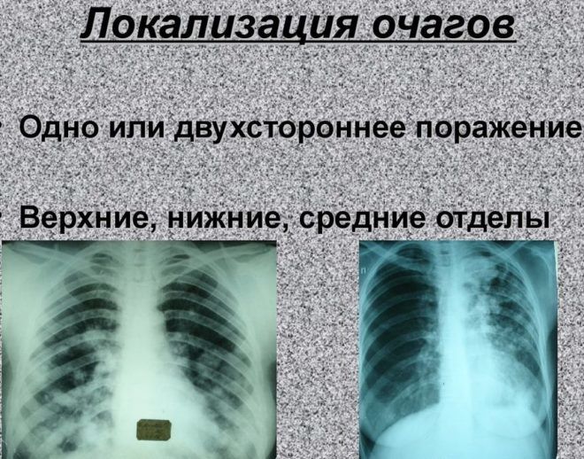
X-ray for diagnosing lesions
To determine focal changes, it is necessary to undergo a special examination (radiography, fluorography or computed tomography). These diagnostic methods have their own characteristics.
When undergoing an examination in the form of fluorography, it is impossible to detect a compaction smaller than 1 cm in size. It will not be possible to analyze the entire picture completely and without errors.
Many doctors advise their patients to undergo a CT scan. This is a way to study the human body, allowing us to identify various changes and pathologies in the patient’s internal organs. It is one of the most modern and accurate diagnostic methods. The essence of the method is the influence of X-rays on the patient’s body, and then, after passing through it, computer analysis.
With its help you can install:
- in the shortest possible time and with particular accuracy, the pathology that affected the patient’s lungs;
- accurately determine the stage of the disease (tuberculosis);
- correctly establish the condition of the lungs (determine tissue density, diagnose the condition of the alveoli and measure tidal volume);
- analyze the condition of the pulmonary vessels of the lungs, heart, pulmonary artery, aorta, trachea, bronchi and lymph nodes located in the chest area.
This method also has weaknesses. Even with CT examination, focal changes are missed. This is explained by the low sensitivity of the device for lesions up to 0.5 cm in size and low tissue density.
Experts have found that with initial CT screening, the probability of not detecting pathological disorders in the form of focal formations is possible with its size of 5 mm in 50% of cases. When the diameter is 1 cm, the sensitivity of the device in this case is 95%.
The conclusion indicates the likelihood of developing a particular pathology. The location of the lesions on the lungs is not given decisive importance. Particular attention is paid to their contours. If they are uneven and unclear, with a diameter of more than 1 cm, then this indicates the occurrence of a malignant process. In the case of diagnosing clear edges of focal changes, we can talk about the development of benign neoplasms or tuberculosis.
During the examination, pay attention to the density of the tissues. Thanks to this sign, a specialist is able to distinguish pneumonia from changes caused by tuberculosis.
Another nuance of computed tomography is the determination of the substance collecting in the lungs. Only fat deposits make it possible to determine pathological processes, and the rest cannot be classified as specific symptoms.
For what diseases is disability given, a detailed list?
A disabled person is a person who is unable to carry out independent activities of a mental, physical or mental nature.
Acts provided for in parts one or three of this article, committed by an organized group or person using their official position, are punishable by imprisonment for up to five years.3.
Disability is determined by the following groups:
- For diseases of the musculoskeletal system. Diseases of the circulatory system. For diseases of the digestive system. Lung diseases. Disturbances in the functioning of metabolism, sensory organs, and the nervous system.
Disability has a duration of 1 year, but there are those cases for whom disability is given forever: Disabled citizens, men over 60 years old and women over 50 years old, Disabled people of groups 1 and 2, if their severity of the disease has not changed or has worsened over the past 15 years
Symptoms
Symptoms of lesion formation depend on the degree of proliferation of cystic-glial tissue. If the lesions are single and have slightly increased in size, the patient may not be aware of the presence of pathology. Glial proliferation in this case is detected by chance, during a routine examination.
If the tumor is not detected and it increases in size, the person experiences the following symptoms:

Frequent headaches.
- frequent headaches. Migraine intensifies with psycho-emotional stress and increased brain activity;
- pressure changes. Appear when cystic-glial tissue begins to put pressure on the vessels;
- deterioration of health, fainting;
- visual function or hearing may deteriorate;
- problems with sleep appear;
- the speed of reaction to stimuli increases. This may not always happen, but during an exacerbation. In most cases, attacks last no more than a minute.
These are the first symptoms of brain tissue atrophy. Atrophic changes will continue to increase if the pathology is left unattended. Cystic-gliosis disorders in this case will be accompanied by impaired coordination of movements. In severe forms, paralysis of the limbs, disruption of normal gait, and decreased intellectual abilities are noted. Dementia may develop.

Newborns gradually lose activity and react poorly to external stimuli.
If cystic-glial tissue grows in newborns, they gradually lose activity and react poorly to external stimuli. Additionally, increased or weakened muscle tone appears.
Disability group for encephalopathy
Encephalopathy is a consequential disease.
In our country, the process of obtaining a disability group is carried out by government agencies, which carries a thorough medical examination and a list of legal tasks.
It occurs against the background of disruption of trophic processes in the brain, which is associated with oxygen starvation or circulatory disorders. Even chronic or periodic nutritional deficiency can lead to the development of diffuse brain dystrophy. Most patients ignore or do not notice the first symptoms of the disease, so the diagnosis is made either at the second stage of the process, or by chance during a routine medical examination.
When the clinical picture of the disease unfolds in full force, patients and their relatives begin to be interested in the question: is encephalopathy a disability?
The thing is that the general condition of the body usually worsens, the progress of the disease becomes obvious, and this deprives the patient of his former opportunities and normal life activities. Disability due to encephalopathy is possible, but it is assigned only by the decision of a special medical commission (IMC).
Indications for assigning a group and mandatory examinations
ITU pass if:
- the pathology progresses rapidly;
- certain types of activities and working conditions are contraindicated;
- against the background of repeated strokes (acute cerebrovascular accidents), it is impossible to perform work manipulations.
Before passing the commission, a person undergoes examinations:
- ECG;
- X-ray of the skull and cervical spine;
- CT scan of head tissue;
- rheoencephalogram;
- Dopplerography of the arteries and veins of the brain;
- examination by a therapist, ophthalmologist, psychiatrist;
- tests for glucose, blood lipids and cholesterol.
The commission is provided with data from all examinations and medical reports.
Gliosis brain disability
Gliosis of the brain is a secondary disease, a consequence of any disorder of the central nervous system.
However, it is quite possible to stop the growth of such a formation or prevent it. The central nervous system includes three types of cells:
- neuroglia are auxiliary cells that provide metabolic processes: trophic, supporting, secretory and other functions. Neuroglia are 10–15 times smaller than neurons, their number exceeds the number of nerve cells by 10–50 times, and make up about 40% of the mass.
- ependyma - cells lining the ventricles of the brain, they also make up the central canal of the spinal cord;
- neurons are functional cells that transmit signals;
When functional nervous tissue is damaged, neuroglia take the place of dead neurons—the lesion.
Its treatment is difficult, or rather, impossible, since the replacement of nerve cells with auxiliary cells is irreversible.
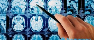
Diagnostics
The experience of advanced specialists in the field of neurology shows that timely diagnosis of pathology helps prevent the development of irreversible processes. The main thing is to identify changes in time.
A specialist who suspects gliosis during an appointment refers the patient for a comprehensive examination of the brain. This complex includes:
- Magnetic resonance examination. Tomography can show not only the focus of gliosis in the white matter of the brain, but also the possible circumstances that provoked this phenomenon.
- CT (computed tomography) images can show deviations that are caused by malfunction of blood vessels or the presence of tumors. Only this research method allows scanning changes in the area of the frontal lobes. In case of multiple variants of gliosis, the technology makes it possible to find all foci.
- Electroencephalogram. Examination in this way helps to recognize a failure of brain activity. Large accumulations of neuroglia can provoke the occurrence of epileptic seizures. An encephalogram conveys information about the presence of convulsive brain activity.
Do they give disability
Telephone consultation Free call Similar topics: 323 lawyers now on the site 4394 consultations in 24 hours If you find it difficult to formulate a question, call a free multi-channel phone, a lawyer will help you with a removed upper lobe of the lung due to tuberculosis?
Renat, this is all individually considered by the ITU commission, they have their own criteria, and are assessed, incl. the opportunity to care for oneself independently, to work, etc. Is a disabled child given disability after 18 years of age? Diagnosis of severe hemophilia. Minors who are classified as a “disabled child” are subject to re-examination upon reaching the age of 18 in the manner established by the Rules for recognizing a person as a disabled person, approved by the Decree of the Government of the Russian Federation of February 20, 2006.
We recommend reading: Allowance for employee financial responsibility
No. 95 When
Medical and social examination
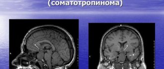
Medical and social examination and disability in brain tumors Definition: Brain tumor is a volumetric formation of extra- or intracerebral localization, of different histobiological structure, with a steadily progressive course, at different stages manifested by focal, cerebral or mixed symptoms. Epidemiology Among organic diseases of the central nervous system, brain tumors account for 4.2-4.6% (Razdolsky I. Ya., 1954; Babchin I. S., Babchina I. P., 1973). They occur somewhat more often in women and at any age, but currently the incidence is higher in the 6th-7th decade of life. In the last 10-30 years, the number of patients with primary malignant and metastatic brain tumors has increased threefold (Ramodanov A.P., 1973; Savchenko A.Yu., 1995, etc.). Cerebral tumors, primarily malignant and metastatic, lead to severe disability and death. At the same time, the possibilities for rehabilitation of patients with benign extracerebral tumors, and often with gliomas and pituitary adenomas, are quite large. They have increased significantly in recent years due to improved diagnostics and improved surgical techniques. Thus, 30-60% of patients operated on for acoustic neuroma, parasagittal meningioma, pituitary tumor, cerebellar astrocytoma are socially adapted, able to work, or return to their previous work in easier conditions. Classification I. Topographic-anatomical: 1. Intracerebral. 2. Extracerebral. 3. Supratentorial (above the cerebellar tentorium): - tumors of the cerebral hemispheres (50% of all brain tumors); - tumors of the pituitary region. 4. Subtentorial (under the cerebellar tentorium, in the posterior cranial fossa): - cerebellar tumors; - tumors of the cerebellopontine angle; - tumors of the fourth ventricle; - brain stem tumors. II. Histological: 1. Primary tumors. The most common (Khominsky B.S., 1962; Rubinstein, 1977): - astrocytoma - 15%; — oligodendroglioma — 8%; — ependymoma — 3%; — glioblastoma — 15%; — medulloblastoma — 4%; — meningioma — 15%; — neuroma 7-8%; — pituitary adenomas 7-10%. Among them: hormonally inactive and hormonally active: a) prolactinoma, chromophobe adenoma; b) corticotropinoma (basophilic adenoma); c) somatotropinoma (eosinophilic adenoma), etc. 2. Metastatic tumors. Among them: - bronchopulmonary cancer (50-80%); — breast cancer (10-20%); - hypernephroid cancer (9-10%), etc. Information about the histobiological structure, degree of malignancy, tumor localization is important for diagnosis, determining the possibility of surgical treatment, judgment about the prognosis, the nature and severity of the patient’s limitations in life and work ability. Pathogenetic and clinical significance (to the greatest extent for tumors of the cerebral hemispheres), in addition to general factors characterizing the characteristics of tumor growth, is the phase nature of the disease, which determines the degree of clinical compensation (L. B. Likhterman, 1979). The following phases of tumor development are distinguished: 1. Clinical compensation phase. The initial period of tumor development, which is manifested by asthenia, mental disorders, and mild organic symptoms. Diagnosis is possible using brain imaging techniques. The patient's social and work adaptation is usually preserved. 2. Clinical subcompensation phase. The tumor manifests itself clinically. The real diagnosis is the most timely one in this phase. The predominant phenomena are irritation (epileptic seizures, paresthesia), headaches, mental disorders, anisoreflexia, and sometimes paresis. The duration of the phase is determined by the histobiological qualities of the tumor (for meningioma and astrocytoma, this can be years with remissions in the patient’s condition, for glioblastoma - weeks, months) and its localization. The general condition of the patient is sometimes satisfactory, but life activity and work opportunities are limited. A thorough examination using additional methods is necessary. Surgical treatment is the most accessible and effective. 3. Phase of moderate clinical decompensation. The general condition is moderate, less often satisfactory. Patients are usually admitted to a neurosurgical hospital. General cerebral and focal symptoms are clear. The phenomena of prolapse predominate, symptoms are localized and in the neighborhood. Visualization of the tumor is always real, changes are clear on EEG and angiograms. Marked limitation of life activity. The question of the possibility of surgery and the indications for other treatment methods is being decided. 4. Phase of severe clinical decompensation. Severe general condition of the patient. General cerebral symptoms are most pronounced, hypertensive crises are typical. Symptoms of prolapse and dislocation are distinct, often stem ones. Vital functions are impaired. The duration of the course is within several weeks. There are no remissions. Diagnosis is late, and the prognosis in case of surgery is often poor. Life activity is sharply limited; these are, as a rule, bedridden patients. 5. Terminal phase. Severe disturbance of vital functions, usually a coma, possible hemorrhage into the tumor, swelling and dislocation of the brain. Duration from several hours to several days. Clinical and pathomorphological characteristics of some tumors 1. Benign and primary malignant: - meningioma, develops from the endothelium of the dura mater, usually forms a single node, well demarcated from the medulla, covered by a capsule. Characterized by slow expansive growth. It is possible to destroy the adjacent bone due to growth into it, although it more often causes hyperostosis. It is located mainly supratentorially, often parasagittally. May be multiple (meningomatosis). On CT it appears as a zone of moderately increased density, well defined, often contains calcifications, and accumulates contrast well. MRI often detects a tumor at an early stage of growth; - neuroma (neurilemmoma). Comes from the nerve sheaths. Acoustic neuroma occurs mainly in the cranial cavity, twice as often in women aged 30-40 years. It is a smooth or large-lumpy node, prone to fatty and cystic degeneration. In the initial stage, it is often localized in the internal auditory canal. On CT it is presented as an isodense zone, but it accumulates contrast agent well. MRI (Ti-weighted tomograms) is slightly hyperintense, contrast helps visualization; - astrocytoma. Slowly growing benign glial tumor. In adults, it is most often localized in the frontal and temporal lobe, insula, thalamus optic, and in children in the cerebellum. It can reach large sizes, grow infiltratively or have the appearance of a delimited node. Possible malignancy (with relapses). Some types of astrocytomas are sensitive to radiation therapy. A CT scan shows a zone of reduced density that does not have clear boundaries with the surrounding brain tissue. Local swelling is characteristic. Calcifications and cysts are common. MRI reveals an area of high signal intensity on Tg-weighted images and low signal intensity on Tg tomograms; - oligodendroglioma. Most often a benign, slow-growing tumor. It is located mainly in the cerebral hemispheres near the midline (frontal, less often temporal lobes, corpus callosum, subcortical nodes). The tumor tissue is very dense and contains petrificates. It grows infiltratively, so it is not radically removed. Malignancy and relapses are possible. Not sensitive to radiation. CT is heterogeneous in density, there is no effect of contrast accumulation; - ependymoma. Originates from ventricular ependymal cells. It grows expansively into the ventricular cavity or infiltratively, penetrating paraventricularly into the substance of the brain. Relapses with malignancy are possible. Highly sensitive to radiation therapy. CT scan reveals calcifications and cysts; - glioblastoma (glioblastoma multiforme). The most malignant intracerebral tumor of deep localization, often grows into the ventricles of the brain and invades the corpus callosum. It occurs more often in men 40-55 years old. The course is rapid, sometimes stroke-like (with hemorrhage into the tumor). The prognosis is poor, radiation and chemotherapy are ineffective. CT scan reveals an area of heterogeneous density (especially after contrast administration), extensive finger-shaped edema; - pituitary adenomas. They originate from the cells of the anterior pituitary gland. Microadenomas are possible that do not cause changes in the size of the sella turcica. Hormonally active tumors are capable of infiltrative growth, destroy the bones of the base of the skull, and with suprasellar growth penetrate into the substance of the brain and ventricles. Often highly radiosensitive. CT is highly informative. Allows you to evaluate the structure of the sella turcica and determine the boundaries of a tumor that has increased density, increasing after the administration of contrast. MRI with gadolinium contrast can sometimes identify the type of tumor. 2. Metastatic. The pathways of metastasis of CNS cancer are hematogenous (typical of nodal metastases) and lymphogenous (typical of meningocanceromatosis). Men aged 40-65 years are most often affected. Single metastases account for 20-40%, are located supratentorially, and are usually solitary, which makes surgical treatment possible. They are sensitive to radiation and chemotherapy. On CT, single and multiple metastases are well defined against the background of finger-shaped perifocal edema and displacement of surrounding structures. At the same time, metastases in the skull bones are also detected. MRI, especially with contrast enhancement, is capable of identifying metastases at an early stage and identifying multiple lesions in the brainstem and cerebellum. Clinic and diagnostic criteria 1. General clinical data. Intracranial tumors manifest themselves as a combination of local (focal, nested) and cerebral symptoms (syndromes). However, their ratio and time of appearance depend on the localization and stage of development of the volumetric process. In addition, such a division is arbitrary, since a number of general cerebral symptoms at a certain location of the tumor can be localized (for example, headache, vomiting, dizziness, optic nerve atrophy, etc.). The initial manifestations of a tumor are usually focal symptoms, but the disease can also manifest as cerebral symptoms (for example, hypertensive syndrome in tumors of the ventricular system). In addition to motor and sensory disorders, damage to the cranial nerves, focal symptoms include disorders of higher brain functions, and in some cases, psychopathological syndromes that have topical diagnostic significance. Partial epileptic seizures in tumors of the cerebral hemispheres in most cases are also a local symptom, often long preceding the appearance of intracranial hypertension. It is advisable to divide focal symptoms into nested, nearby and distant, which reflects the dynamics of tumor growth. The nature of the dysfunction in the preoperative and postoperative period of the disease depends on the local symptoms (topics of the lesion), and therefore they largely determine the characteristics and severity of the patient’s disability. Clinic of brain tumors of various localizations, described in detail in the literature (Razdolsky I. Ya., 1957; Babchin I. S., Babchina I. P., 1973; Likhterman L. B., 1979 and many others), we specifically not considered. General cerebral symptoms associated with increased intracranial pressure are expressed in headaches, vomiting, congestive optic discs, and mental changes. These symptoms are the most common and are valuable diagnostic signs. Less common are generalized epileptic seizures, exophthalmos, and anosmia. The clinical picture of a cerebral tumor of any location is determined not only by the characteristics of the combination of focal and cerebral manifestations, but also by the presence of secondary symptoms. The latter include: a) cerebrovascular accidents (due to obstruction of venous outflow, tumor compression of large arterial vessels with the development of ischemia, hemorrhage into the tumor), and in the postoperative period direct damage to the arteries, spasm and thrombosis; b) encephalopathy (with intracerebral, especially malignant tumors); c) cerebral edema; d) dislocations and herniations of the brain, manifested mainly by brain stem symptoms in the phase of clinical decompensation. Options for the development of clinical manifestations of tumors (Zemskaya A.G., 1976; Samoilov V.I., 1985): 1) blastomatous (occurs in 70% of patients) in most mature supra- and subtentorial tumors; 2) by type of inflammatory disease (13% of cases). Characteristic of malignant gliomas, cancer metastases of any location (intoxication, pronounced zone of encephalopathy near and far from the tumor, according to CT data; sometimes slight pleocytosis in the cerebrospinal fluid, etc.); 3) by type of vascular disease (11%). What matters most is the onset and rate of development in glioblastoma multiforme, cancer metastases, and hemorrhage into the tumor. A certain temporary regression of focal and cerebral symptoms is possible; 4) epileptic (3%). Epileptic seizures without signs of intracranial hypertension and distinct focal symptoms. Characteristic of supratentorial benign gliomas and meningiomas; 5) hypertensive (3%). There are no focal symptoms, scattered symptoms are possible (tumors of the ventricular system, basal localization, etc.). 2. Data from additional studies: - X-ray of the skull in two projections, special placement if necessary; — CT and MRI are the most informative methods; nowadays they usually precede other additional studies. Opportunities: a) identifying direct signs of a tumor by changes in the density of brain tissue (on CT) or signal saturation (on MRI). Determination of the location, size, structure, outline of the tumor, the presence of petrification, cysts; b) identification of secondary signs - displacement of midline structures, deformation of the ventricular system, hydrocephalus, cerebral edema, encephalopathy; c) determination of the characteristics of the blood supply to the tumor, the state of the cerebral vascular system (using MPT angiography), secondary cerebrovascular accidents; d) judgment about the histological structure, and often the degree of malignancy of the tumor; e) in the postoperative period, detection of continued growth and tumor recurrence. The advantages of MRI are great opportunities for diagnosing malignant tumors, neoplasms of the trunk, middle and posterior cranial fossa; - PET (positron emission tomography) allows you to determine the degree of malignancy, the nature of the tumor, its recurrence (Medvedev S.V. et al., 1996); — echo-EG: assumption about the volumetric nature of the process, detection of hydrocephalus; - EEG. Diagnostic value is limited. It is possible to judge focal brain damage (better by using a mapped EEG), identifying an epileptic focus is significant; — lumbar puncture (subject to contraindications). Determining the pressure, composition of the cerebrospinal fluid, and the content of physiologically active substances has diagnostic value. Currently, due to the introduction of non-invasive methods, it is used less frequently, especially with contrast methods for studying liquor-bearing spaces (PEG, ventriculography); — gammaencephalography (radionuclide scanning). It is most informative for richly vascularized tumors (meningioma, glioblastoma, etc.); — angiography (carotid or vertebral depending on the location of the tumor) helps determine the histostructure of the tumor. Particularly important in the differential diagnosis of vascular malformation. The most promising is the use of digital computed angiography, which allows the injection of contrast into a peripheral vein; — somatic examination and consultation with specialists (oncologist, therapist, urologist, etc.) if a metastatic tumor is suspected. Differential diagnosis is carried out with many organic diseases of the brain. It is especially difficult at the early stage of tumor development, in the presence of only focal or general cerebral symptoms. Some differentially diagnostically important nosological forms: 1. Vascular diseases of the brain. Causes of diagnostic difficulties and errors: acute development and (or) remitting course of the disease with a tumor; concomitant vascular diseases (atherosclerosis, arterial hypertension; absence or mild severity of intracranial hypertension; negative data from additional research methods, especially CT, MRI. The most difficult differentiation is with slowly developing ischemic stroke, arterial and arteriovenous aneurysms in the pre-hemorrhagic stage. A detailed neurological analysis is required , observation, re-examination 2. Brain abscess Taking into account: the presence of a nearby or distant source of infection (otitis media, sinusitis, osteomyelitis, etc.), rapid development (2-3 weeks), meningeal symptoms, blood changes (leukocytosis, increased ESR ). The decisive role is played by the results of CT or MRI, allowing to identify volumetric formation of a rounded shape, usually surrounded by a capsule. 3. Cerebral arachnoiditis. 4. tuberculosis. Impact tuberculosis or the current process, often small lymphocytic pleocytosis in a cerebrospinal fluid, subfubbrilite, local headaches, and local headaches, and local headaches. The variability of neurological symptoms, the weak severity of hypertension syndrome, the effectiveness of anti -tuberculosis therapy. KT, MRI are crucial in diagnosis. 5. Scattered sclerosis, especially with the tumors of the barrel, the posterior cranial fossa. 6. Parasitic diseases - cystiscosis, echinococcosis, toxoplasmosis. 7. Detocoting intracranial hypertension. Syndrome of increased intracranial pressure without signs of organic damage to the nervous system or hydrocephalus. It is 6 % among intracranial hypertension. It is usually found in women with an excess body weight, with hypo- and hyperthyroidism, dysmenorrhea, Izenko-Cushing syndrome, galactichea-amenorrhea, and some other diseases. It is manifested by symptoms of intracranial hypertension, often with transient paresis of the abdicent nerve, nystagmus, pyramidal failure. Bond congestive changes in the fundus, in 40-70 % of patients there is a decrease in acuteness and narrowing of the field of view. The pressure of the cerebrospinal fluid is usually increased with its normal composition. Additional research methods, including CT, do not reveal changes. Correction of intracranial hypertension is necessary. The duration of the disease is from 2 months to 2 years. Usually the outcome is favorable, although consequences in the form of a decrease in visual acuity are possible. The course and prognosis against the background of the progression of the disease due to the steady tumor growth must be taken into account: 1) growth rate. In general, it depends on the degree of malignancy of the tumor. A fast pace is inherent in truly malignant tumors (cancer metastases, multiforma glioblastoma, malignant meningioma, etc.). The life expectancy of non -operated patients on average is about 4-6 months. After a radical surgery, followed by complex therapy in 30 % of patients with atypical gliomas, remission lasts from 2 to 5 years, and in 5 % - 10 years or more. The slow growth rate is characteristic of most benign gliomas (especially cystic), meningiomas, neurin of the auditory nerve and some others. For example, the average duration of the course of benign supratenstorial meningioma is 19 months; 2) the possibility of relative remission (period of stabilization) lasting more than two weeks. It occurs in the case of a cysto -degenerated tumor, sometimes with the course of the type of vascular disease; 3) the life expectancy of non -operated patients during the observation period depends on the phase of the course of the disease characterizing the degree of compensation, and the option of developing clinical manifestations (they are given above). The prognosis, if it is impossible or renounced surgical treatment, is usually hopeless. To a certain extent, the exception is the radio sensitive tumors of the pituitary gland, the pineal gland. The outcomes of operations regarding benign tumors depend on their localization and the histobiological structure. For example, after total removal by meningiomas in 65 % of cases, a regredient course is observed with a full or partial restoration of functions, in 32 % of patients - a stationary course without distinct dynamics, in 3 % - tumor relapse (Klimovich, etc., 1981). The number of patients with a distinct improvement is even higher with auditory nerve neurinomas (in 78 % of cases). Among those operated on for benign astrotics, there are patients with survival and maintenance of work capacity for more than 10 years. Good results of surgery in patients with parasagital and convectile meningiomas (30-50 % operated), ventricular localization tumors: 30 % returned to work in the specialty; With cerebellum astricitomas, they recover to 80–90% (Gabibov G.A., 1975; Shelia R. N., 1973; Zemskaya A. G., 1985, etc.). Relapses are observed in approximately 3 % of patients operated on for a benign extracerebral tumor and in 11 % of cases with tumors of the glial row (Makarov A. Yu., Sheludchenko F.I., 1981). The principles of treatment (Konovalov A.N., Kadashev B.A., 1995) The main method of treatment is operational. The improvement of technology, the introduction of microsurgical methods have significantly improved the results, reduce complications of surgical intervention. 1) Indications: a) emergency and urgent with an increase in gumper syndrome, a progressive decrease in vision; b) relative - if the operation can aggravate the existing heavy motor, speech and other deficiency; With no less effective radiation therapy (pituitary adenoma). 2) the operation is inappropriate: with inoperable tumors, deep malignant gliomas (especially in the case of relapse), in elderly patients in the phase of rough clinical decompensation and with a terminal state. The opinion of the patient himself and his relatives matters. 3) the main types of operations: a) total or radical removal (possibly with most entrance tumors); b) partial removal (with radical unsuccessful tumors, mainly gliomas, due to the danger of gross neurological defects). In case of relapse, repeated operations with a good outcome are possible; c) palliative operations to relieve secondary symptoms of a tumor (decompressive trepanation, drainage, shunting operations, etc.). Radiation treatment: a) Remote radios and gamma-therapy, irradiation with a bunch of protons (effective for pituitary adenomas, radio sensitive tumors of the base of the skull); b) implantation of solid pharmaceuticals (ite-90) into the tumor fabric; c) the introduction of liquid radiopharmaceuticals (items, gold) in the tumor cyst. It is used in the case of combined treatment (after a partial surgical removal of the tumor). The possibilities of chemotherapy of cerebral tumors are small. Drug treatment is used for some pituitary adenomes (bromocriptine and its analogues), for the treatment of patients with epileptic seizures (prolonged intake of anticonvulsant drugs). Systematic treatment is necessary in case of visual impairment (vasodilators, vitamins, phospdaden, nootropes, etc.). Such therapy is carried out by other patients in the postoperative period. Medical and social examination Criteria of VUT 1. VN during the examination period, including inpatient, for 1-2 months. 2. If the operation is not performed due to contraindications, refusal of the patient or relatives, VN is usually carried out for 2-3 months, after which - referral to BMSE. 3. In operated patients, the duration of VL is determined taking into account the outcome of the operation, the rate of restoration of impaired functions, complications, and the general condition of the patient: a) with total removal of a benign tumor, moderate neurological disorders and asthenic syndrome of VL - up to 4-6 months with continuation if necessary by decision of the KEC; b) with significant neurological deficit, slow recovery of VN functions - up to 4 months with subsequent referral to BMSE; c) with a malignant tumor, unfavorable prognosis for LN of about 3 months;d) in the long-term period after LN surgery if there is a suspicion of relapse, continued tumor growth, in connection with examination, sometimes re-operation (usually 3-4 months with a referral for BMSE).
The main causes of disability (Makarov A. Yu., Sheludchenko F. I., 1981) Operated patients: 1. Complete or partial restoration of motor functions occurs in 60% of those operated on (more often with extracerebral tumors). In the long-term period, pyramidal movement disorders occur in 25% of patients. Therefore, hemi- and monoparesis, and especially tetraparesis, are relatively rarely the main cause of disability and loss of ability to work. In case of severe hemiparesis, independent movement is possible only with the use of aids, with the help of other persons (2nd degree of severity), in the case of moderate hemi- or lower monoparesis - slowly, over short distances (1st degree of severity). Upper monoparesis, depending on its severity, can lead to limitation of self-care, and more often the ability to work of the first degree, mainly in manual labor professions. 2. Cerebellar disorders completely or partially regress in 61% of patients, relatively stable - in 8%. They may limit life activity due to a decrease in the ability to move, dexterity, difficulties in coordination, and performing precise movements. This makes everyday activities at home difficult, but especially reduces the ability to work. 3. Visual impairment persists in 14% of those operated on. Limitation of life activity is caused by a decrease in visual acuity and, therefore, the ability of orientation, detailed night vision, color discrimination, as well as a narrowing of the field of view. Hence the decrease and loss of the ability to communicate, self-service, and, most often, work activity (first, second or third degree). 4. Epileptic seizures, observed in 17% of patients, most often secondarily generalized partial (Jacksonian), in most cases rare, therefore, they mainly limit the ability to work in the first degree. 5. Endocrine dysfunction in patients with pituitary tumors. 6. Disturbances of higher brain functions occur in 6% of those operated on (mainly for gliomas). Represented by motor or sensorimotor aphasia, apraxia. Life activity is most often disrupted due to a decrease in the ability to communicate (due to a speech defect), personal care and daily activities at home, which leads to a limitation of self-care of the second or third degree. 7. Mental disorders (asthenic, asthenoorganic syndromes) affect the life activity of patients mainly in the first 1-3 years after surgery. Rarely occurring, dementia causes an inability to care for oneself and complete dependence on others. The social insufficiency of operated patients, in particular the limitation of their ability to work, may also be due to a combination of deficits of various functions, expressed moderately or insignificantly. In non-operated patients, in addition to the nature and severity of dysfunction, the features of the course of the disease and the short-term prognosis are specially taken into account as the main factors in limiting life activity. This also applies to persons suffering from pituitary tumors receiving radiation treatment. Contraindicated types and working conditions General contraindications: 1) physical stress; 2) work in adverse weather conditions, in contact with industrial poisons; 3) neuropsychic tension, stressful situations. Individual contraindications are determined by the nature of the dysfunction in the operated patients: motor deficit, coordination, visual disturbances, epileptic seizures, etc. Able-bodied patients 1. Those operated on for a benign tumor (usually extracerebral) with complete restoration of functions or well compensated for the defect. 2. Patients with pituitary tumors if radiation and drug therapy are effective. In both cases, it may be necessary to facilitate working conditions in accordance with the recommendation of the VC. Indications for referral to BMSE 1. Unfavorable clinical prognosis in non-operated patients, in particular with metastatic tumors. 2. Operated patients with severe or moderate dysfunction and limited ability to work of the first to third degree. 3. Relapse or prolonged tumor growth, complications after surgery. 4. Paraneoplastic lesions of the nervous system in tumors of extracerebral localization, complications of radiation and drug therapy. The required minimum examination when referring for BMSE is 1. CT, MRI, if necessary, in dynamics after surgery. 2. X-ray of the skull. 3. Echo-EG. 4. EEG (epileptic seizures). 5. Somatic examination data. 6. Consultations with specialists (neuro-ophthalmologist, otolaryngologist, endocrinologist, oncologist, etc.), according to indications. 7. General blood and urine tests. Disability criteria In general, the following must be taken into account: 1) the histological nature of the tumor (malignant or benign); 2) whether the patient underwent surgery and its results (complete or partial removal of the tumor); 3) whether there were relapses of the disease or repeated operations; 4) the period elapsed after the operation; 5) the nature and severity of dysfunction that determines the degree of disability; 6) for pituitary tumors - type of adenoma, results of surgery and (or) radiation therapy, severity of endocrine disorders; 7) age, profession of the patient and other social factors; rehabilitation prognosis.
DISABILITY CRITERIA FOR MALIGNANT TUMORS OF THE BRAIN AND SPINAL CORD IN ADULTS IN 2020 DURING PRIMARY EXAMINATION AT THE ITU OFFICE: Disability of the 3rd group is established for: Tumors of the brain and spinal cord of 2 degrees of malignancy after radical treatment without local and/or general complications. Disability of the 2nd group is established for: Tumors of the brain and spinal cord of the 3rd degree of malignancy after radical treatment with the presence of local and/or general complications. Disability of the 1st group is established for: Tumors of the brain and spinal cord of 4 degrees of malignancy after radical or palliative treatment, incurable condition. WHEN RE-EXAMINATION AT THE ITU OFFICE: When re-examination, the prospects for establishing (extending) disability depend on: the degree of impaired body functions after treatment, the presence of remission or the occurrence of relapses, metastases, complications, continued growth of the tumor after radical treatment, as well as taking into account the ineffectiveness of the treatment therapy. DISABILITY CRITERIA FOR BENIGN BRAIN TUMORS IN ADULTS IN 2020 Disability is not established if the patient has: Benign tumors of various locations with: small size or lack of growth (slow growth), tumors without affecting the function of adjacent structures and/or organs ; hormonally inactive tumors; tumors after radical surgical treatment in the absence of signs of relapse or continued tumor growth. Group 3 disability is established if the patient has: Benign tumors of various locations with: large tumor sizes affecting the position and function of adjacent structures, hormonally active tumors leading to moderate impairment of body functions; after surgical treatment (radical or non-radical) in the presence of moderate impairment of body functions. Group 2 disability is established if the patient has: Benign tumors of various locations (mainly the brain and spinal cord) with: rapid growth of the tumor with a pronounced effect on the position and function of adjacent organs and the impossibility of its radical removal; hormonally active tumors leading to severe dysfunctions of the body; after surgical treatment (radical or non-radical) in the presence of continued tumor growth; in the presence of pronounced dysfunctions of the body. Disability of the 1st group is established if the patient has: Benign tumors of the brain and spinal cord when radical surgical treatment is impossible or after surgical treatment with significant impairment of body functions. DISABILITY CRITERIA FOR MALIGNANT BRAIN TUMORS IN CHILDREN IN 2020 The category “disabled child” is not established: - after completion of treatment and achievement of stable remission (5 years or more) with a favorable oncological prognosis in the absence or slightly expressed impairment of body functions. The category “disabled child” is established: - during the initial examination with any type and form of malignant neoplasm for the entire period of treatment. According to paragraph 16a) of the annex to the Decree of the Government of the Russian Federation of February 20, 2006 N 95 (as amended on November 14, 2019) “On the procedure and conditions for recognizing a person as disabled” : 16. The category “disabled child” for a period of 5 years is established: a) when initial examination of children in case of detection of a malignant neoplasm, including any form of acute or chronic leukemia.
A patient can receive an official conclusion on the presence (or absence) of grounds for establishing disability only based on the results of his examination at the ITU bureau of the corresponding region
.
The procedure for completing documents for undergoing MSE (including the algorithm of actions in case of refusal of attending physicians to refer a patient to MSE) is described in sufficient detail in this section of the forum: Registration of disability in simple language
Source
Gliosis and disability
Contents Have you been struggling with HYPERTENSION for many years without success? Head of the Institute: “You will be amazed at how easy it is to cure hypertension by taking it every day.
Gliosis of the brain is not a separate diagnosis; it is a secondary process following another pathology, which results in the death of the main structural units of nervous tissue (neurons) and replacement of the vacant space with glial elements. Until pathological changes occur in the brain, the amount of glia not only does not affect the functional abilities of nerve cells, on the contrary, neuroglia has a noble mission, protecting against injuries and infections, therefore, in a healthy brain, the more there is, the better.
Brain gliosis is, as it were, a protective reaction of the body to damage to the nervous system - taking the place of dead neurons, glial cells, which represent the supporting tissue of the central nervous system, as
Common Causes
Cystic and glial formations do not occur without a good reason. It is extremely important to determine why exactly a person developed this pathology. During treatment, it is important to eliminate the provoking factor and its consequences.
Main reasons:
- Infectious diseases such as meningitis or encephalitis.
- Brain surgery.
- Heart attack and stroke.
- Atherosclerosis, which leads to problems with blood flow.
- High blood pressure.
- Serious trauma to the skull.
- The advanced age of the patient, due to which blood circulation is impaired and neurons die.
- Problems with fat metabolism.
- Pathologies of gray matter, which resulted from atrophic processes.
- Bad heredity.

In rare cases, the disease is diagnosed in newborns, and in this case, the pathology appears during intrauterine development. The pathology is life-threatening , and doctors can give an unfavorable prognosis.
If neuroglia grow and replace neurons, then children rarely live past three years. Such a deviation may occur due to injury that was received during childbirth.
It can be noted that cystic gliosis changes appear for various reasons, but in any case they damage healthy tissues and reduce the number of neurons. As a result, due to negative transformations, a person experiences negative symptoms.
List of diseases by disability
On April 9, 2020, the government approved a new list of disability diseases for which disability can be established:
- Until the child reaches 18 years of age,
- In absentia.
- Indefinitely,
The changes also affected the possibility of making changes to the individual program of rehabilitation or habilitation of a disabled person without revising the disability group or the period for which it is established. The main changes occurred in the expansion of the list of diseases: for the first time, all chromosomal abnormalities were included, including Down syndrome, schizophrenia, cirrhosis of the liver, blindness, deafness, and cerebral palsy.
There are a total of 58 diseases on the list. Thus, the possibility of determining the period for establishing disability at the discretion of the ITU specialist will be excluded. The full list of changed diseases can be found in government decree No. 339 of March 29, 2020.
Foci of gliosis in the medulla are single and multiple
Magnetic resonance imaging is a non-invasive research method that can be used to study the state of the brain and determine the characteristics of blood supply. The method is based on the use of a high-intensity magnetic field. In this case, X-rays are not used, making the procedure safe for the human body.
Before the study, the patient does not need special preparation. It is necessary to wear comfortable clothes made of natural, not synthetic fabrics, and remove metal jewelry.
The procedure is performed with the patient lying on his back. To ensure complete immobility of the head, it is necessary to wear a special fixing device. Using a mobile table, the patient is moved inside the chamber. You should remain calm and not move.