Focal neurological symptoms
(
focal neurological deficit
) is a term that refers to neurological symptoms characteristic of local damage to certain structures of the central or peripheral nervous system. Focal neurological symptoms in many cases are combined with general cerebral symptoms, which are a manifestation of diffuse damage [1]. Focal neurological symptoms allow not only to identify the presence of local damage, but also to carry out topical diagnostics, that is, to fairly reliably determine the area of damage based on knowledge of neuroanatomy.
Focal neurological symptoms are characteristic of a number of diseases, including traumatic brain injury[2], brain tumors, strokes, etc.[3]
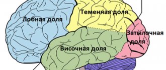
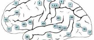
Lesions of the cerebral cortex
Frontal cortex lesion
Left frontal lobe highlighted in red
Frontal lobe damage usually involves the motor system and can present with a variety of symptoms, depending on which part of the frontal lobe is affected:
- unsteady gait (unsteadiness when walking);
- muscle rigidity, difficulty in passive movements in the limbs (hypertonicity);
- paralysis of one limb (monoparesis) or two limbs on one side of the body (hemiparesis);
- paralysis of the head and eye movements
- a speech disorder in which a person has difficulty finding words, synonyms, cases, sound order, grammatical tense, that is, motor aphasia (Broca's aphasia);
- focal epileptiform Jacksonian seizures, that is, tonic or clonic spasms of the fingers or toes that are not accompanied by loss of consciousness;
- grand mal epileptic or tonic-clonic seizures;
- the appearance of “frontal psyche”, that is, personality changes such as disinhibition, foolishness (inappropriate jocularity), causeless rage, lack of initiative and indifference, apathy, akinetic mutism (“waking coma”, in which the patient does not speak, does not answer questions and does not comes into contact with others while maintaining consciousness), general lethargy, tendency to antisocial acts (arson, attacks);
- “frontal signs”, that is, a return to primitive reflexes such as the proboscis, grasping and palmo-oral reflexes;
- unilateral loss of smell (anosmia).
Damage to the parietal lobe cortex
The left parietal lobe is highlighted in red.
Damage to the parietal lobe is manifested by disturbances in sensitivity and perception, including:
- impaired tactile sensitivity;
- violation of kinesthesia, that is, postural sensations (the sensation of changing the position of the body in space) and the sensation of passive movement;
- syndromes of sensory and visual neglect, that is, the inability to pay attention to things in certain parts of the sensory or spatial environment of a person, in its extreme form this can be “abandonment of a limb”;
- loss of the ability to read, write or count (dyslexia, dysgraphia, dyscalculia);
- loss of ability to find a specific place (geographic agnosia);
- loss of the ability to recognize familiar objects when feeling them with closed eyes (astereognosia - a type of tactile agnosia).
Damage to the temporal lobe cortex
The left temporal lobe is highlighted in red.
Signs of damage to the temporal lobe are manifested by defects in auditory perception, convulsive syndrome, hallucinations, etc., in particular, the following may be observed:
- deafness without damage to ear structures (cortical deafness);
- tinnitus, auditory hallucinations;
- loss of the ability to understand music or language - sensory aphasia or Wernicke's aphasia;
- amnesia (loss of long-term and/or short-term memory);
- other memory disturbances such as déjà vu;
- complex multimodal hallucinations;
- complex partial seizures (temporal lobe epilepsy).
Damage to the occipital lobe
The left occipital lobe is highlighted in red.
When the occipital lobe is damaged, the cortical part of the visual analyzer usually suffers, as evidenced by:
- complete loss of vision (cortical blindness);
- loss of vision with denial of loss (Anton-Babinsky syndrome);
- loss of perception of the same right or left halves of the visual field (homonymous hemianopsia);
- visual agnosia - inability to recognize familiar objects, colors or faces;
- visual illusions such as micropsia (objects appear smaller) and macropsia (objects appear larger);
- visual hallucinations displaying elementary forms, such as zigzags and flashes in one half of the visual field for each eye separately; they should be distinguished from temporal visual hallucinations, which display complex shapes and fill the entire visual field.
Cerebral and cortical atrophy - causes and signs
Brain atrophy, or cerebral atrophy (Latin “atrophia” - starvation), is a violation of the nutrition of brain tissue and a lifetime decrease in its size. Trophic disorder affects nerve cells and processes of the nervous system. As it progresses, brain functions become impaired.
Atrophy of the cortex is observed mainly in older people, which is associated with impaired blood circulation in the brain. The disease ends with profound damage to mental functions: memory deteriorates, the pace of thinking decreases, stability of attention is lost, motivation and will disappear.
Types and symptoms of atrophy
The types of pathology are determined by the location and degree of death of brain cells.
Atrophic changes in the cerebellum
The area of cell destruction is located in the cerebellum, the coordination center. The disease is accompanied by changes in muscle tone, the inability to hold the head straight and impaired coordination of body position.
People with cerebellar atrophy lose the ability to care for themselves: movements are often uncontrollable, and limbs tremble when performing actions.
Speech is disrupted: it slows down and becomes chanted. In addition to specific symptoms, destruction of the cortex causes headaches, dizziness, drowsiness and apathy.
As atrophy occurs, pressure inside the skull rises. Often the cranial nerves are paralyzed, which can immobilize the eye muscles. Basal reflexes also disappear.
Cortical atrophy
Pathology is manifested by personality degradation. A sick person loses the ability to control his behavior, and criticism of his condition decreases.
Cognitive abilities decrease: thinking, memory, attention - the quantitative properties of these mental processes (speed, pace, concentration, volume) are disrupted.
Memory regresses according to Ribot's law: first, recent events are forgotten, then events of several years ago, after which memories of ten years ago and early youth are forgotten.
Atrophy of the cortex entails the development of infantilism. The patient’s psyche degenerates to the previous stage of development: “adulthood” disappears, decisions are made with difficulty, childish qualities appear in the personality pattern.
Interest in social issues is lost, and hobbies include entertainment. Emotions also degrade: egocentrism, capriciousness, and restlessness develop.
People with an atrophied cortex do not want to take into account the interests and opinions of family, team or friends.
Intellectual deficiency is increasing. With the dynamics of atrophy, the ability for abstract logical thinking decreases. Difficulties in understanding professional terminology, hampering the ability to solve standard and everyday problems.
Violation of trophism affects the sphere of higher skills. Patients forget how to tie shoelaces and cook food. Musicians forget chords, artists forget how to use a brush correctly, writers forget what order the words in a sentence should be in.
As the pathology deepens, patients lose the ability to perform basic actions: brushing their teeth, holding a spoon, looking both ways when crossing the road.
The outcome of the disease is social degradation, deep infantilism and dementia. Such people are hospitalized in a psychiatric hospital and then sent to boarding schools.
Cortical subatrophy
Cortical subatrophy refers to a partial disruption of the nutrition of the brain matter, in which the cognitive abilities of the nervous system are only partially lost. We can say that this is mild atrophy of the entire brain.
Diffuse atrophy
The pathology begins with damage to the substance of the cerebellum: coordination and accuracy of movements are impaired. As you progress, organic changes appear. This includes cerebrovascular accident. Symptoms most often have no specificity, mainly the cognitive sphere of the psyche worsens.
Cystic-atrophic changes
The disease appears mainly after traumatic brain injuries and hemorrhages in the brain. Signs of atrophy on visual examination methods: the cortex is smoothed, its area is reduced.
The disease has a relatively favorable prognosis with constant monitoring by a neurologist.
At the first stages of atrophic changes, the brain activates compensatory capabilities, so higher functions do not change.
Generalized cerebral atrophy
This is a systemic progressive atrophy of all parts of the human telencephalon. This form of pathology includes atrophy of the cortex and cerebellum. The brain decreases in size over time. As it progresses, most intellectual abilities are lost.
The severity of atrophy is determined by its degree:
Brain nutrition disorder 1st degree.
Characterized by minimal manifestations of the disease. People become forgetful, think more slowly, their attention is distracted, and their vocabulary decreases. Proposals are hard to write. Difficulties arise in choosing words.
The first degree is most often asymptomatic. The first signs are regarded as fatigue, lack of sleep, stress. Hypochondriacal patients begin to look for illnesses within themselves that could provoke something wrong.
By consulting a doctor, you can slow down the dynamics of the disease, prevent the progression of the clinical picture and partially restore impaired functions.
Second degree.
The clinical picture is characterized by an increase in intellectual defects. The ability to remember new information deteriorates, and it becomes more difficult to master new skills. Signs of degree 2: decreased attention, deterioration of short-term memory, inability to make decisions independently.
Mental illnesses accompanied by brain atrophy
Malnutrition of nervous tissue provokes neurodegenerative diseases:
- Alzheimer's disease. Pathology is diagnosed after 65 years. It starts with reducing the amount of RAM. People cannot remember yesterday's events, or their breakfast food. As it progresses, speech becomes disordered and long-term memory deteriorates. People lose the ability to take care of themselves and forget the area: old people easily get lost in previously familiar surroundings.
- Pick's disease. Diagnosed at 50-60 years of age. Characterized by damage to the frontal and temporal lobes. Patients with this diagnosis do not live more than 10 years from the moment it is made. The disease is accompanied by total dementia. Speech disintegrates, the sequence of thinking is disrupted. Memory and attention are severely impaired.
A distinctive feature of patients is anosognosia: patients do not have a critical assessment of their illness and consider themselves healthy. Their behavior is characterized by passivity and predictability. Swear words are often used in speech. Pick's disease resembles Alzheimer's disease, but the former progresses much faster and is more malignant.
Diagnosis and treatment
The disease is diagnosed comprehensively: an objective examination, a conversation with a doctor, instrumental examination and psychodiagnostics.
- An objective examination involves studying elementary nervous activity: the activity of tendon reflexes, coordination of the eyes and limb movements, performing simple actions (tying shoelaces).
- During the conversation, the doctor finds out the patient’s vocabulary and his criticism of his disease. The general condition is assessed: the presence of consciousness, satisfaction with health in general.
- The task of instrumental methods is to visualize atrophic disorders in the brain using MRI, CT or vasography. In the resulting drawings, organic changes in the telencephalon are studied.
- Using psychodiagnostics, a medical psychologist studies the degree of loss of intellectual functions. The doctor determines the patient’s ability to remember, consistency of thinking, attention span, IQ and emotional state.
Treatment of GM atrophy is symptomatic. To correct emotional disorders, mood stabilizers are prescribed - drugs that stabilize mood. Lost intellectual functions are not restored, so the patient requires constant care: hygiene, feeding, ensuring comfort and coziness.
Drug treatment acts only as an auxiliary method. The best thing that loved ones can give is caring for the sick.
The patient should be provided with maximum comfort of life, ease of household chores, support, stimulation and praise.
To prevent the progression of pathology, you should engage in light physical activity, walk in the fresh air, read, and, if possible, solve simple problems and puzzles such as Sudoku or crosswords.
Damage to the extrapyramidal nervous system
| This section is not completed. You will help the project by correcting and expanding it. |
Cerebellar lesion
Main article: Cerebellum § Symptoms of lesions
Cerebellar lesions usually cause problems with balance and motor coordination and may present with the following symptoms:
- ataxia - unsteady and clumsy movements of the limbs or torso;
- inability to coordinate fine motor skills (tremor, poor finger-nose test);
- dysdiadochokinesia - the inability to perform rapid alternating movements, for example, quickly bend and straighten fingers; voluntary eye movements are inhibited in extreme positions and lead to saw-tooth movements (nystagmus).
Types of atrophy
Let's look at what types of brain atrophy there are:
- Cortical atrophy of the brain is the process of death of tissue of the cerebral cortex associated with age-related changes in the structure of nervous tissue or with general disorders occurring in the patient’s body. Most often the frontal lobes are damaged, but it is possible that other parts of the process may also be involved.
- Multisystem brain atrophy is an increasing neurodegenerative disease with damage to the basal ganglia, brain stem, cerebellum, spinal cord, expressed by parkinsonism, cerebellar ataxia, autonomic failure and pyramidal syndrome in different proportions.
- Diffuse brain atrophy appears in many processes of different origins, the course is very variable. Initially, the disease occurs as a disorder of the cerebellum, and only later are special signs observed that make it possible to identify the primary pathological process.
- Cerebellar atrophy is an increase in cerebellar disorders in combination with manifestations of damage to other parts of the nervous system.
- Posterior cortical - deposits in the form of plaques and neurofibrillary tangles that cause the death of nerve cells in the parieto-occipital parts of the brain.
A fracture of the base of the skull can also cause brain atrophy and other severe consequences. What is acoustic neuroma - treatment, symptoms and signs, diagnosis of the disease and other necessary information about the pathological condition.
Limbic system damage
The left half of the limbic system is highlighted in red.
Damage to the limbic system includes memory loss or impairment and may include the following symptoms:
- loss or confusion of long-term memory to focal neuropathy (retrograde amnesia);
- inability to form new memories (anterograde amnesia);
- loss or decrease in emotionality (apathy);
- loss of smell (anosmia);
- loss of ability to make decisions and learn new skills.
Causes of the disease
One of the main causes of brain atrophy is a hereditary predisposition to this disease. But a violation may appear for other reasons:
- Poisonous effects of alcohol , some drugs and medications. In this case, damage to both the cortex and subcortical formations of the brain can be observed.
- Injuries , including those received during neurosurgical intervention. The damaging effect on brain tissue occurs when blood vessels are compressed and ischemic abnormalities occur. In addition, this can also appear in the presence of benign formations that compress the blood passages.
- Ischemic manifestations can also occur due to significant damage to blood vessels by atherosclerotic plaques, which is typical for older people, which causes deterioration in the nutrition of nervous tissue and its death.
- Chronic anemia with a significant decrease in the number of red blood cells or hemoglobin in them. This deviation causes a decrease in the blood’s ability to attach oxygen molecules and carry them to the body’s tissues, and to the nerves too. Ischemia and atrophy appear.
However, there is a list of conditions favorable to such a violation:
- low mental stress;
- excessive smoking;
- hydrocephalus;
- chronic low blood pressure;
- long-term use of substances that constrict blood vessels.
Notes
- Lovell MK, Franzen MD.
Neuropsychological assessment // Neuropsychiatry of Traumatic Brain Injury / Silver JM, Yudofsky SC, Hales RE. - Washington, DC: American Psychiatric Press, 1994. - P. 152–3. — “Although brain injuries are often described as diffuse or focal in nature, in reality many traumatic brain injuries have both focal and diffuse components.” — ISBN 0-88048-538-8. - Thiruppathy SP, Muthukumar N.
Mild head injury: revisited // Acta Neurochir (Wien). - 2004. - Vol. 146. - P. 1075-1082. - PMID 15744844. - Thal GD, Szabo MD, Lopez-Bresnahan M. et al.
Exacerbation or unmasking of focal neurologic deficits by sedatives // Anesthesiology. - 1996. - Vol. 85. - P. 21-25. - PMID 8694368.
| This is a draft article on neurology. You can help the project by adding to it. |
Therapy for brain atrophy
When treating brain atrophy, it is important for a person to provide good care, as well as increased attention from family. To alleviate the symptoms of cerebral cortex atrophy, only treatment of manifestations is prescribed.
When the first signs of atrophy processes are detected, it is necessary to create a calm environment for the patient.
He should not change his standard lifestyle. The best thing is to do ordinary household chores, support and care from loved ones.
It is extremely harmful to keep a patient in a medical institution, because this will only worsen his condition and accelerate the progression of the disease.
Other treatment methods include:
- use of sedatives;
- use of light tranquilizers;
- taking antidepressants.
These remedies help a person maintain a calm state. The patient certainly needs to create all the conditions for active movement; he must regularly engage in simple daily activities.
Among other things, a person with such a disorder should not sleep during the day.