When the disease affects young patients, it is scary, and diseases that develop during pregnancy, which include cerebellar vermis hypoplasia, are doubly scary. Therefore, expectant mothers need to seriously think about their lifestyle during pregnancy, but first things first...
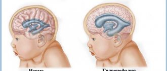
So, hypoplasia of the cerebellar vermis in the fetus, or simply microcephaly, is characterized by a decrease in the size of the cerebellum itself, or one of its lobes, which ultimately entails irreversible consequences for the body and its full development.
The dash indicates the cerebellum with hypoplasia
At risk are girls who, during pregnancy:
- drink alcohol;
- smoke;
- take drugs;
- exposed to radioactive radiation;
- suffered an infectious disease (rubella, toxoplasmosis, etc.).
In addition to the above, you can add expectant mothers who eat food that is harmful to the health of the fetus during pregnancy, but, as a rule, junk food does not have such an effect compared to alcohol, but in the aggregate it can lead to various anomalies.
Alcohol
Abuse of alcoholic beverages affects the fetus through ethanol. This substance independently penetrates into the central nervous system of the fetus (CNS), where, as a result of prolonged exposure, it can influence the formation of tumors and also affect the blood-brain barrier, making it weaker and thereby exposing the child’s body to even greater risk.
The blood-brain barrier is a kind of wall that protects the central nervous system from harmful infections that can affect it through the blood.
Smoking
In the process of smoking, the greatest danger for the formation of such an ailment as fetal hypoplasia is caused by toxic substances, and not nicotine. These substances affect the very process of formation of the spinal cord and brain, in particular, the formation of the neural tube. For this reason, cerebellar hypoplasia in a newborn may not be the only anomaly.
Addiction
Many films have already been made about the dangers of drug addiction and even more books have been written, so we can confidently say that cerebellar hypoplasia in children can develop as a result of drug use, as well as in the case of uncontrolled use of medications. It is no coincidence that some tablets contain a note stating that their use is possible only in extreme cases, if the positive effect for the mother is greater than the potential harm for the baby.
Radioactive substances
Radiation is dangerous for an adult, but what about an immature baby. Hypoplasia of the cerebellar vermis in the fetus can develop due to excessive exposure of the pregnant mother to radiation or excessive time spent in places with an increased radioactive background. The radioactive isotopes themselves accumulate in the amniotic fluid and placenta, and can change the structure of DNA, and it is easy to guess that with excessive irradiation of the body, fetal hypoplasia can be the mildest diagnosis of all possible consequences.
Infectious diseases
Cerebellar hypoplasia in a newborn can develop as a result of such a harmless disease as rubella. However, it is harmless only in childhood, and during pregnancy it is the number one enemy for a young mother. If this disease is diagnosed in the first trimester, doctors in 90% of cases send the girl for an artificial termination of pregnancy; in the second and third trimester, drug treatment is used, but it is usually ineffective.
Toxoplasmosis, in turn, is no less dangerous, but it causes missed abortion. It is transmitted from cats, rodents, birds, however, if the pathogen has not passed through the cat’s body, it does not pose a danger to humans. Dangerous processes are launched in the cat’s body and an ordinary virus turns into a serious disease.
Symptoms
Such a serious anomaly as hypoplasia of the cerebellar vermis in the fetus has special symptoms, since the cerebellum in the brain is one of the most important organs, responsible for many functions.
Main signs of the disease:
- tremor of the limbs;
- declarative speech (the child speaks as if shouting slogans);
- lack of smooth movement;
- inconsistency of limbs when moving;
- developmental problems (such children may begin to sit and walk later than their peers);
- inability to move without adult support;
- deafness or blindness.
Since the cerebellum has a rather complex structure, damage to its individual parts can lead to certain consequences, for example, hypoplasia of the medial parts of the cerebellar hemispheres entails disorders associated with the organs of vision (the child may remember visual information worse).
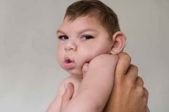
In addition to disorders associated with motor and physical changes, parents may notice that with hypoplasia of the cerebellar vermis, the baby’s head is smaller than that of his peers. This occurs as a result of a smaller brain volume, and since the brain is smaller, the formation of a bone plate around it does not occur as usual. Over time, the skull will enlarge and various cranial deformities may develop.
Cerebellar lesions in children
Cerebellar insufficiency in children is a normal condition that develops in the first year of a newborn’s life. It is characterized by a shaky, unstable gait, frequent falls, and poor coordination. As you grow older, your movements become clearer and more precise. And the number of falls decreases by 2 years.
If the symptoms of ataxia do not go away by 2-3 years, this is a reason to consult a neurologist. At the same time, the baby lags behind in mental and motor development. He lacks the skills typical of his peers. Cerebellar insufficiency is formed as a result of hereditary or congenital diseases, with ischemia during childbirth and with a previous neuroinfection.
An enlarged cerebellum in a child appears as a result of active tumor growth. At the same time, other symptoms of ataxia are observed. Normally, the size of the small brain should not exceed the norms specified for imaging.
Consequences
Tissue changes as a result of injury, tumor or infection lead to serious consequences for the young body. For example, children may never master motor skills (walking, complex coordinated movements). With minimal consequences, the patient continues to have an unsteady gait, which leads to frequent falls.
When the small brain is damaged, there is a slight retardation of intellectual activity. But it can be compensated for by an adapted training program for such children. After all, their cognitive abilities are not reduced. They are simply limited in their capabilities.
Features of treatment
Cerebellar hypoplasia in infants is a serious disease and there are cases of death at an early age, however, this is not a dogma and people live with this disease.
Undoubtedly, such children need the care and support of adults more than usual, since some functions are inaccessible to them or are difficult to perform.
Unfortunately, an effective treatment for a disease such as fetal hypoplasia has not been found to date, so it is of a therapeutic nature.
Therapy for hypoplasia in a child:
- occupational therapy (instilling work skills in the child);
- speech therapy classes;
- massage courses;
- social development;
- physiotherapy;
- therapy aimed at restoring the child’s balance.
The treatment options listed above will help the baby live painlessly in society in the future.
Among other things, there are cases of cerebellar hypoplasia in adults. This happens, as a rule, if the affected area was insignificant and did not entail any special changes in a person’s life.
Treatment in adulthood, in the absence of other pathologies, may differ slightly, but the treatment program listed above remains unchanged.
When a child’s fontanel overgrows - physiology, norms and care for the crown
New parents know that newborn babies have a special place on their heads that requires careful care - this is the fontanel.
What is it? What is its function? What size is considered normal - large or small? When does the fontanel overgrow? Is there a danger of injuring him and disrupting his brain function? How to properly care for it? The fontanelle is soft fibrous membrane tissue that connects two or more bones of the skull. Based on its location in the crown area, it is also called the crown.
Newborn babies have 6 fontanelles:
- Front
- Rear
- Two nipple-like
- Two wedge-shaped
When examining a newborn by pediatricians, a neonatologist, pediatrician and neurologist feel the size of the crown and monitor the rate of overgrowth of the anterior (large) fontanel throughout the year. It is the most visible, and it is by the large crown that pediatricians assess the normal development of the child. The last 5 close almost immediately or are invisible due to their small size.
The large crown should be diamond-shaped, and its normal size is from 0.5 to 4 cm. It is located in the middle of the crown (on the crown). Normally, the fontanel overgrows when the baby is one to one and a half years old. The skin on the soft part of the head pulsates to the rhythm of the baby’s heart. It's quite normal.
Function of the large fontanelle:
- Facilitating the passage of the baby through the mother's birth canal. With the help of fontanelles, the skull is elastic and tends to shrink. In the first weeks after birth, the baby's head has a slightly elongated shape.
- A child's brain grows and develops very rapidly. The fontanelles provide normal spatial conditions for this (so that there is room to grow)
- The crown is a kind of thermostat on the baby's head. When the body temperature rises above 38 C, the cooling mode “turns on” on the surface of the fontanel, thereby cooling the brain tissue.
- The crown is a kind of shock absorber that protects the baby’s brain from injury during falls and impacts.
It is advisable for all children under one year of age to undergo neurosonography - ultrasound of the brain.
The procedure is painless and safe, but very informative, so it is recommended for all children, even in the absence of complaints and neurological symptoms.
What should you be wary of?
Up to a year, parents should come to the pediatrician for a routine examination once a month. The process of overgrowth of the fontanel is controlled by a pediatrician. As parents spend more time with their baby, there are certain signs that should alert them. With such symptoms, it is advisable to contact a pediatric neurologist as soon as possible:
- Slow overgrowth of the crown (poor overgrowth in children born with low body weight)
- Soft fabric size more than 4 cm
- Closing too early (between 1 and 8 months of age)
- Sunkenness
Sometimes, the skin in the soft part of the head sinks (due to hot weather or elevated temperature, exicosis of the body occurs). The child needs to be given boiled water to drink and the crown of his head to recover. If it is tense and sticks out, the baby should be shown to a doctor. Only a neurologist can diagnose whether this is normal or a symptom of pathology.
Periods of crown overgrowth
By systematically measuring the crown, you can determine an increase in its size and delayed tightening, which is a sign of rickets or hydrocephalus of the brain. In the latter case, the crown remains wide open for two years and closes very slowly.
Tension and bulging of the anterior fontanelle is observed when the baby screams loudly. Or with increased intracranial pressure, with diseases such as meningitis, meningoencephalitis and others.
If the crown overgrows too quickly and early, this may indicate a calcium oversaturation in the child’s body. In this case, it is necessary to reduce the amount of calcium and vitamin D.
How to care for a fontanel?
The presence of a fontanel becomes a cause of fear in some parents, who are afraid of injuring the child’s head through it. Normally, the crown can pulsate, be sunken when thirsty, and even swell when the baby is crying. If the baby’s general condition is not disturbed and he behaves as usual, then there is no reason to worry.
There are no special rules for caring for the crown. By repeating normal daily routines, such as bathing your baby, brushing your baby's hair, or stroking the head, the risk of injury is reduced. Of course, you need to make sure that the baby doesn’t fall and hit his head. But this rule applies to children of any age and is not related specifically to the presence of a fontanel.
In order not to miss neurological problems, you need to monitor the baby’s development and see a pediatric neurologist. For children under one year of age, the schedule of visits to this doctor is as follows: 1, 3, 6, 9 and 12 months.
And if everything is in order, this will give you confidence that the baby is developing normally and his state of health is Healthy!!!
Source: https://detivse.com/razvitie/kogda-zarastaet-rodnichok-u-rebenka
Prevention
The main preventive measures to help prevent the development of cerebellar hypoplasia during pregnancy are maintaining a healthy lifestyle and being attentive to your health.
Undoubtedly, it is not worth taking things to the point of absurdity, but it is important to remember that balance is necessary in everything. Even half a glass of good red wine once a month will not only not harm the child’s body, but will even have a beneficial effect.
But half a cigarette smoked once a month will already have a negative effect on the fetus, so you need to approach everything headlong and with caution.
Take care of your health and the health of your little ones.
The cerebellum is located in the posterior region of the brain. Its role is undeniable - it contributes to coordinating effects on muscle movements and ensuring the maintenance of balance. He also participates in the management of human organs.
In the middle zone of the cerebellum is its vermis, which maintains communication between the two lobes and allows a person to maintain a specific posture. In some cases, at the intrauterine stage of development, a serious pathology is formed - hypoplasia of the cerebellar vermis, leading to failures in the implementation of the regulatory function.
Microcephaly is a decrease in the volume of the cerebellum or a specific part, leading to serious irreversible consequences and disruption of the normal course of development of the body. The pathology occurs at the stage of intrauterine development, so hypoplasia of the cerebellar vermis in the fetus occurs.
The severity of the disease varies among patients, so the disease is completely comprehensive or partial, affecting one or both lobes. Half of the cases of cerebellar hypoplasia are associated with a genetic predisposition, which is caused by the factor of combinative variability (hereditary variability in the body).
The second group of provoking factors is represented by teratogenic effects (disturbances in the development of the embryo) to which a woman is exposed during pregnancy. This happens during the first trimester, when the basic systems of the unborn child are laid. Alcohol, smoking, and infectious diseases lead to problems.
Pathological changes in the cerebellar vermis cause loss of regulatory function, resulting in chaotic walking with asymmetrical movements. This is why a child’s illness is noticed immediately - he often loses his balance and falls for no apparent reason.
. This disease is associated with permanent developmental delays.
Sick children require constant attention from loved ones. Adaptation in a team is difficult for them. As a rule, the disease is progressive in nature, so mental insufficiency, deterioration of vision and hearing are added to the main problems, although the consciousness continues to perceive all information.
Pathogenesis
Dandy-Walker syndrome was first described by Dandy and Blackfan in 1914. After the initial description, additional studies were conducted that described the various morphological features of this defect.
Studies by D'Agostino in 1963 and Hart in 1972 identified the characteristic triad of Dandy-Walker syndrome, namely:
- Complete or partial agenesis of the worm
- Cystic dilatation of the fourth ventricle
- Enlargement of the posterior cranial fossa with upward movement of the lateral paranasal sinuses and cerebellum.
This triad is usually combined with supratentorial hydrocephalus, which are complications and not part of the defect complex.
Classically, cystic defects of the posterior cranial fossa are divided into Dandy-Walker syndrome, Dandy-Walker variant, mega cisterna magna, arachnoid cyst of the posterior fossa - these are the defects that make up the Dandy-Walker complex.
The Dandy-Walker variant combines vermis hypoplasia and cystic dilatation of the fourth ventricle, without enlargement of the posterior fossa.
This syndrome is a congenital pathology, which is expressed in the fact that the CSF pathways and the cerebellum do not fully develop. Most of all, the anomaly concerns the openings that connect the two ventricles (third and fourth) with the cistern magna of the brain, and those that connect these same ventricles with the subarachnoid space of the meninges. Due to impaired cerebrospinal fluid outflow, the formation of a cerebrospinal fluid cystic formation occurs in the posterior cranial fossa and obstructive hydrocephalus as a result of secondary cystic dilatation of the fourth ventricle (70-90% of cases).
Malformations of the central nervous system associated with Dandy-Walker anomaly (70% of cases):
Other rare associated CNS malformations (20-33% of cases):
- Orofacial deformities and palate (6%).
- Polydactyly and syndactyly.
- Anomalies of heart development.
- Abnormalities of the urinary tract (polycystic kidney disease).
- Retinal cataract, choroid dysgenesis, coloboma.
- Hemangioma on the face.
- Hypertelorism.
- Meckel-Gruber syndrome.
- Neurocutaneous melanosis.
Causes of the disease
Cerebellar hypoplasia, which affects certain areas of this part of the brain, develops at the intrauterine stage of the formation of future life. In addition to hereditary predisposition, the following factors lead to the occurrence of the disease.
Alcohol
. Ethanol supplied with alcoholic beverages easily passes through the placental barrier. It not only affects the fetus, but begins to accumulate in its central nervous system. During the decomposition process, it breaks down into carcinogenic acetaldehyde. Penetrating through the BBB (blood-brain barrier), this substance has a detrimental effect on the baby’s central nervous system structures.
Smoking
, in which the greatest influence is not from nicotine, but from the toxic components contained. They have a detrimental effect on the process of the formation of the neural tube, the construction of the spinal cord and brain as a whole. This effect is complex, therefore, along with hypoplasia of the cerebellar vermis, other congenital problems appear in the child.
Addiction
. Regular or periodic use of narcotic drugs means that cerebellar vermis hypoplasia has become a common disease. Similar harm is caused by uncontrolled use of medications and the use of drugs prohibited during pregnancy and lactation.
Radiation
– excessive exposure of the mother is associated not only with unauthorized diagnostic procedures, but also with being in areas where the radioactive situation has deteriorated. Isotopes can accumulate in amniotic fluid, as well as the placenta, destructively affecting the structural characteristics of DNA.
Intoxication with chemical components with a toxic effect, volatile mixtures, biological substances.
Infectious diseases
. For example, rubella, which is not dangerous in its normal state, poses a great threat to pregnant women and leads to hypoplasia of the child’s cerebellum.
That is why this disease becomes an indication for artificial termination of pregnancy in the first trimester. However, even in the 2-3 trimesters, drug treatment will not be particularly effective. ARVI and influenza pose a similar risk.
To prevent risks, you need to take a responsible approach to your health when planning to conceive and while carrying a baby.
Hypoplasia of the cerebellar vermis in a child: symptoms, treatment, prevention
Vermis hypoplasia is a defect in the intrauterine development of the cerebellum, leading to a partial impairment of motor function; symptoms appear in the first days of the baby’s life.
The cerebellum (in Latin – “Cerrebelum”, which literally translates as “small brain”) is a part of the brain.
Located in the back of the brain, it is responsible for coordinating human muscle movements, maintaining balance in space and muscle tension, or tone, and also controls internal organs. The small brain itself belongs to the autonomic central nervous system - the work of the cerebellum occurs unconsciously.
The following definition applies to the cerebellar vermis - it is its middle part. It communicates between opposite lobules. The worm is responsible for a person’s ability to maintain a posture.
Hypoplasia of the cerebellar vermis leads to loss of the ability to stand and walk normally.
- 1 Reasons
- 2 Symptoms
- 3 Treatment
- 4 Prevention
Causes
Hypoplasia of the cerebellar vermis in the fetus is manifested as a consequence of the influence of both hereditary factors of the parents and harmful factors on the fetus during the prenatal period. Hypoplasia develops more often if the following factors are present:
- Smoking during pregnancy;
- Consumption of alcohol, toxic or narcotic substances;
- Radiation exposure;
- Diseases suffered by the mother during the first months of development.
The danger in smoking is not so much nicotine, but rather toxic substances that affect the process of laying down the neural tube - the embryo of the spinal cord and brain, as well as oxygen starvation - the abundant intake of carbon dioxide into the mother's blood.
Alcoholic beverages contain ethanol, which easily penetrates the placenta and concentrates in the fetus in the central nervous system.
Ethanol decomposes to acetaldehyde, which is a mutagen and carcinogen, influencing genetic mutations and tumor formation.
And cetaldehyde easily damages the blood-brain barrier, which protects the central nervous system from the effects of harmful substances and its own immune system.
Toxic substances can be classified as those that enter the body through the consumption of unhealthy foods or those that contain components of medications that are harmful to the baby.
For the safety of the child, before consuming this or that product, you must make sure of the composition and harmfulness of its components to the fetus, and secondly, under no circumstances take medications on your own without a doctor’s prescription.
Radiation received by a mother during pregnancy is a serious threat to the unborn child, as it affects the DNA structure.
Radioactive isotopes, entering the body, are concentrated in the placenta and amniotic fluid, destroying the immune system, reproductive function of the fetus and its hormonal secretion glands. The type of isotope and the intensity of its radiation have a strong influence.
The health of the fetus will be affected by both radiation and radioactive isotopes that were exposed to or entered the woman’s body before pregnancy, since radiation tends to accumulate in tissues. Radiation can lead to various pathologies in the thyroid gland, which will affect future pregnancies.
A striking example of a disease is rubella called rubelliosis, which is caused by the corresponding virus.
A relatively harmless childhood disease can cause irreparable harm to the fetus in the first trimester of pregnancy. Hypoplasia in this case will be acute, spreading to both lobules.
Flu is also a threat. Toxoplasmosis is extremely dangerous. It is caused by the parasite Toxoplasma gondi. It is transmitted to humans from cats and other domestic animals through feces on the fur. Through unwashed hands they enter the body, where they sometimes go unnoticed for many years.
Symptoms
Cerebellar hypoplasia is symptomatically associated with neurological signs. Underdevelopment or reduction in size of its individual sections or the cerebellum as a whole negatively affects the entire brain and the central nervous system. This pathology manifests itself in the following:
- Inhibition in the development of motor type functions. Sick children are delayed in their development, they later learn to sit and move, and suffer from speech problems.
- Minimum level of coordination in the movement of the muscles of the trunk and limbs.
- Children's arms and legs cannot move smoothly.
- Speech takes on the character of “chanting.” Sick children place stress in accordance with the rhythm, without connection with the meaning of the sentences and phrases being formed.
- Limbs and head are shaking. Tremor can manifest itself from an early age with cerebellar hypoplasia - almost from the first days of life.
- Difficulty maintaining balance in any position - both sitting and standing.
- Difficulty in moving independently, which requires constant support from an accompanying person.
- Malfunctions in the functioning of the smooth muscles of internal organs.
- Problems with hearing and vision, including deafness and blindness. This situation may be congenital, but can develop as one grows older.
- Malfunctions of the respiratory and cardiac organs.
- Increased aggressiveness and hysteria.
- Difficulties in social adaptation.
Signs appear from birth and become most clearly noticeable before the age of ten. Although the pathology after this age limit does not progress very quickly, problems with the functioning of internal organs and systems can have a negative impact on the baby’s growth.
Sometimes it is possible to detect cerebellar hypoplasia in adulthood. This is evidence of the insignificance of damage to the cerebellum. With a mild form of the disease, its signs are often not noticeable in childhood; there is only a slight impairment of fine motor skills and coordination.
Symptoms of cerebellar damage
We list the characteristic cerebellar signs below.
- Ataxia is static;
- Locomotor ataxia;
- Nystagmus;
- Chanted speech;
- Intentional trembling;
- Adiadochokinesis;
- Dysmetria;
- Muscle hypotension;
- Dysmetria;
- Missing;
- Megalography;
- Asynergy;
- Unsteady gait.
These symptoms do not always appear when the cerebellum is affected. The clinic also resembles disorders of the extrapyramidal system. Therefore, each sign should be described in detail.
Gait changes
When the structure is damaged, gait becomes unsteady. Patients with symptoms of the disease are similar to drunk people. They move vaguely and spread their legs wide. In this case, the patient sways when walking. The rolling intensifies towards the affected hemisphere. Similar problems arise with cerebellar ataxia.
Fact! The patient cannot quickly change direction of movement. Turning is difficult.
Nystagmus
Nystagmus is the twitching of the eyeballs. It manifests itself clearly when the patient looks in one direction. Nystagmus can be horizontal, vertical and circular. Moreover, with cerebellar problems, horizontal nystagmus is more common. It is expressed when the patient looks towards the lesion.
Muscle tone in cerebellar lesions
The tone in such diseases is reduced until complete muscle atony. Disorders are especially pronounced when the “worm” is affected. With hypotension, the patient quickly gets tired and exhausted. Excessive passive movements appear in the joints. Superficial and deep tendon reflexes disappear.
Treatment
Medicine at the present stage of development does not have effective methods of therapy to get rid of this serious pathology. The main efforts of doctors and parents are aimed at curbing the development of the disease and alleviating the baby’s condition. Supportive measures for cerebellar hypoplasia include:
- visiting a speech therapist;
- working with a psychologist;
- physiotherapeutic procedures that provide increased skills in maintaining balance, coordinating one’s movements, and eliminating chaotic behavior;
- massages with therapeutic and relaxing effects;
- facilitating social adaptation;
- vitamin therapy.
Handicrafts and construction kits help develop fine motor skills, and thinking skills improve with regular solving of logic problems.
Unfortunately, even now, many babies diagnosed with cerebellar hypoplasia die during the first months of life. With parental efforts, love and care, the condition of young patients can be improved. Such children will constantly need your care.
Diagnostics
To diagnose cerebellar hypoplasia, DNA testing of both parents should be performed. But first of all, it is worth examining in a similar way a parent in whose family there was cerebellar hypoplasia, because a person does not necessarily have to suffer from this pathology. He may be its carrier, which means it is quite possible that he passes it on to his offspring.
If the diagnosis of cerebellar hypoplasia is in question, during an external examination, the neurologist conducts a series of motor, speech, and mental exercises that can help identify signs of pathology. By conducting these tests, the doctor evaluates the child’s ability to maintain balance, ability to think, visual and hearing acuity, and the level of speech development according to age. The diagnosis can be made by a group of doctors, including not only a neurologist, but also a therapist, cardiologist, pulmonologist, ophthalmologist, and psychologist. To clarify the diagnosis, more accurate research methods are prescribed, such as magnetic resonance and computed tomography. After these studies, the diagnosis can be made very clearly.
Prevention
The main efforts in the prevention of cerebellar vermis hypoplasia should be aimed at organizing a healthy lifestyle for a woman at the stage of pregnancy planning and in the process of bearing a child.
It is imperative to eliminate bad habits -
drinking alcohol, smoking, avoid taking drugs, avoid radiation exposure. It is important to monitor your health, eliminating risk factors such as dangerous infectious diseases.
You should not get carried away with medications. It is recommended to maintain a balanced diet and undergo regular and timely examinations by a specialist. In this case, you will be able to minimize the risk of cerebellar vermis hypoplasia in the unborn child.
Major defects of cerebellar development that damage predominantly the hemispheres (Robain et al. 1987) or almost equally the hemispheres and vermis constitute a heterogeneous group of rare abnormalities with varying pathology and clinical manifestations (Sarnat and Alcala, 1980; Robain et al., 1987; Altman et al., 1992). Cases of probable atrophy and congenital pontocerebellar atrophy are discussed in a separate article on the website.
Diagnosis is controversial, as many apparent malformations are actually acquired diseases resulting from destructive or metabolic processes (Sener, 1995; Boltshauser, 2004). Correct classification of cerebellar developmental defects is difficult. Friede (1989) argues that the classic division of neocerebellar versus paleocerebellar hypoplasia is artificial and in many cases it is more appropriate to speak of aplasia of the anterior or posterior lobe of the cerebellum.
Total cerebellar aplasia is an exception (Glickstein, 2004). The interesting thing is that it does not always manifest itself with clinical symptoms. In reality, cerebellar remnants are probably always present (Gardner, 2001) and the term “hypoplasia” or “atrophy” is more accurate than “agenesis.” Cerebellar agenesis has been reported in association with diabetes insipidus (Zafeiriou et al., 2004). Hypoplasia is less common and may be predominantly unilateral.
In some cases, it is limited to specific structures, such as the lobule of the hemisphere. Often accompanied by anomalies of the deep cerebellar and brain stem nuclei and dysplasia of the inferior olive. In some cases, there is hydrocephalus (Friede, 1989), and other pathological changes are possible, for example, agenesis of the corpus callosum or arenencephaly.
Unilateral aplasia (Boltshauser et al., 1996; Kilickesmez et al., 2004) is less common and is often asymptomatic. May be accompanied by an ipsilateral facial angioma and various cerebral and/or peripheral vascular abnormalities in PHACES syndrome (Pascual-Castroviejo et al. 2003, Bhattacharya et al. 2004).
Clinical manifestations are extremely unstable.
They may be completely absent or manifest as ataxia, imbalance and other cerebellar symptoms. Characterized by mental retardation to varying degrees. Some cases are represented by ataxic cerebral palsy. In most cases, movement disorders predominated, but some patients showed signs of autism or microcephaly. Cerebellar atrophy of unknown origin in an 8-year-old girl, predominantly affecting the vermis.
The lower part of the worm may be completely missing. The diagnosis of cerebellar hypoplasia is usually based on radiological methods. Diffuse hypoplasia can be difficult to distinguish from the Dundee-Walker variant on CT, and megacistern is often mistaken for cerebellar hypoplasia. Differential diagnosis includes visualization of the cerebellar falx, which characterizes the cisternae pattern, and the complete presence of the cerebellar vermis on sagittal MRI images.
Some cases of cerebellar hypoplasia are part of genetic syndromes, but complete definition is still a long way off. Some features are due to congenital ataxia with different types of transmission, while additional features are present in other cases (Al Shahwan et al., 1995). An association with chromosomal aberration has been reported (de Azevedo-Moreira et al., 2005, Harper et al., 2007).
Other unusual cerebellar malformations:
1. Tectocerebellar dysraphism
(Mori et al., 1994, Dehdashti et al., 2004) is a complex developmental anomaly that includes an inverted cerebellum developing within the fourth ventricle, vermis hypoplasia, and an occipital cephalocele.
2. Rhombencephalosynapsis
(Toelle et al., 2002) refers to congenital fusion of the cerebellar hemispheres with midline fusion of the dentate nuclei and aqueductal atresia. Patients with mild symptoms live long.
Complex cerebellar malformations due to cephalocele may result from ischemic destruction of a previously normally formed cerebellum due to arterial compression by a diverticulum of the fourth ventricle.
3. Dentato-olivar dysplasia
is responsible for neonatal tonic seizures resembling Ohtahara syndrome found in several patients (Harding and Boyd, 1991, Robain and Dulac, 1992). A genetic origin of the pathology is suggested (Harding and Sorr, 1997).
Brain hypoplasia
(microcephaly) is a pathology in which the brain is reduced in size and underdeveloped. This disease is often combined with other developmental defects - hypoplasia of the spinal cord, limbs and some internal organs.
Neurosonography is a “lifeline” for neonatologists
My child is 6 months old. Ultrasound results revealed dilation of the ventricles of the brain. Is this scary? I am very worried. We were prescribed Pantogam, Diakarb, Asparkam. We drank them according to the plan for 2 months. But nothing improved. How long does it take to cure? But the final diagnosis and treatment plan can only be established by a qualified neurologist based on both NSG and clinical manifestations of neurological abnormalities.
It should be noted that enlargement of the ventricles of the brain is often observed in children under one year of age. But the size of the ventricles must be compared with the position of other liquor systems. Consultation with a neurologist and the use of modern research methods are vital for this. Dilatation of the ventricles may be a sign of developing hydrocephalus, but the volumes of the ventricles only become larger over time.
Do not panic ahead of time - in many children, such anomalies disappear by the age of one year. The main thing is to contact a reliable and experienced neurologist. Subscribe to the weekly newsletter so you don't miss interesting materials:. Mobile applications: iPad iPhone Android. E-books. Close menu Here are all the sections of the site. TVNZ. Change text size: A A. Send questions to doctors to mama kp. Subscribe to news:.
Found an error in the text? Did you like the material?
Brain hypoplasia - causes
Brain hypoplasia is a congenital pathology. Genetic disorders, as well as the influence of various harmful factors on the body of a pregnant woman, can lead to its occurrence, for example:
- smoking;
- consumption of alcoholic beverages, drugs or toxic substances;
- ionizing radiation;
- some diseases (rubella, influenza, toxoplasmosis).
The impact of these factors is especially dangerous in the early stages of pregnancy, when the baby is actively forming and developing various organs, including the brain.
What could be the reasons?
So, hypoplasia of the cerebellar vermis in the fetus, or simply microcephaly, is characterized by a decrease in the size of the cerebellum itself, or one of its lobes, which ultimately entails irreversible consequences for the body and its full development.
The dash indicates the cerebellum with hypoplasia
At risk are girls who, during pregnancy:
- drink alcohol;
- smoke;
- take drugs;
- exposed to radioactive radiation;
- suffered an infectious disease (rubella, toxoplasmosis, etc.).
In addition to the above, you can add expectant mothers who eat food that is harmful to the health of the fetus during pregnancy, but, as a rule, junk food does not have such an effect compared to alcohol, but in the aggregate it can lead to various anomalies.
Alcohol
Abuse of alcoholic beverages affects the fetus through ethanol. This substance independently penetrates into the central nervous system of the fetus (CNS), where, as a result of prolonged exposure, it can influence the formation of tumors and also affect the blood-brain barrier, making it weaker and thereby exposing the child’s body to even greater risk.
Smoking
In the process of smoking, the greatest danger for the formation of such an ailment as fetal hypoplasia is caused by toxic substances, and not nicotine. These substances affect the very process of formation of the spinal cord and brain, in particular, the formation of the neural tube. For this reason, cerebellar hypoplasia in a newborn may not be the only anomaly.
Addiction
Many films have already been made about the dangers of drug addiction and even more books have been written, so we can confidently say that cerebellar hypoplasia in children can develop as a result of drug use, as well as in the case of uncontrolled use of medications. It is no coincidence that some tablets contain a note stating that their use is possible only in extreme cases, if the positive effect for the mother is greater than the potential harm for the baby.
Radiation is dangerous for an adult, but what about an immature baby. Hypoplasia of the cerebellar vermis in the fetus can develop due to excessive exposure of the pregnant mother to radiation or excessive time spent in places with an increased radioactive background. The radioactive isotopes themselves accumulate in the amniotic fluid and placenta, and can change the structure of DNA, and it is easy to guess that with excessive irradiation of the body, fetal hypoplasia can be the mildest diagnosis of all possible consequences.
Cerebellar hypoplasia in a newborn can develop as a result of such a harmless disease as rubella. However, it is harmless only in childhood, and during pregnancy it is the number one enemy for a young mother. If this disease is diagnosed in the first trimester, doctors in 90% of cases send the girl for an artificial termination of pregnancy; in the second and third trimester, drug treatment is used, but it is usually ineffective.
Toxoplasmosis, in turn, is no less dangerous, but it causes missed abortion. It is transmitted from cats, rodents, birds, however, if the pathogen has not passed through the cat’s body, it does not pose a danger to humans. Dangerous processes are launched in the cat’s body and an ordinary virus turns into a serious disease.
Brain hypoplasia - symptoms
With microcephaly, there is a sharp decrease in brain mass. In addition, other gross disturbances occur in it - the structure of the convolutions and internal structures changes. As a rule, the temporal and frontal lobes are more underdeveloped. The convolutions become flat - only large grooves remain, and small ones almost completely disappear. The pyramids of the medulla oblongata, cerebellum, brain stem and thalamus often decrease in size. With cerebral hypoplasia, the skull circumference is reduced. As a child grows, the bones of the facial skeleton grow faster than the brain. Almost all patients have signs of intellectual impairment. In addition, there is also a delay in physical development - children with microcephaly begin to hold their heads, sit and walk very late.
Brain hypoplasia - treatment
Unfortunately, to date this disease has no cure. Therapy for microcephaly is symptomatic - it is aimed at “smoothing”, reducing the manifestations of some neurological symptoms. Life expectancy in patients with cerebral hypoplasia is reduced. And, unfortunately, there is practically no hope that the child’s brain will develop normally. Only a small proportion of children with microcephaly are able to study in auxiliary schools. The rest need constant care and supervision.
Typical lesions of the fetal nervous system: correlation between MRI and ultrasound findings
A benchmark for new standards! Unparalleled clarity, resolution, ultra-fast data processing, and a comprehensive suite of advanced ultrasound technologies to solve the most challenging diagnostic problems. To assess the state of the brain in children with an open anterior fontanelle, a sector or microconvex sensor with a frequency of .5 MHz is used. If the fontanel is closed, then you can use sensors with a lower frequency - 1.5 MHz, but the resolution will be low, which gives worse quality echograms. When studying premature babies, as well as to assess the surface structures of the grooves and convolutions on the convexital surface of the brain and extracerebral space, sensors with a frequency of 7. MHz are used.
Cerebellar hypoplasia
Hypoplasia does not always affect the entire brain. In some cases, only some of its parts, such as the cerebellum, are affected. Cerebellar hypoplasia is a congenital malformation in which some functional areas of the cerebellum and/or its cortex are underdeveloped. Like any other type of hypoplasia, cerebellar hypoplasia occurs during fetal development. In more than half of all cases, the development of the disease is caused by genetic abnormalities. In other cases, the reason lies in the impact of certain negative factors on the body of a pregnant woman.
Cerebellar hypoplasia - symptoms and treatment
Symptoms of cerebellar hypoplasia are:
- Delayed physical and mental development. Children start sitting and walking late. Their speech development is delayed;
- Tremor (shaking) of the head and limbs, which often appears in the first year of life;
- Difficulty maintaining balance;
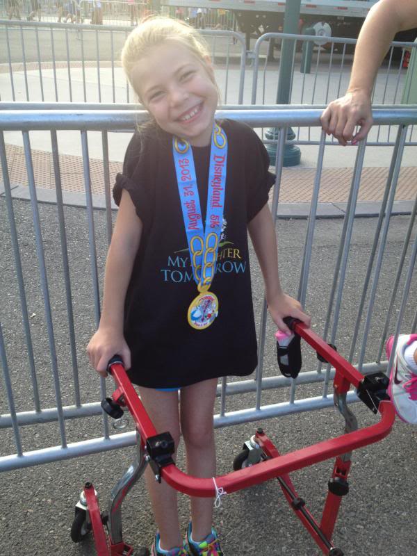
- Clumsy gait. Often, children with cerebellar hypoplasia can only move with assistance.
With age, the symptoms of the disease stabilize. And usually after 10 years of age, no further growth is observed. In some patients, cerebellar hypoplasia leads to the development of mental deficiency, accompanied by deafness and/or blindness. Unfortunately, medicine is currently unable to cure children with this disease. However, long-term social and motor rehabilitation - for example, classes with a speech therapist, occupational therapy, balance therapy, massage, physical therapy - can improve the condition of patients and instill in them self-care skills.
Even if a child has been given such a terrible diagnosis, there is no point in giving up. Such a baby, much more than other children, needs affection, attention and care - and mother’s love sometimes works wonders!