Symptoms
The change is often communicated by the family as “this is not the father I know” and is difficult to detect during normal conversation.
Characteristic features are:
- Decreased spontaneous activity - the patient does not feel the desire to do anything, cannot plan actions, and has periods of anxiety.
- Loss of attention – shows lack of interest and is easily distracted.
- Memory is normal.
- Loss of abstract thinking - cannot understand proverbs.
- Perseveration is the tendency to continue one form of behavior when the situation requires its change.
- Changes in affect - depending on the nature of the brain damage, the person either becomes apathetic or overly active, possibly with unusual sexual behavior.
A little about the terms
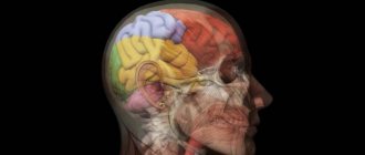
This is one of the youngest parts of the human brain, accounting for about 30%. And it is located in the front part of our head, which is where the name “frontal” comes from (in Latin it sounds like lobus frontalis, and lobus is “lobe”, not “frontal”). It is separated from the parietal lobe by the central sulcus (sulcus centralis). In each frontal lobe there are four gyri: one vertical and three horizontal - the superior, middle and inferior frontal gyri (that is, gyrus frontalis superior, medius and inferior, respectively - you can simply find these Latin terms in English texts).
The frontal lobes regulate the system of distribution of voluntary movements, motor processes of speech, regulation of complex forms of behavior, thinking functions, and even controls urination.
At the temples there is a part of the lobes “responsible” for intellectual processes.
The left lobe forms the qualities that determine a person’s personality: attention, abstract thinking, the desire for initiative, the ability to solve problems, self-control and critical self-assessment. For most people, the center of speech is located here, but there are approximately 2-5 inhabitants of the planet for whom it is based in the right frontal lobe. But in reality, the ability to speak does not change depending on the location of the “control cabin”.
The convolutions, of course, also have their own unique functions. The anterior central gyrus is responsible for the motor abilities of certain parts of the body. In essence, it turns out to be an “inverted person”: the face is controlled by the lower third of the gyrus, the one closer to the forehead, and the legs are controlled by the upper third, the one closer to the parietal region.
In the posterior parts of the superior frontal gyrus there is an extrapyramidal center, that is, the extrapyramidal system. It is responsible for the function of voluntary movements, the “readiness” of the central motor apparatus to perform movements for the redistribution of muscle tone when performing actions. She also takes part in maintaining normal posture. In the posterior part of the middle frontal gyrus there is the frontal oculomotor center, which is responsible for the simultaneous rotation of the head and eyes. Irritation of this center turns the head and eyes in the opposite direction.
The main function of the frontal lobe is “legislative”. She controls behavior. Only this part of the brain gives a command that does not allow a person to carry out socially undesirable impulses. For example, if emotions dictate to hit your boss, the frontal lobes signal: “Stop or you will lose your job.” Of course, they only notify you that you don’t need to do this, but they cannot stop actions and turn off emotions. What's interesting is that the frontal lobes work even when we sleep.
In addition, they are also a conductor, helping all areas of the brain work in harmony.
And it was in the frontal lobes that neurons were discovered, which have been called the most outstanding event in neuroscience in recent decades. In 1992, a resident of Kiev by birth, an Italian by passport, Giacomo Rizzolati discovered and in 1996 published the so-called mirror neurons. They are excited both when performing a certain action and when observing the execution of this action. It is believed that it is to them that we owe the ability to learn. Later, such neurons were found in other lobes, but it was in the frontal lobes that they were found first.
Damage to the frontal lobes results in carelessness, unhelpful goals, and a tendency to make inappropriate, funny jokes. A person loses the meaning of life, interest in his surroundings and can sleep all day long. So if you know such a person, perhaps he is not a lazy person and a quitter, but his frontal lobe cells are dying off!
Disruption of the activity of these cortical zones subordinates a person's actions to random impulses or stereotypes. At the same time, noticeable changes affect the patient’s very personality, and his mental abilities inevitably decrease. Such injuries have a particularly difficult impact on individuals whose life is based on creativity. They are no longer able to create something new.
Damage to this area of the brain can be detected using pathological reflexes that are normally absent: for example, grasping (Yanishevsky-Bekhterev reflex), when a person’s hand closes when any object touches the hand. Less commonly, this phenomenon manifests itself as obsessive grasping of objects that appear before the eyes. There are other similar violations: closing of the lips, jaw and even eyelids.
Diagnostics
The mental status test does not correctly measure frontal lobe damage. The following are more accurate.
Not really
- Tell the patient to show two fingers if you show one and vice versa. Give 10 tries.
- Typically, a person with frontal syndrome will copy you (echopraxia).
Fluency in writing
Ask them to name different words that start with F in one minute (without proper names). Must be at least 8.
Motor test
- Perseveration can be seen by asking a person to perform a series of three movements: make a fist, place your palm on the table, and then place the edge of your hand on the table (fist-edge-palm).
Find out more ADHD in children is a problem, is there a solution?
Neglect most often occurs after damage to the right hemisphere, involving either the right parietal lobe or the right frontal lobe. Patients with right-sided brain lesions neglect the left hemisphere.
This can be assessed by asking them to draw or read. People neglect the left half of a picture or the left half of words (rule out dyslexia).
Causes
Pathology appears as a result of the following reasons:
- Tumor.
- Neurodegenerative diseases of the central nervous system: Pick's disease, Alzheimer's disease.
- Genetic diseases, such as Tourette's syndrome.
- Stroke.
- Atherosclerosis and vascular disorders in the cerebral cortex.
- Mechanical injuries of the brain and skull.
Classic frontal syndromes
There are three classically characterized syndromes that result from damage to the frontal lobes.
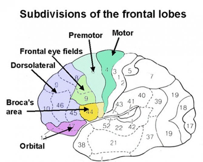
Dorsolateral frontal injury
- Function: Monitoring and adjustment using "working memory", executive function, planning, strategy formation
- Dysfunction: lack of ability to plan, consistently carry out actions or tasks, poor working memory for verbal or spatial information (depending on the left or right cortex), disinterest, decreased attention to stimuli, lack of abstract thinking, mental flexibility, apathy.
Orbitofrontal injury, limbic, reticular areas
- Function: Emotional input, arousal, suppression of distracting signals.
- Dysfunction: disinhibition, emotionally labile memory impairment, impulsivity, lack of concern for others, distractibility, hyperkinesis, restlessness, explosive behavior.
Dorsomedial frontal injury
- Function: motivation, initiation of action.
- Dysfunction: apathy, lack of anxiety, abulia (lack of motivation), decreased awareness, spontaneous movements, may be akinetic or exhibit mutism.
Trauma in other areas of the frontal cortex
- Primary cortex: weakness, impaired fine movements.
- Pretort cortex: weakness of the proximal muscle (rougher movements).
- Broca's area: motor, expressive aphasia on the dominant hemisphere, deficit of expressive prosody.
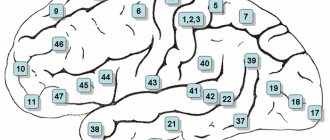
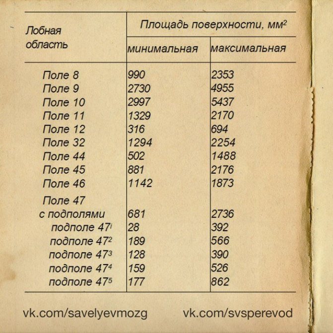
What fields are included?
Fields and subfields are responsible for specific functions that are generalized under the frontal lobes. Because The polymorphism of the brain is enormous; the combination of the sizes of different fields makes up a person’s individuality. Why do they say that over time a person changes. Throughout life, neurons die, and the remaining ones form new connections. This introduces an imbalance in the quantitative ratio of connections between different fields that are responsible for different functions.
Not only do different people have different margin sizes, but some people may not have these margins at all. Polymorphism was identified by Soviet researchers S.A. Sarkisov, I.N. Filimonov, Yu.G. Shevchenko. They showed that the individual ways in which the cerebral cortex is structured within one ethnic group are so great that no common features can be seen.
- Field 8 - located in the posterior parts of the middle and superior frontal gyri. Has a center for voluntary eye movements
- Area 9 - dorsolateral prefrontal cortex
- Area 10 - Anterior prefrontal cortex
- Area 11 - olfactory area
- Area 12 - control of the basal ganglia
- Field 32 - Receptor area of emotional experiences
- Area 44 - Broca's Center (processing information about the location of the body relative to other bodies)
- Field 45 - music and motor center
- Field 46 - motor analyzer of head and eye rotation
- Field 47 - nuclear zone of singing, speech motor component Subfield 47.1
- Subfield 47.2
- Subfield 47.3
- Subfield 47.4
- Subfield 47.5
Differential diagnosis
- injury;
- dementia;
- tumor;
- epilepsy;
- Abscess, infection;
- Ischemic hemorrhagic stroke.
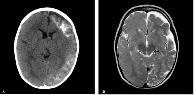
classic presentation of orbitofrontal injury.
Although personality and behavioral disturbances have been described following frontal lobe lesions since the middle of the last century, pathological conditions in frontal lobe syndrome often go undetected clinically.
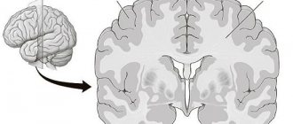
Structure and location
The frontal lobes are divided into two main areas: the motor cortex and the prefrontal cortex. The area of the brain involved in language and speech, known as Broca's area, is located in the left frontal lobe.
The prefrontal cortex is the front part of the frontal lobes and controls complex cognitive processes such as memory, planning, reasoning, and problem solving.
This area of the frontal lobe helps set and maintain goals, control negative impulses, organize events in a temporal order, and form individual personalities.

Problems
One of the specific behavioral disorders after damage to the frontal lobe is attention disorder; patients exhibit distractibility. They are characterized by poor memory, sometimes called “forgetting to remember.” Their thinking is concrete, they show persistence and stereotyping.
Find out more 10 brilliant people with Savant syndrome
Persistence, with an inability to move from one line of thinking to another, leads to difficulty with arithmetic calculations such as successive sevens or subtraction.
Aphasia is sometimes observed, but it is different from Wernicke's and Broca's aphasia. Luria called it dynamic aphasia.
Other features of frontal syndrome include decreased activity, inability to plan, and lack of restlessness. Sometimes this is associated with bouts of restless, aimless, uncoordinated behavior.
The patient shows indifference to the outside world. Clinically, the picture resembles a serious affective disorder with psychomotor retardation.
Euphoria and disinhibition are sometimes described. Euphoria does not refer to a manic state. Disinhibition leads to pronounced deviations in behavior, sometimes associated with outbursts of irritability and aggression.
How does the brain work?
The brain is an organ that has been studied rather poorly due to the complexity of its design. Its structure is still the subject of debate in scientific circles.
Nevertheless, there are these basic facts:
- The adult human brain consists of twenty-five billion neurons (approximately). This mass makes up the gray matter.
- There are three shells:
- Solid;
- Soft;
- Arachnoid (cerebrospinal fluid circulation channels);
They perform protective functions, responsible for safety during impacts and any other damage.
Next, controversial issues begin in choosing a position of consideration.
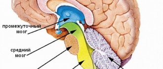
In the most common aspect, the brain is divided into three sections such as:
- Two cerebral hemispheres;
- Cerebellum;
- Trunk;
It is impossible not to highlight another common view of this organ:
- Final (hemisphere);
- Intermediate;
- Posterior (cerebellum);
- Average;
- Oblong;
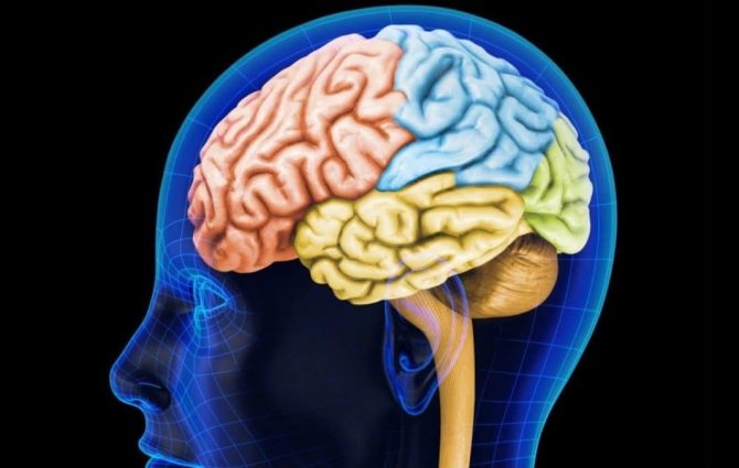
In addition, it is necessary to mention the structure of the telencephalon and the united hemispheres:
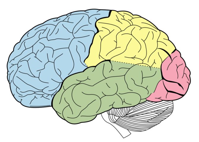
- Frontal lobe;
- Occipital;
- Parietal;
- Temporal;
Table 1. Clinical characteristics of the three main frontal lobe syndromes
Orbitofrontal syndrome (disinhibited)
- Impulsive behavior (pseudopsychopathic);
- Inappropriate humorous affect, euphoria;
- Emotional lability;
- Poor judgment and insight.
Apathetic syndrome
- Apathy (occasional brief angry or aggressive outbursts);
- Indifference;
- Psychomotor retardation;
- Motor perseveration, improvisation;
- Loss of sensation;
- Stimulated behavior;
- Dispersive motor and verbal behavior;
- Motor programming deficits;
- Three-step hand sequence;
- Program Variables;
- Mutual programs;
- Rhythm overlay;
- Several cycles;
- Poor word list formation;
- Poor abstraction and categorization;
- A segmented approach to visuoscopic analysis.
Medial frontal syndrome (akinetic)
- Neglect of spontaneous movement and gesture;
- Weak verbal output (repetition may persist);
- Weakness of the lower limb and loss of sensation;
- Incontinence.

What is each part of the brain responsible for?
As mentioned earlier, the number of functions performed by the brain is very, very extensive. Some of them are very important because they are noticeable, some are the opposite. However, it is not always possible to accurately determine which part of the brain is responsible for what. The imperfection of even modern medicine is obvious. However, those aspects that have already been sufficiently researched are presented below.
In addition to the various departments, which are highlighted in separate paragraphs below, there are just a few departments that need to be mentioned, without which your life would become a real nightmare:
- The medulla oblongata is responsible for all the body's defensive reflexes. This includes sneezing, vomiting and coughing, as well as some important reflexes.
- The thalamus is a translator of information received by receptors about the environment and the state of the body into signals understandable to humans. Thus, it controls pain, muscle, auditory, olfactory, visual (partial), temperature and other signals entering the brain from various centers.
- The hypothalamus simply controls your life. Keeps his finger on the pulse, so to speak. It regulates heart rhythm. In turn, this also affects the regulation of blood pressure and thermoregulation. In addition, the hypothalamus can influence the production of hormones in case of stress. It also controls feelings such as hunger, thirst, sexuality and pleasure.
- Epithalamus - controls your biorhythms, that is, it makes it possible to fall asleep at night and feel alert during the day. In addition, he is also responsible for metabolism, “in charge.”
This is not a complete list, even if you add what you read below. However, most of the functions are displayed, while others are still under debate.
Left hemisphere
The left cerebral hemisphere is the controller of such functions as:
- Oral speech;
- Analytical activities of various kinds (logic);
- Mathematical calculations;
In addition, this hemisphere is also responsible for the formation of abstract thinking, which distinguishes humans from other species of animals. It also controls the movement of the left limbs.
Right hemisphere
The right cerebral hemisphere is a kind of human hard drive. That is, it is there that memories of the world around you are stored. But such information by itself is of rather little use, which means that along with the preservation of this knowledge, algorithms for interaction with various objects of the surrounding world, based on past experience, are also preserved in the right hemisphere.
The cerebellum is, to a certain extent, a branch from the connection of the spinal cord and the cerebral cortex. This location is quite logical, since it makes it possible to receive duplicate information about the position of the body in space and the transmission of signals to various muscles.
The cerebellum is mainly engaged in constantly adjusting the position of the body in space, being responsible for automatic, reflexive movements, and for conscious actions. Thus, it is the source of such a necessary function as coordination of movements in space. You might be interested in reading about how to test your motor coordination.
In addition, the cerebellum is also responsible for regulating balance and muscle tone, while also working with muscle memory.
Also interesting is the ability of the cerebellum to adapt to any changes in the perception of information in the shortest possible time. It is implied that even with visual impairment (experiment with an invertoscope), a person adapts to the new state in just a few days and can again coordinate the position of the body, relying on the cerebellum.
Frontal lobes
The frontal lobes are like the dashboard of the human body. It supports him in an upright position, allowing him to move freely.
In addition, it is through the frontal lobes that a person’s curiosity, initiative, activity and independence are “calculated” at the time of making any decisions.
Also, one of the main functions of this department is critical self-assessment. Thus, this makes the frontal lobes something of a conscience, at least in relation to social markers of behavior. That is, any social deviations that are unacceptable in society do not pass the control of the frontal lobe, and, accordingly, are not carried out.
Any injuries to this part of the brain are fraught with:
- behavioral disorders;
- mood changes;
- general inadequacy;
- the meaninglessness of actions.
Another function of the frontal lobes is voluntary decisions and their planning. Also, the development of various skills and abilities depends on the activity of this department. The dominant share of this department is responsible for the development of speech and its further control. Equally important is the ability to think abstractly.
Pituitary
The pituitary gland is often called the medullary appendage. Its functions are reduced to the production of hormones responsible for puberty, development and functioning in general.
Essentially, the pituitary gland is something like a chemical laboratory in which it is decided what kind of person you will become as your body grows.
Coordination
Coordination, as the skill of navigating in space and not touching objects with different parts of the body in a random order, is controlled by the cerebellum.
In addition, the cerebellum controls a brain function such as kinetic awareness - in general, this is the highest level of coordination that allows you to navigate the surrounding space, noting the distance to objects and calculating opportunities to move in free zones.
Such an important function as speech is managed by several departments at once:
- The dominant part of the frontal lobe (mentioned above), which is responsible for the control of spoken language.
- The temporal lobes are responsible for speech recognition.
The interesting thing here is that both lobes are involved at once - the left one deciphers a set of sounds into understandable meanings, and the right one complements this with an understanding of intonations and analysis of the interlocutor’s facial expressions, thus also considering the attitude towards us.
Basically, we can say that the left hemisphere of the brain is responsible for speech, if we do not take into account the division of the telencephalon into various lobes and sections.
Emotions
Emotional regulation is an area controlled by the hypothalamus, along with a number of other important functions.
Strictly speaking, emotions are not created in the hypothalamus, but it is there that the human endocrine system is influenced. Already after a certain set of hormones has been produced, a person feels something, however, the gap between the orders of the hypothalamus and the production of hormones can be completely insignificant.
Prefrontal cortex
The functions of the prefrontal cortex lie in the area of mental and motor activity of the body, which correlates with future goals and plans.
In addition, the prefrontal cortex plays a significant role in the creation of complex thought patterns,
action plans and algorithms.
The main feature is that this part of the brain does not “see” the difference between regulating the internal processes of the body and following the social framework of external behavior.
When you find yourself faced with a difficult choice that was created largely by your own conflicting thoughts, thank your prefrontal cortex for that. It is there that differentiation and/or integration of various concepts and objects is carried out.
Also in this department, the result of your actions is predicted and adjustments are made in comparison with the result that you want to get.
Thus, we are talking about volitional control, concentration on the subject of work and emotional regulation. That is, if you are constantly distracted while working and cannot concentrate, it means that the conclusion made by the prefrontal cortex was disappointing, and you will not be able to achieve the desired result this way.
The last proven function of the prefrontal cortex to date is one of the substrates of short-term memory.
Memory
Memory is a very broad concept that includes descriptions of higher mental functions that allow one to reproduce previously acquired knowledge, skills and abilities at the right time. All higher animals possess it, however, it is most developed, naturally, in humans.
It is almost impossible to determine exactly which part of the brain is responsible for memory (long-term or short-term). Physiological studies show that the areas responsible for storing memories are distributed over the entire surface of the cerebral cortex.
The mechanism of memory action is as follows: in the brain, a certain combination of neurons is excited in a strict sequence. These sequences and combinations are called neural networks. Previously, the more common theory was that individual neurons were responsible for memories.
Each section of the complex structure that is the human brain performs certain functions. At the same time, the holistic activity of the most important organ of the nervous system is aimed at determining consciousness, forming character, temperament and important psychological aspects of behavior.
A feature of the interaction of all brain structures is the fact that despite the clear distribution of the functional load between its lobes, there is a possibility of redistribution of these functions. For example, when an injury or disease develops in one of the lobes, its function is performed by another part of the brain.
The coordinated activity of all brain structures becomes a prerequisite for the performance of its functions and the harmonious maintenance of balance throughout the human body.