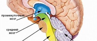
The cerebral cortex is divided into ancient ( archicortex
), old (
paleocortex
) and new (
neocortex
) according to phylogenetic characteristics, that is, according to the order of occurrence in animals during the process of evolution. These cortical areas form extensive connections within the limbic system. In more phylogenetically ancient animals, the ancient and old cortex, like the entire Limbic system, was primarily responsible for the sense of smell. In humans, the limbic system performs much broader functions related to the emotional and motivational sphere of behavior regulation. All three areas of the cortex are involved in performing these functions.
Ancient bark
along with other functions, it is related to smell and ensuring the interaction of brain systems. The ancient cortex includes the olfactory bulbs, which receive afferent fibers from the olfactory epithelium of the nasal mucosa; olfactory tracts located on the lower surface of the frontal lobe, olfactory tubercles in which secondary olfactory centers are located. This is phylogenetically the earliest part of the cortex, occupying adjacent areas of the frontal and temporal lobes on the lower and medial surfaces of the hemispheres.
old bark
includes the cingulate cortex, hippocampus and amygdala.
Cingulate gyrus. It has numerous connections with the cortex and brainstem centers and acts as the main integrator of various brain systems that form emotions.
The amygdala also forms extensive connections with the olfactory bulb. Thanks to these connections, the sense of smell in animals is involved in the control of reproductive behavior.
In primates, including humans, damage to the amygdala reduces the emotional coloring of reactions, in addition, aggressive affects completely disappear in them. Electrical stimulation of the amygdala causes predominantly negative emotions - anger, rage, fear. Bilateral removal of tonsils dramatically reduces the aggressiveness of animals. Calm animals, on the contrary, can become uncontrollably aggressive. In such animals, the ability to evaluate incoming information and correlate it with emotional behavior is impaired. The amygdala is involved in the process of identifying dominant emotions and motivations and choosing behavior in accordance with them. The amygdala is a powerful emotion modifier.
The hippocampus is located in the medial temporal lobe. The hippocampus receives afferent inputs
from the hippocampal gyrus (receives inputs from almost all areas of the neocortex and other parts of the brain), from the visual, olfactory and auditory systems.
Damage to the hippocampus results in characteristic deficits in memory and learning
. The activity of the hippocampus is to consolidate memory - the transition of short-term memory to long-term memory. Damage to the hippocampus causes a sharp disruption in the assimilation of new information and the formation of short-term and long-term memory. Consequently, the hippocampus, as well as other structures of the limbic system, significantly influences the functions of the neocortex and learning processes. This influence is carried out primarily through the creation of an emotional background, which is largely reflected in the rate of formation of any conditioned reflex.
Pathways from the temporal lobe of the cortex reach the amygdala and hippocampus, transmitting information from the visual, auditory and somatic sensory systems. Connections between the limbic system and the frontal lobes of the forebrain cortex have been established.
At the neocortex
The greatest development of size and differentiation of functions is observed in humans. The thickness of the neocortex ranges from 1.5 to 4.5 mm and is maximum in the anterior central gyrus. In the limbic system and in nervous activity in general, the cortex is involved in the highest functions of organizing activity.
Frontal lobe lesion
causes emotional dullness and difficulty changing emotions. It is when this area is damaged that the so-called frontal syndrome occurs. The prefrontal region and associated subcortical structures (the head of the caudate nucleus, the mediodorsal nucleus of the thalamus) form the prefrontal system, which is responsible for complex cognitive and behavioral functions. In the orbitofrontal cortex, pathways from the association cortical areas, paralimbic cortical areas, and limbic cortical areas converge. Thus, this is where the prefrontal system and the limbic system intersect. This organization determines the involvement of the prefrontal system in complex forms of behavior where coordination of cognitive, emotional and motivational processes is necessary. Its integrity is necessary for assessing the current situation, possible actions and their consequences, and thereby for making decisions and developing behavioral programs.
Removal of temporal lobes
causes hypersexuality in monkeys, and their sexual activity can be directed even towards inanimate objects.
Finally, postoperative syndrome is accompanied by so-called mental blindness
. Animals lose the ability to correctly evaluate visual and auditory information, and this information is in no way connected with the monkeys’ own emotional state.
The temporal lobes are closely connected to the structures of the hippocampus and amygdala and are also responsible for storing information and long-term memory and play a key role in the process of transferring short-term memory to long-term memory. The temporal lobe cortex is also responsible for combining stored memory traces.
The cerebral cortex is a multi-level brain structure in humans and many mammals, consisting of gray matter and located in the peripheral space of the hemispheres (the gray matter of the cortex covers them). The structure controls important functions and processes occurring in the brain and other internal organs.
(hemispheres) of the brain in the cranium occupy about 4/5 of the total space. Their component is white matter, which includes the long myelinated axons of nerve cells. On the outer side, the hemisphere is covered with the cerebral cortex, which also consists of neurons, as well as glial cells and unmyelinated fibers.
It is customary to divide the surface of the hemispheres into certain zones, each of which is responsible for performing certain functions in the body (for the most part these are reflexive and instinctive activities and reactions).
There is such a thing as “ancient bark”. This is the evolutionarily most ancient structure of the telencephalon of the cerebral cortex in all mammals. They also distinguish the “new cortex,” which in lower mammals is only outlined, but in humans forms the majority of the cerebral cortex (there is also the “old cortex,” which is newer than the “ancient” one, but older than the “new one”).
General information
The neocortex (new cortex, isocortex or lat. neocortex) is an area of the cerebral cortex, occupying about 96% of the surface of the hemispheres and having a thickness of 1.5 - 4 mm, which are responsible for the perception of the surrounding world, motor skills, thinking and speech.
The neocortex consists of three main types of neurons - pyramidal, stellate and fusiform. The first, the most numerous group, which makes up about 70-80% of the total amount in the brain. The proportion of stellate neurons is at the level of 15-25%, and fusiform neurons - about 5%.
In its structure, the neocortex is almost homogeneous and consists of 6 horizontal layers and vertical columns of the cortex. The layers of the new cortex have the following structure:
- Molecular, consisting of fibers and a small number of small stellate neurons. The fibers form a tangential plexus.
- The outer granular layer is formed by small neurons of various shapes, which are connected to the molecular layer over the entire area. At the very end of the layer there are small pyramidal cells.
- External pyramidal, consisting of small, medium and large pyramidal neurons. The processes of these cells can be associated with both layer 1 and the white matter.
- Internal granular, which consists mainly of stellate cells. This layer is characterized by a loose arrangement of neurons.
- Internal pyramidal, formed by medium and large pyramidal cells, the processes of which are connected with all other layers.
- Polymorphic, the basis of which is made up of spindle-shaped neurons, connected by processes with layer 5 and white matter.
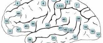
In addition, the neocortex is divided into areas, which in turn are subdivided into Brodmann areas. The following areas are distinguished:
- Occipital (17,18 and 19 fields).
- Superior parietal (5 and 7).
- Inferior parietal (39 and 40).
- Postcentral (1, 2, 3 and 43).
- Precentral (4 and 6).
- Frontal (5, 9, 10, 11, 12, 32, 44, 45, 46 and 47).
- Temporal (20, 21, 22, 37, 41 and 42).
- Limbic (23, 24, 25 and 31).
- Ostrovkovaya (13 and 14).
The cortical columns are a group of neurons that are located perpendicular to the cerebral cortex. Within a small column, all cells perform the same task. But a hypercolumn, consisting of 50-100 mini-columns, can have one or many functions.
Functions of neocortex
The new cortex is responsible for performing higher nervous functions (thinking, speech, processing information from the senses, creativity, etc.). Clinical trials have shown that each area of the cerebral cortex is responsible for strictly defined functions. For example, human speech is controlled by the left frontal gyrus. However, if any of the areas is damaged, the neighboring one can take over its function, although this requires a long period of time. Conventionally, there are three main groups of functions performed by the neocortex - sensory, motor and associative.
Sensory
This group includes a set of functions with the help of which a person is able to perceive information from the senses.
Each sense is analyzed by a separate area, but signals from others are also taken into account.
Signals from the skin are processed by the posterior central gyrus. Moreover, information from the lower extremities goes to the upper part of the gyrus, from the body to the middle part, from the head and hands to the lower part. In this case, the posterior central gyrus processes only pain and temperature sensations. The sense of touch is controlled by the superior parietal region.
Vision is controlled by the occipital region. Information is received in field 17, and in fields 18 and 19 it is processed, that is, color, size, shape and other parameters are analyzed.
Hearing is processed in the temporal region.
Charm and taste sensations are controlled by the hippocampal gyrus, which, unlike the general structure of the neocortex, has only 3 horizontal layers.
It is worth noting that in addition to the zones of direct reception of information from the senses, next to them there are secondary ones, in which the relationship between the received images and those stored in memory occurs. When these areas of the brain are damaged, a person completely loses the ability to recognize incoming data.
Motor
This group includes the functions of the neocortex, with the help of which any movement of the human limbs is carried out. Motor skills are controlled and controlled by the precentral region. The lower limbs depend on the upper parts of the central gyrus, and the upper limbs depend on the lower ones. In addition to the precentral, the frontal, occipital and superior parietal regions are involved in movement. An important feature of the performance of motor functions is that they cannot be performed without constant connections with sensory areas.
Associative
This group of neocortical functions is responsible for such complex elements of consciousness as thinking, planning, emotional control, memory, empathy and many others.
Associative functions are performed by the frontal, temporal and parietal regions.
In these areas of the brain, a reaction to data coming from the senses is formed and command signals are sent to the motor and sensory areas.
To receive and control, all sensory and motor areas of the cerebral cortex are surrounded by associative fields, in which the received information is analyzed. But at the same time, it is worth considering that the data coming into these fields is already primarily processed in the sensory and motor areas. For example, if there is a disruption in the functioning of such an area in the visual area, a person sees and understands that there is an object, but cannot name it and, accordingly, make a decision about his further behavior.
In addition, the frontal lobe of the cortex is very tightly connected to the limbic system, which allows it to control and manage emotional messages and reflexes. This makes it possible for a person to develop as a person.
The performance of associative functions in the neocortex is possible due to the fact that the neurons of this part of the central nervous system are able to retain traces of excitation based on the feedback principle and can persist for a long time (from several years to a lifetime). This ability is memory, with the help of which associative connections of the information received are built.
Prefrontal cortex
A large section of the cerebral cortex, which is represented in the form of the anterior sections of the frontal lobes. With its help, control, management, and focusing of any actions that a person performs are carried out. This department allows us to properly distribute our time. The famous psychiatrist T. Galtieri described this area as a tool with the help of which people set goals and develop plans. He was confident that a properly functioning and well-developed prefrontal cortex was the most important factor in a person’s effectiveness.
The main functions of the prefrontal cortex also include:
- Concentration, focusing on obtaining only the information a person needs, ignoring other thoughts and feelings.
- The ability to “reboot” consciousness, directing it in the right thinking direction.
- Perseverance in the process of performing certain tasks, the desire to achieve the intended result, despite the emerging circumstances.
- Analysis of the current situation.
- Critical thinking, which allows you to create a set of actions to search for verified and reliable data (checking the information received before using it).
- Planning, development of certain measures and actions to achieve set goals.
- Forecasting events.
The ability of this department to control human emotions is especially noted. Here, the processes occurring in the limbic system are perceived and translated into specific emotions and feelings (joy, love, desire, grief, hatred, etc.).
Different functions are attributed to different structures of the cerebral cortex. There is still no consensus on this issue. The international medical community now comes to the conclusion that the cortex can be divided into several large zones, including cortical fields. Therefore, taking into account the functions of these zones, it is customary to distinguish three main sections.
Area responsible for processing pulses
Impulses entering through the receptors of the tactile, olfactory, and visual centers go precisely to this zone. Almost all reflexes associated with motor skills are provided by pyramidal neurons.
This is also where the department is located, which is responsible for receiving impulses and information from the muscular system and actively interacts with different layers of the cortex. It receives and processes all impulses that come from the muscles.
If for some reason the scalp cortex is damaged in this area, then the person will experience problems with the functioning of the sensory system, problems with motor skills and the functioning of other systems that are associated with sensory centers. Externally, such disorders will manifest themselves in the form of constant involuntary movements, convulsions (of varying degrees of severity), partial or complete paralysis (in severe cases).
Sensory zone
This area is responsible for processing electrical signals entering the brain. There are several departments located here that ensure the human brain’s sensitivity to impulses coming from other organs and systems.
- Occipital (processes impulses coming from the visual center).
- Temporal (processes information coming from the speech-hearing center).
- Hippocampus (analyzes impulses coming from the olfactory center).
- Parietal (processes data received from taste buds).
In the sensory perception zone there are departments that also receive and process tactile signals. The more neural connections there are in each department, the higher its sensory ability to receive and process information will be.
The sections noted above occupy about 20-25% of the entire cerebral cortex. If the sensory perception area is damaged in some way, a person may have problems with hearing, vision, smell, and the sensation of touch. The received impulses will either not arrive or will be processed incorrectly.
Not always violations of the sensory zone will lead to the loss of some sense. For example, if the auditory center is damaged, this will not always lead to complete deafness. However, a person will almost certainly have some difficulties with the correct perception of the sound information received.
Association zone
The structure of the cerebral cortex also contains an associative zone, which ensures contact between the signals of neurons in the sensory zone and the motor center, and also provides the necessary feedback signals to these centers. The associative zone forms behavioral reflexes and takes part in the processes of their actual implementation. It occupies a significant (comparatively) part of the cerebral cortex, covering sections included in both the frontal and posterior parts of the cerebral hemispheres (occipital, parietal, temporal).
The human brain is designed in such a way that in terms of associative perception, the posterior parts of the cerebral hemispheres are especially well developed (development occurs throughout life). They control speech (its understanding and reproduction).
If the anterior or posterior parts of the association zone are damaged, this can lead to certain problems. For example, if the departments listed above are damaged, a person will lose the ability to competently analyze the information received, will not be able to make simple forecasts for the future, will not be able to build on facts in the thinking process, or will not be able to use previously acquired experience stored in memory. There may also be problems with spatial orientation and abstract thinking.
The cerebral cortex acts as a higher integrator of impulses, while emotions are concentrated in the subcortical zone (hypothalamus and other departments).
Different areas of the cerebral cortex are responsible for performing specific functions. You can examine and determine the difference using several methods: neuroimaging, comparison of electrical activity patterns, study of cellular structure, etc.
At the beginning of the 20th century, K. Brodmann (a German researcher of human brain anatomy) created a special classification, dividing the cortex into 51 sections, basing his work on the cytoarchitecture of nerve cells. Throughout the 20th century, the fields described by Brodmann were discussed, refined, and renamed, but they are still used to describe the cerebral cortex in humans and large mammals.
Many Brodmann fields were initially defined based on the organization of neurons within them, but later their boundaries were refined in accordance with correlations with various functions of the cerebral cortex. For example, the first, second and third fields are defined as the primary somatosensory cortex, the fourth field is the primary motor cortex, and the seventeenth field is the primary visual cortex.
However, some Brodmann fields (for example, area 25 of the brain, as well as fields 12-16, 26, 27, 29-31 and many others) have not been fully studied.
Speech motor area
A well-studied area of the cerebral cortex, which is also commonly called the speech center. The zone is conventionally divided into three large sections:
- Broca's speech motor center. Forms a person's ability to speak. Located in the posterior gyrus of the anterior part of the cerebral hemispheres. Broca's center and the motor center of the speech motor muscles are different structures. For example, if the motor center is damaged in some way, then a person will not lose the ability to speak, the semantic component of his speech will not suffer, but speech will cease to be clear, and the voice will become poorly modulated (in other words, the quality of pronunciation of sounds will be lost). If Broca's center is damaged, the person will not be able to speak (just like a baby in the first months of life). Such disorders are commonly called motor aphasia.
- Wernicke's sensory center. Located in the temporal region, it is responsible for the functions of receiving and processing oral speech. If Wernicke's center is damaged, sensory aphasia will form - the patient will not be able to understand speech addressed to him (and not only from another person, but also his own). What the patient says will be a collection of incoherent sounds. If simultaneous damage to Wernicke's and Broca's centers occurs (usually this occurs during a stroke), then in these cases the development of motor and sensory aphasia is observed simultaneously.
- Center for Comprehension of Written Speech. Located in the visual part of the cerebral cortex (field No. 18 according to Brodmann). If it turns out to be damaged, then the person experiences agraphia - loss of the ability to write.
The role of the neocortex in emotions and stereogynesis
Emotions in humans initially appear in the limbic system of the brain. But in this case they are represented by primitive concepts, which, once in the new cortex, are processed using the associative function. As a result, a person can operate with emotions at a higher level, which makes it possible to introduce concepts such as joy, sadness, love, anger, etc.
The neocortex also has the ability to dampen strong outbursts of emotion in the limbic system by sending calming signals to areas of high neuronal excitability. This leads to the fact that in a person the dominant role in behavior is played by the mind, and not by instinctive reflexes.
Thickness
All mammals that have relatively large brains (in a general sense, not in comparison with body size) have a fairly thick cerebral cortex. For example, in field mice its thickness is about 0.5 mm, and in humans it is about 2.5 mm. Scientists also highlight a certain dependence of the thickness of the bark on the weight of the animal.
With modern examinations (especially MRI), it is possible to accurately measure the thickness of the cerebral cortex in any mammal. However, it will vary significantly in different areas of the head. It is noted that in the sensory areas the cortex is much thinner than in the motor (motor) areas.
Research shows that the thickness of the cerebral cortex largely depends on the level of human intelligence. The smarter the individual, the thicker the cortex. Also, a thick cortex is recorded in people who constantly and for a long time suffer from migraine pain.
Differences from old bark
The old cortex (archicortex) is an earlier emerging part of the cerebral cortex than the neocortex. But in the process of evolution, the new cortex became more developed and extensive. In this regard, the archicortex ceased to play a dominant role and became one of the constituent parts.
If we compare the old one in terms of the functions performed, then the first is assigned the role of fulfilling innate reflexes and motivation, and the second - managing emotions and actions at a higher level.
In addition, the neocortex is significantly larger in size than the old cortex. So the first occupies about 96% of the total surface of the hemispheres, and the size of the second is no more than 3%. This ratio shows that the archicortex cannot perform higher nervous functions.
NEOCORTEX NEOCORTEX
(from neo... and lat. cortex - bark, shell), new bark, neopallium, basic. part of the cerebral cortex. N. carries out the highest level of coordination of brain function and the formation of complex forms of behavior. In the process of evolution, N. first appears in reptiles, in which it is small in size and has a relatively simple structure (the so-called lateral cortex). The N. has a typical multilayer structure only in mammals, in which it consists of 6-7 layers of cells (pyramidal, stellate, fusiform) and is divided into lobes: frontal, parietal, temporal, occipital and mediobasal. In turn, the lobes are divided into regions, subregions and fields, differing in their cellular structure and connections with the deep parts of the brain. Along with projection (vertical) fibers, N.'s neurons form associative (horizontal) fibers, which in mammals and especially in humans are collected in anatomically distinct bundles (for example, the occipital-frontal bundle), providing simultaneous coordinated activity of various types. zones N. The N. consists of the most complexly constructed associative cortex, the edges in the process of evolution experience the greatest increase, while the primary sensory fields of the N. are relatively reduced. (see CEREBRAL CORTICAL HEMISPHERES).
.(Source: “Biological Encyclopedic Dictionary.” Editor-in-chief M. S. Gilyarov; Editorial Board: A. A. Babaev, G. G. Vinberg, G. A. Zavarzin and others - 2nd ed., corrected - M.: Sov. Encyclopedia, 1986.)
See what “NEOCORTEX” is in other dictionaries:
- Neocortex...
New cortex (synonyms: neocortex, isocortex) (lat. neocortex) new areas of the cerebral cortex, which in lower mammals are only outlined, but in humans they form the main part of the cortex. The new cortex is located in the upper layer of the hemispheres... ... Wikipedia
neocortex
— 3.1.15 neocortex: The new cerebral cortex, which ensures the implementation of intellectual mental activity by human thinking. 3.1.16 Source... Dictionary-reference book of terms of normative and technical documentation
- (neocortex; neo + lat. cortex bark) see New bark ... Big medical dictionary
neocortex
- y, part. Evolutionary innovation and complexity of the nerve tissues that form the forehead, thus the cerebral cortex and other parts of the brain... Ukrainian Tlumachny Dictionary
NEOCORTEX (NEW CORTEX)
— Evolutionarily the newest and most complex of the nervous tissues. The frontal, parietal, temporal and occipital lobes of the brain consist of the neocortex ... Explanatory Dictionary of Psychology
Arches, paleo, neocortex ... Spelling dictionary-reference book
cortex
- cerebrum: cortex (cerebral cortex) the upper layer of the cerebral hemispheres, consisting primarily of nerve cells with a vertical orientation (pyramidal cells), as well as bundles of afferent (centripetal) and efferent... ... Great psychological encyclopedia
The term cortex refers to any outer layer of brain cells. The mammalian brain has three types of cortex: the pyriform cortex, which has olfactory functions; the old cortex (archicortex), which makes up the main. part... ...Psychological encyclopedia
Neocortex -
evolutionarily the youngest part of the cortex, occupying most of the surface of the hemispheres. Its thickness in humans is approximately 3 mm.
The cellular composition of the neocorhex is very diverse, but approximately three-quarters of the cortical neurons are pyramidal neurons (pyramids), and therefore one of the main classifications of cortical neurons divides them into pyramidal and non-pyramidal (fusiform, stellate, granular, chandelier cells, Martinotti cells, etc. .). Another classification is related to axon length (see paragraph 2.4). Long-axon Golgi I cells are mainly pyramids and spindles, their axons can exit the cortex, the remaining cells are short-axon Golgi II.
Cortical neurons also differ in the size of the cell body: the size of ultra-small neurons is 6x5 microns, the size of giant ones is more than 40 x 18. The largest neurons are Betz's pyramids, their size is 120 x 30-60 microns.
Pyramidal neurons (see Fig. 2.6, d)
have a body shape in the form of a pyramid, the top of which is directed upward. An apical dendrite extends from this apex and ascends into the overlying cortical layers. Basal dendrites extend from the remaining parts of the soma. All dendrites have spines. A long axon extends from the base of the cell, forming numerous collaterals, including recurrent ones, which bend and rise upward. Stellate cells do not have an apical dendrite, and in most cases there are no spines on the dendrites. In spindle cells, two large dendrites extend from opposite poles of the body; there are also small dendrites extending from the rest of the body. Dendrites have spines. The axon is long and has few branches.
During embryonic development, the new cortex necessarily goes through a stage of a six-layer structure; during maturation, in some areas the number of layers may decrease. The deep layers are phylogenetically more ancient, the outer layers are younger. Each layer of the cortex is characterized by its neural composition and thickness, which may differ from each other in different areas of the cortex.
Let's list the layers of the new crust
(Fig. 9.8).
Layer I - molecular
- the outermost, contains a small number of neurons and mainly consists of fibers running parallel to the surface. The dendrites of neurons located in the underlying layers also rise here.
II layer - outer granular
, or
external granular
, - consists mainly of small pyramidal neurons and a small number of medium-sized stellate cells.
Layer III - outer pyramidal -
the widest and thickest layer, contains mainly small and medium-sized pyramidal and stellate neurons. In the depths of the layer there are large and gigantic pyramids.
IV layer - internal granular
, or
internal granular
, - consists mainly of small neurons of all varieties, there are also a few large pyramids.
Layer V - inner pyramidal
, or
ganglionic,
a characteristic feature of which is the presence of large and in some areas (mainly in fields 4 and 6; Fig. 9.9; subsection 9.3.4) - giant pyramidal neurons (Betz's pyramids). The apical dendrites of the pyramids, as a rule, reach layer I.
Layer VI - polymorphic
, or
multiform,
contains predominantly spindle-shaped neurons, as well as cells of all other forms. This layer is divided into two sublayers, which a number of researchers consider as independent layers, speaking in this case of a seven-layer cortex.
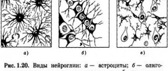
Rice. 9.8.
A
— neurons are stained entirely;
b
— only the neuron bodies are stained;
c
- painted
only neuron processes
Main functions
Each layer is also different. Layers I and II carry out connections between neurons of different layers of the cortex. Callosal and associative fibers mainly come from the pyramids of layer III and come to layer II. The main afferent fibers entering the cortex from the thalamus end on neurons of layer IV. Layer V is mainly associated with the system of descending projection fibers. The axons of the pyramids of this layer form the main efferent pathways of the cerebral cortex.
In most cortical fields, all six layers are equally well expressed. Such a cortex is called homotypic.
However, in some fields the expression of the layers may change during development.
This cortex is called heterotypic.
It comes in two types:
granular (zeros 3, 17, 41; Fig. 9.9), in which the number of neurons in the outer (II) and especially in the inner (IV) granular layers is greatly increased, as a result of which layer IV is divided into three sublayers. Such a cortex is characteristic of the primary sensory areas (see below);
Agranular (fields 4 and 6, or motor and premotor cortex; Fig. 9.9), in which, on the contrary, there is a very narrow layer II and practically no IV, but very wide pyramidal layers, especially the inner one (V).
The cerebral cortex is the center of higher nervous (mental) activity in humans and controls the performance of a huge number of vital functions and processes. It covers the entire surface of the cerebral hemispheres and occupies about half of their volume.
The cerebral hemispheres occupy about 80% of the volume of the cranium, and consist of white matter, the basis of which consists of long myelinated axons of neurons. The outside of the hemisphere is covered by gray matter or the cerebral cortex, consisting of neurons, unmyelinated fibers and glial cells, which are also contained in the thickness of the sections of this organ.
The surface of the hemispheres is conventionally divided into several zones, the functionality of which is to control the body at the level of reflexes and instincts. It also contains the centers of higher mental activity of a person, ensuring consciousness, assimilation of received information, allowing adaptation in the environment, and through it, at the subconscious level, through the hypothalamus, the autonomic nervous system (ANS) is controlled, which controls the organs of circulation, respiration, digestion, excretion , reproduction, and metabolism.
In order to understand what the cerebral cortex is and how its work is carried out, it is necessary to study the structure at the cellular level.
Functions
The cortex occupies most of the cerebral hemispheres, and its thickness is not uniform over the entire surface. This feature is due to the large number of connecting channels with the central nervous system (CNS), which ensure the functional organization of the cerebral cortex.
This part of the brain begins to form during fetal development and is improved throughout life, by receiving and processing signals coming from the environment. Thus, it is responsible for performing the following brain functions:
- connects the organs and systems of the body with each other and the environment, and also ensures an adequate response to changes;
- processes incoming information from motor centers using mental and cognitive processes;
- consciousness and thinking are formed in it, and intellectual work is also realized;
- controls speech centers and processes that characterize the psycho-emotional state of a person.
In this case, data is received, processed, and stored thanks to a significant number of impulses passing through and generated in neurons connected by long processes or axons. The level of cell activity can be determined by the physiological and mental state of the body and described using amplitude and frequency indicators, since the nature of these signals is similar to electrical impulses, and their density depends on the area in which the psychological process occurs.
It is still unclear how the frontal part of the cerebral cortex affects the functioning of the body, but it is known that it is little susceptible to processes occurring in the external environment, therefore all experiments with the influence of electrical impulses on this part of the brain do not find a clear response in the structures . However, it is noted that people whose frontal part is damaged experience problems communicating with other individuals, cannot realize themselves in any work activity, and they are also indifferent to their appearance and outside opinions. Sometimes there are other violations in the performance of the functions of this body:
- lack of concentration on everyday objects;
- manifestation of creative dysfunction;
- disorders of a person’s psycho-emotional state.
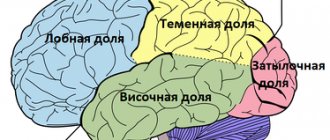
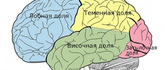
The surface of the cerebral cortex is divided into 4 zones, outlined by the most distinct and significant convolutions. Each part controls the basic functions of the cerebral cortex:
- parietal zone - responsible for active sensitivity and musical perception;
- the primary visual area is located in the occipital part;
- the temporal or temporal is responsible for speech centers and the perception of sounds coming from the external environment, in addition, it is involved in the formation of emotional manifestations, such as joy, anger, pleasure and fear;
- The frontal zone controls motor and mental activity, and also controls speech motor skills.
Features of the structure of the cerebral cortex
The anatomical structure of the cerebral cortex determines its characteristics and allows it to perform the functions assigned to it. The cerebral cortex has the following number of distinctive features:
- neurons in its thickness are arranged in layers;
- nerve centers are located in a specific place and are responsible for the activity of a certain part of the body;
- the level of activity of the cortex depends on the influence of its subcortical structures;
- it has connections with all underlying structures of the central nervous system;
- the presence of fields of different cellular structure, which is confirmed by histological examination, while each field is responsible for performing some higher nervous activity;
- the presence of specialized associative areas makes it possible to establish a cause-and-effect relationship between external stimuli and the body’s response to them;
- the ability to replace damaged areas with nearby structures;
- This part of the brain is capable of storing traces of neuronal excitation.
The large hemispheres of the brain consist mainly of long axons, and also contain in their thickness clusters of neurons that form the largest nuclei of the base, which are part of the extrapyramidal system.
As already mentioned, the formation of the cerebral cortex occurs during intrauterine development, and at first the cortex consists of the lower layer of cells, and already at 6 months of the child all structures and fields are formed in it. The final formation of neurons occurs by the age of 7, and the growth of their bodies is completed at 18 years.
An interesting fact is that the thickness of the cortex is not uniform over its entire length and includes a different number of layers: for example, in the area of the central gyrus it reaches its maximum size and has all 6 layers, and sections of the old and ancient cortex have 2 and 3 layers. x layer structure, respectively.
The neurons of this part of the brain are programmed to restore the damaged area through synoptic contacts, so each of the cells actively tries to restore damaged connections, which ensures the plasticity of neural cortical networks. For example, when the cerebellum is removed or dysfunctional, the neurons connecting it with the terminal section begin to grow into the cerebral cortex. In addition, the plasticity of the cortex also manifests itself under normal conditions, when the process of learning a new skill occurs or as a result of pathology, when the functions performed by the damaged area are transferred to neighboring areas of the brain or even hemispheres.
The cerebral cortex has the ability to retain traces of neuronal excitation for a long time. This feature allows you to learn, remember and respond with a certain reaction of the body to external stimuli. This is how the formation of a conditioned reflex occurs, the neural pathway of which consists of 3 series-connected apparatuses: an analyzer, a closing apparatus of conditioned reflex connections and a working device. Weakness of the closure function of the cortex and trace manifestations can be observed in children with severe mental retardation, when the formed conditioned connections between neurons are fragile and unreliable, which entails learning difficulties.
The cerebral cortex includes 11 areas consisting of 53 fields, each of which is assigned its own number in neurophysiology.
Regions and zones of the cortex
The cortex is a relatively young part of the central nervous system, developing from the terminal part of the brain. The evolutionary development of this organ occurred in stages, so it is usually divided into 4 types:
- The archicortex or ancient cortex, due to the atrophy of the sense of smell, has turned into the hippocampal formation and consists of the hippocampus and its associated structures. With its help, behavior, feelings and memory are regulated.
- The paleocortex, or old cortex, makes up the bulk of the olfactory area.
- The neocortex or new cortex has a layer thickness of about 3-4 mm. It is a functional part and performs higher nervous activity: it processes sensory information, gives motor commands, and also forms conscious thinking and human speech.
- The mesocortex is an intermediate version of the first 3 types of cortex.
Physiology of the cerebral cortex
The cerebral cortex has a complex anatomical structure and includes sensory cells, motor neurons and internerons, which have the ability to stop the signal and be excited depending on the received data. The organization of this part of the brain is built according to the columnar principle, in which the columns are divided into micromodules that have a homogeneous structure.
The basis of the micromodule system is made up of stellate cells and their axons, while all neurons react equally to the incoming afferent impulse and also send an efferent signal synchronously in response.
The formation of conditioned reflexes that ensure the full functioning of the body occurs due to the connection of the brain with neurons located in various parts of the body, and the cortex ensures synchronization of mental activity with the motor skills of organs and the area responsible for analyzing incoming signals.
Signal transmission in the horizontal direction occurs through transverse fibers located in the thickness of the cortex, and transmit the impulse from one column to another. Based on the principle of horizontal orientation, the cerebral cortex can be divided into the following areas:
- associative;
- sensory (sensitive);
- motor.
When studying these zones, various methods of influencing the neurons included in its composition were used: chemical and physical stimulation, partial removal of areas, as well as the development of conditioned reflexes and registration of biocurrents.
The associative zone connects incoming sensory information with previously acquired knowledge. After processing, it generates a signal and transmits it to the motor zone. In this way, it is involved in remembering, thinking, and learning new skills. Association areas of the cerebral cortex are located in proximity to the corresponding sensory area.
The sensitive or sensory area occupies 20% of the cerebral cortex. It also consists of several components:
- somatosensory, located in the parietal zone, is responsible for tactile and autonomic sensitivity;
- visual;
- auditory;
- taste;
- olfactory.
Impulses from the limbs and organs of touch on the left side of the body enter along afferent pathways to the opposite lobe of the cerebral hemispheres for subsequent processing.
Neurons of the motor zone are excited by impulses received from muscle cells and are located in the central gyrus of the frontal lobe. The mechanism of data receipt is similar to the mechanism of the sensory zone, since the motor pathways form an overlap in the medulla oblongata and follow to the opposite motor zone.
Convolutions, grooves and fissures
The cerebral cortex is formed by several layers of neurons. A characteristic feature of this part of the brain is a large number of wrinkles or convolutions, due to which its area is many times greater than the surface area of the hemispheres.
Cortical architectonic fields determine the functional structure of areas of the cerebral cortex. All of them are different in morphological characteristics and regulate different functions. In this way, 52 different fields are identified, located in certain areas. According to Brodmann, this division looks like this:
- The central sulcus separates the frontal lobe from the parietal region; the precentral gyrus lies in front of it, and the posterior central gyrus lies behind it.
- The lateral groove separates the parietal zone from the occipital zone. If you separate its side edges, you can see a hole inside, in the center of which there is an island.
- The parieto-occipital sulcus separates the parietal lobe from the occipital lobe.
The core of the motor analyzer is located in the precentral gyrus, while the upper parts of the anterior central gyrus belong to the muscles of the lower limb, and the lower parts belong to the muscles of the oral cavity, pharynx and larynx.
The right-sided gyrus forms a connection with the motor system of the left half of the body, the left-sided one - with the right side.
The posterior central gyrus of the 1st lobe of the hemisphere contains the core of the tactile sensation analyzer and is also connected to the opposite part of the body.
Cell layers
The cerebral cortex carries out its functions through neurons located in its thickness. Moreover, the number of layers of these cells may differ depending on the area, the dimensions of which also vary in size and topography. Experts distinguish the following layers of the cerebral cortex:
- The surface molecular layer is formed mainly from dendrites, with a small inclusion of neurons, the processes of which do not leave the boundaries of the layer.
- The external granular consists of pyramidal and stellate neurons, the processes of which connect it with the next layer.
- The pyramidal layer is formed by pyramidal neurons, the axons of which are directed downward, where they break off or form associative fibers, and their dendrites connect this layer with the previous one.
- The internal granular layer is formed by stellate and small pyramidal neurons, the dendrites of which extend into the pyramidal layer, and its long fibers extend into the upper layers or descend down into the white matter of the brain.
- The ganglion consists of large pyramidal neurocytes, their axons extend beyond the cortex and connect various structures and sections of the central nervous system with each other.
The multiform layer is formed by all types of neurons, and their dendrites are oriented into the molecular layer, and axons penetrate the previous layers or extend beyond the cortex and form associative fibers that form a connection between gray matter cells and the rest of the functional centers of the brain.
Layers of the cerebral cortex
The cerebral cortex is formed by several layers, each of which has a unique structure and is responsible for performing specific functions. They all interact with each other, doing a common job. It is customary to distinguish several main layers of the cortex:
- Molecular. In this layer, a huge number of dendritic formations are formed, which are woven together in a chaotic manner. The neurites are parallel oriented and form a layer of fibers. There are relatively few nerve cells here. It is believed that the main function of this layer is associative perception.
- External. Many nerve cells with processes are concentrated here. Neurons vary in shape. Nothing is known yet about the exact functions of this layer.
- The outer one is pyramidal. Contains many nerve cells with processes that vary in size. Neurons are predominantly conical in shape. The dendrite is large.
- Internal grainy. It includes a small number of small neurons that are located at some distance. Between the nerve cells there are fibrous grouped structures.
- Internal pyramidal. Nerve cells with processes that enter into it are large and medium in size. The upper part of the dendrites may be in contact with the molecular layer.
- Cover. Includes spindle-shaped nerve cells. It is characteristic of neurons in this structure that the lower part of the nerve cells with processes reaches all the way to the white matter.
The cerebral cortex includes various layers that differ in shape, location, and functional components of their elements. The layers contain pyramidal, spindle, stellate, and branched neurons. Together they create more than fifty fields. Despite the fact that the fields do not have clearly defined boundaries, their interaction with each other makes it possible to regulate a huge number of processes associated with receiving and processing impulses (that is, incoming information), creating a response to the influence of stimuli.
The structure of the cortex is extremely complex and not fully understood, so scientists cannot say exactly how some elements of the brain work.
The level of a child’s intellectual abilities is related to the size of the brain and the quality of blood circulation in the brain structures. Many children who have had hidden birth injuries in the spinal area have a noticeably smaller cerebral cortex than their healthy peers.