Neuroglia, its functions. Types of Glial Cells
In addition to neurons, the central nervous system contains cells .
Glial cells are smaller than neurons, but make up 10% of the brain volume. Depending on the size and number of processes, astrocytes, oligodendrocytes, and microgliocytes .
Neurons and glial cells are separated by a narrow (20 nM) intercellular gap. These slits are interconnected and form the extracellular space of the brain, filled with interstitial fluid. Due to this space, neurons and glia are provided with oxygen and nutrients.
Glial cells rhythmically increase and decrease at a frequency of several oscillations per hour. This promotes the flow of axoplasm along the axons and the movement of intercellular fluid. Thus, glions serve as a supporting apparatus of the central nervous system, ensure metabolic processes in neurons, and absorb excess neurotransmitters and their decay products.
It is assumed that glia are involved in the formation of conditioned reflexes and memory.
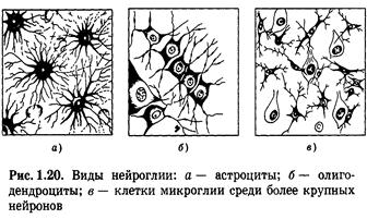
In addition to neurons, nervous tissue also includes cells that are companions of neurons - neuroglia (Fig. 1.20). Neuroglial cells (astrocytes, oligodendrocytes, microglia) fill the entire space between neurons, protecting them from mechanical damage (support function). There are about 10 times more of them than neurons, and, unlike them, glial cells retain the ability to divide throughout life. In addition, they form myelin sheaths around nerve fibers. During this process, the oligodendrocyte (in the central nervous system) or its variant, the Schwann cell (in the peripheral nervous system), wraps around a section of the nerve fiber. Then it forms an outgrowth in the form of a tongue, which twists around the fiber, forming layers of myelin (the cytoplasm is squeezed out). Thus, the layers of myelin are, in fact, a tightly compressed cytoplasmic membrane.
Neuroglia also perform a protective function. It lies, firstly, in the fact that glial cells (mainly astrocytes) together with the epithelial cells of the capillaries form a barrier between the blood and neurons, preventing unwanted (harmful) substances from passing through to the latter. This barrier is called the blood-brain barrier. Secondly, microglial cells perform the function of phagocytes in the nervous system. Carrying out a trophic function, neuroglia supplies neurons with nutrients, controls water-salt metabolism, etc.
Rudolf Virchow. 1856. Nerve glue.
Types of neuroglia:
A - protoplasmic astrocytes (in gray matter),
B - fibrous astrocytes (in the white matter),
B - microglia,
G - oligodendrocytes.
Neuroglia. Astrocytes. Astrocytes:largest&most numerous
A silvered preparation of astrocytes, showing their many fine cytoplasmic processes. Notice their close association with the capillaries (the heavy black structures). Since astrocytes touch both capillaries and neurons they are thought to play an intermediary role in the nutrition and metabolism of neurons.
Functions of astrocytes:
- support of nerve cells,
- restoration of nerve fibers when damaged,
- isolation and union of nerve fibers,
- participation in metabolic processes between capillaries and neurons,
— participation in the processes of neuron migration in ebryogenesis.
Features of the structure and types of nerve fibers
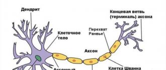
The nerve fiber - the axon - is covered with a cell membrane.
There are 2 types of nerve fibers.
Unmyelinated nerve fibers are one layer of Schwann cells, with slit-like spaces between them. The cell membrane is in contact with the environment throughout. When irritation is applied, excitation occurs at the site of action of the irritant. Unmyelinated nerve fibers have electrogenic properties (the ability to generate nerve impulses) throughout their entire length.
Myelinated nerve fibers are covered with layers of Schwann cells, which in some places form nodes of Ranvier (areas without myelin) every 1 mm. The duration of the node of Ranvier is 1 µm. The myelin sheath performs trophic and insulating functions (high resistance). Areas covered with myelin do not have electrogenic properties. They are possessed by nodes of Ranvier. Excitation occurs at the node of Ranvier closest to the site of action of the stimulus. At the nodes of Ranvier there is a high density of Na channels, so at each node of Ranvier there is an increase in nerve impulses.
The nodes of Ranvier function as relays (generate and amplify nerve impulses).
The mechanism of excitation along the nerve fiber
1885 - L. Herman - circular currents arise between excited and non-excited sections of the nerve fiber.
When a stimulus acts, there is a potential difference between the outer and inner surfaces of the tissue (areas carrying different charges). An electric current arises between these areas (the movement of Na+ ions). Inside the nerve fiber, a current arises from the positive pole to the negative pole, i.e., the current is directed from the excited area to the non-excited one. This current exits through the unexcited area and causes it to recharge. On the outer surface of the nerve fiber, the current flows from the unexcited area to the excited one. This current does not change the state of the excited area, since it is in a state of refractoriness.
Proof of the presence of circular currents: the nerve fiber is placed in a NaCl solution and the speed of excitation is recorded. Then the nerve fiber is placed in oil (resistance increases) - the conduction speed decreases by 30%. After this, the nerve fiber is left in the air - the speed of excitation is reduced by 50%.
Features of the conduction of excitation along myelinated and non-myelinated nerve fibers:
1. myelin fibers - have a sheath with high resistance, electrogenic properties only in the nodes of Ranvier. Under the influence of a stimulus, excitation occurs in the nearest node of Ranvier. The neighboring interception is in a state of polarization. The resulting current causes depolarization of the adjacent interception. At the nodes of Ranvier there is a high density of Na channels, therefore, at each subsequent node a slightly larger (in amplitude) action potential arises, due to this the excitation spreads without decrement and can jump over several nodes. This is Tasaki's saltatory theory. Proof of the theory - drugs were injected into the nerve fiber, blocking several interceptions, but the conduction of excitation was recorded even after that. This is a highly reliable and profitable method, since minor damage is eliminated, the speed of excitation is increased, and energy costs are reduced;
2. unmyelinated fibers - the surface has electrogenic properties throughout. Therefore, small circular currents arise at a distance of several micrometers. Excitation has the appearance of a constantly traveling wave.
This method is less profitable: higher energy costs (for the operation of the Na-K pump), lower excitation speed.
Classification of nerve fibers
Nerve fibers are classified according to:
1. action potential duration;
2. structure (diameter) of the fiber;
3. speed of excitation.
The following groups of nerve fibers are distinguished:
1. group A (alpha, beta, gamma, delta) - the shortest action potential, the thickest myelin sheath, the highest speed of excitation;
2. group B - the myelin sheath is less pronounced;
3. group C - without myelin sheath.
Synapses of the central nervous system
Synapse
is a morphofunctional formation of the central nervous system, which ensures signal transmission from a neuron to another neuron or from a neuron to an effector cell (muscle fiber, secretory cell).
Synapse structure:
1) presynaptic membrane (electrogenic membrane in the axon terminal, forms a synapse on the muscle cell); 2) postsynaptic membrane (electrogenic membrane of the innervated cell on which the synapse is formed); 3) synaptic cleft (the space between the presynaptic and postsynaptic membrane, filled with fluid, which composition resembles blood plasma).
2) There are several classifications of synapses .
By localization:
1) central synapses; 2) peripheral synapses.
Central synapses
lie within the central nervous system and are also found in the ganglia of the autonomic nervous system. Central synapses are contacts between two nerve cells, and these contacts are heterogeneous and, depending on the structure on which the first neuron forms a synapse with the second neuron, they are distinguished:
1) axosomatic, formed by the axon of one neuron and the body of another neuron; 2) axodendritic, formed by the axon of one neuron and the dendrite of another; 3) axoaxonal (the axon of the first neuron forms a synapse on the axon of the second neuron); 4) dendrodentrite (the dendrite of the first neuron forms a synapse on dendrite of the second neuron).
There are several types of peripheral synapses
:
1) myoneural (neuromuscular), formed by the axon of a motor neuron and a muscle cell; 2) neuroepithelial, formed by the axon of a neuron and a secretory cell.
Functional classification of synapses: 1) excitatory synapses; 2) inhibitory synapses.
According to the mechanisms of excitation transmission in synapses: 1) chemical; 2) electrical.
Features of chemical synapses
lies in the fact that the transfer of excitation is carried out using a special group of chemicals - mediators.
There are several types of chemical synapses: 1) cholinergic. They transmit excitation with the help of acetylcholine; 2) adrenergic. They transmit excitation with the help of three catecholamines; 3) dopaminergic. They transmit excitation with the help of dopamine; 4) histaminergic. They transmit excitation with the help of histamine; 5) GABAergic. In them, excitation is transmitted with the help of gamma-aminobutyric acid, i.e., the process of inhibition develops.
Features of electrical synapses
lies in the fact that the transfer of excitation is carried out using electric current. Few such synapses have been found in the body.
Synapses have a number of physiological properties : 1) the valve property of synapses, i.e. the ability to transmit excitation in only one direction from the presynaptic membrane to the postsynaptic; 2) the property of synaptic delay, associated with the fact that the rate of transmission of excitation decreases; 3) the property of potentiation ( each subsequent impulse will be conducted with a shorter postsynaptic delay). This is due to the fact that the transmitter from the previous impulse remains on the presynaptic and postsynaptic membrane; 4) low lability of the synapse (100–150 impulses per second).
The speed of excitation through the synapse is much less than along the nerve fiber, since time is spent here on the activation of the presynaptic membrane, the passage of calcium through it, the release of acetylcholine into the synaptic cleft, the depolarization of the postsynaptic membrane, and the development of PPP. Synaptic transmission of excitation has a number of properties:
1) The presence of a mediator in the presynaptic part of the synapse; 2) The relative mediator specificity of the synapse, i.e., each synapse has its own dominant mediator; 3) The transition of the postsynaptic membrane under the influence of mediators into a state of de- or hyperpolarization; 4) The possibility of action of specific blocking agents on receptive structures of the postsynaptic membrane; 5) Increasing the duration of the postsynaptic membrane potential when suppressing the action of enzymes that destroy the synaptic transmitter; 6) Development in the postsynaptic membrane of PSP from miniature potentials caused by quanta of the mediator; 7) Dependence of the duration of the active phase of the action of the mediator in the synapse on the properties of the mediator; 8) Unilateral conduction of excitation; 9) The presence of chemosensitive receptor-controlled channels of the postsynaptic membrane; 10) An increase in the release of transmitter quanta into the synaptic cleft is proportional to the frequency of impulses arriving along the axon; 11) Dependence of the increase in the efficiency of synaptic transmission on the frequency of use of the synapse (“training effect”); 12 ) Fatigue of the synapse, which develops as a result of prolonged high-frequency stimulation. In this case, fatigue may be caused by exhaustion and untimely synthesis of the transmitter in the presynaptic part of the synapse or by deep, persistent depolarization of the postsynaptic membrane (pessimal inhibition).
Neuron. Structural features, meaning, types
Basic principles of the functioning of the central nervous system. Structure, functions, methods of studying the central nervous system
The basic principle of the functioning of the central nervous system is the process of regulation, management of physiological functions, which are aimed at maintaining the constancy of the properties and composition of the internal environment of the body. The central nervous system ensures optimal relationships between the body and the environment, stability, integrity, and the optimal level of vital activity of the body.
There are two main types of regulation: humoral and nervous.
The humoral control process involves changing the physiological activity of the body under the influence of chemicals that are delivered by body fluids. The source of information transfer is chemicals - utilizons, metabolic products (carbon dioxide, glucose, fatty acids), informons, hormones of the endocrine glands, local or tissue hormones.
The nervous process of regulation involves controlling changes in physiological functions along nerve fibers using excitation potential under the influence of information transfer.
Characteristics:
1) is a later product of evolution;
2) provides quick regulation;
3) has an exact target of impact;
4) implements an economical method of regulation;
5) ensures high reliability of information transmission.
In the body, the nervous and humoral mechanisms work as a single system of neurohumoral control. This is a combined form where two control mechanisms are used simultaneously; they are interconnected and interdependent.
The nervous system is a collection of nerve cells, or neurons.
According to localization they distinguish:
1) central section – brain and spinal cord;
2) peripheral - processes of nerve cells of the brain and spinal cord.
According to functional features they are distinguished:
1) somatic department, regulating motor activity;
2) vegetative, regulating the activity of internal organs, endocrine glands, blood vessels, trophic innervation of muscles and the central nervous system itself.
Functions of the nervous system:
1) integrative-coordination function. Provides the functions of various organs and physiological systems, coordinates their activities with each other;
2) ensuring close connections between the human body and the environment at the biological and social levels;
3) regulation of the level of metabolic processes in various organs and tissues, as well as in oneself;
4) ensuring mental activity by the higher departments of the central nervous system.
The structural and functional unit of nervous tissue is the nerve cell – neuron.
A neuron is a specialized cell that is capable of receiving, encoding, transmitting and storing information, establishing contacts with other neurons, and organizing the body’s response to stimulation.
Functionally, a neuron is divided into:
1) the receptive part (dendrites and membrane of the soma of the neuron);
2) integrative part (soma with axon hillock);
3) transmitting part (axon hillock with axon).
Perceiving part.
Dendrites are the main The dendrite membrane is capable of responding to mediators. A neuron has several branching dendrites. This is explained by the fact that a neuron as an information formation must have a large number of inputs. Through specialized contacts, information flows from one neuron to another. These contacts are called "spines".
The neuron soma membrane is 6 nm thick and consists of two layers of lipid molecules. The hydrophilic ends of these molecules face the aqueous phase: one layer of molecules faces inward, the other outward
The hydrophilic ends are turned towards each other - inside the membrane. The lipid bilayer of the membrane contains proteins that perform several functions:
1) pump proteins - move ions and molecules in the cell against a concentration gradient;
2) proteins embedded in the channels provide selective membrane permeability;
3) receptor proteins recognize the desired molecules and fix them on the membrane;
4) enzymes facilitate the occurrence of a chemical reaction on the surface of the neuron.
In some cases, the same protein can function as both a receptor, an enzyme, and a pump.
Integrative part.
The axon hillock is where the axon exits the neuron.
The neuron soma (neuron body) performs, along with an informational and trophic function, relative to its processes and synapses. Soma ensures the growth of dendrites and axons. The neuron soma is enclosed in a multilayer membrane, which ensures the formation and propagation of electrotonic potential to the axon hillock.
Transmitting part.
An axon is an outgrowth of the cytoplasm adapted to carry information that is collected by dendrites and processed in a neuron. The axon of a dendritic cell has a constant diameter and is covered with a myelin sheath, which is formed from glia; the axon has branched ends that contain mitochondria and secretory formations.
Functions of neurons:
1) generalization of the nerve impulse;
2) receiving, storing and transmitting information;
3) the ability to summarize excitatory and inhibitory signals (integrative function).
Types of neurons:
1) by localization:
a) central (brain and spinal cord);
b) peripheral (cerebral ganglia, cranial nerves);
2) depending on the function:
a) afferent (sensitive), carrying information from receptors to the central nervous system;
b) intercalary (connector), in the elementary case providing communication between afferent and efferent neurons;
c) efferent:
– motor – anterior horns of the spinal cord;
– secretory – lateral horns of the spinal cord;
3) depending on the functions:
a) stimulating;
b) inhibitory;
4) depending on the biochemical characteristics, on the nature of the mediator;
5) depending on the quality of the stimulus that is perceived by the neuron:
a) monomodal;
b) multimodal.