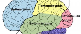
Classification of brain cysts in adults.
By localization:
- Sylvian fissure (temporal lobe) 49%.
- Cerebellopontine angle 11%.
- Craniovertebral junction 10%.
- Cerebellar vermis (retrocerebellar cyst of the brain) 9%.
- Sellar and parasellar 9%.
- Interhemispheric fissure 5%.
- The convexital surface of the cerebral hemispheres is 4%.
- Slope areas 3%.
A retrocerebellar cyst of the brain can simulate Dandy-Walker syndrome, but the cerebellar vermis is preserved and the cerebrospinal fluid does not drain into the fourth ventricle.
Types of Sylvian fissure cyst.
I. is small in size, located in the region of the pole of the temporal lobe, does not cause compression, the cerebrospinal fluid flows into the subarachnoid space. II. covers the proximal and middle parts of the Sylvian fissure, almost rectangular in shape, the cerebrospinal fluid does not completely drain into the subarachnoid space. III. covers the entire Sylvian fissure, there may be a protrusion of the temporal bone, minimal drainage into the subarachnoid space, the operation often does not lead to straightening of the brain (a transition to type 2 is possible).
Certain types of congenital cysts.
- Transparent partition;
- Verge;
- Intermediate sail.
A cyst of the transparent septum of the brain is a cavity between the sheets of the transparent septum filled with cerebrospinal fluid.
It is a stage of normal development that does not last long after birth. Therefore, almost all premature babies have a cyst. Occurs in approximately 10% of adults as a congenital asymptomatic developmental anomaly that does not require treatment. A cyst of the cerebral septum can communicate with the third ventricle.
Verge's cyst is localized behind the cavity of the transparent septum and often communicates with it. Very rare.
The intermediate velum cyst forms between the thalami above the third ventricle as a result of separation of the crura of the fornix. Present in 60% of children under 1 year of age and 30% between 1 and 10 years of age. As a rule, it does not cause any changes in the clinical condition, but a large cyst can lead to occlusive hydrocephalus. In most cases, no treatment is required.
Dimensions and norm
Patients are often interested in the question of the normal size of the cyst and the volume at which surgical intervention is necessary.
It must be said that we can speak of normality only in the absence of a cystic neoplasm; if there is a cyst, then this is already an anomaly, and its size essentially does not matter much.
This type can have different sizes, but the cyst still cannot grow very much. Brain fluid resists its growth. If the size of the cyst is 1-2 mm, it is considered small; if the neoplasm grows to 1 cm, it is considered medium; cysts larger than 1 cm are considered large.
When diagnosing small cysts, doctors, as a rule, do not prescribe treatment, but choose a wait-and-see approach and simply monitor changes in the volume of the tumor. Large cysts are treated surgically; surgery is also indicated for medium-sized tumors when symptoms appear.
Clinical signs.
Clinical manifestations usually occur in early childhood. In adults, symptoms appear much less frequently. They depend on the location. Often a cyst in the brain in an adult is asymptomatic, is an accidental finding during examination and does not require treatment.
Typical clinical manifestations of a brain cyst in an adult:
- General cerebral symptoms: headache, nausea, vomiting, drowsiness.
- Epileptic seizures.
- Protrusion of the skull bones.
- Focal symptoms: monoparesis, hemiparesis, sensitivity disorders of the mono- and hemitype, speech disorders in the form of sensory, motor or mixed sensorimotor aphasia, loss of visual fields, paresis of cranial nerves.
- Sudden deterioration, which is accompanied by depression of consciousness up to coma:
- Due to hemorrhage into the cyst;
- Due to cyst rupture.
Causes
Primary (congenital) cysts are formed due to abnormalities in the developing fetus, most often this occurs under the influence of the following factors:
- intoxication of the fetus with narcotic substances, alcohol, nicotine, medications that have a teratogenic effect;
- intrauterine infection of the fetus with viruses;
- irradiation;
- severe overheating of the body during pregnancy.
In addition, the formation can occur with Marfan syndrome - this is an anomaly of connective tissue, as well as with pathologies of the corpus callosum.
Secondary pathologies can develop when:
- brain injuries;
- pathological processes in the brain, for example, degenerative;
- in the presence of infections;
- deterioration of cerebral circulation.
In addition, pathology can be observed after surgery.
The mechanism of development of this neoplasm is quite complex and is associated with impaired circulation of the cerebrospinal fluid. As a result, a cavity filled with cerebrospinal fluid is formed.
Diagnosis of a cyst in the brain in an adult.
Computed tomography or magnetic resonance imaging are mandatory research methods.
Additional diagnostic methods:
- Cisternography and ventriculography are contrast studies of the cerebrospinal fluid pathways. They are rarely required, for example, when examining median suprasellar cysts and when affecting the posterior fossa for the purpose of differential diagnosis with Dandy-Walker anomaly.
- Examination of the fundus by an ophthalmologist for hypertension syndrome.
- Electroencephalography (EEG) if there was an epileptic seizure.
Retrocerebellar type formation
This is a fairly rare pathology, which is diagnosed in 5% of the population.
This neoplasm combines several types of neoplasms that differ in location:
- arachnoid,
- intracerebral - can form in any part,
- inferior retrocerebellar.
This cyst is spoken of when the tumor is localized only in the cerebellum. In this case we observe:
- migraine;
- vomit;
- paralysis and paresis;
- problems with balance;
- the image before your eyes becomes unclear;
- decreased concentration;
- deterioration of character;
- loss of consciousness;
- twitching of the eyeballs;
- deterioration of thinking;
- violation of orientation in space and time;
- decreased muscle tone.
Brain cyst treatment.
Most congenital cysts are asymptomatic and do not require treatment. Sometimes a neurosurgeon may recommend dynamic monitoring of the size of the cyst. To do this, you need to periodically perform CT or MRI.
In rare cases, when an arachnoid cyst is accompanied by symptoms and compresses nerve structures, surgical treatment is resorted to.
In some cases with a sharp deterioration, due to rupture of the arachnoid cyst or hemorrhage into it, urgent surgical treatment is resorted to.
Indications for surgery are determined taking into account the location and symptoms, and not just the size.
Absolute indications for surgery.
- intracranial hypertension syndrome caused by an arachnoid cyst or concomitant hydrocephalus;
- the appearance of neurological deficit.
Relative indications for surgery.
- large “asymptomatic arachnoid cysts” that cause deformation of neighboring lobes of the brain;
- progressive increase in size;
- deformation of the cerebrospinal fluid pathways, leading to disruption of the cerebrospinal fluid circulation.
Contraindications for surgery.
- decompensated state of vital functions (unstable hemodynamics, breathing), terminal coma (coma III);
- the presence of an active inflammatory process.
Methods of surgical treatment of brain cysts.
- Evacuation of a brain cyst in an adult through a burr hole in the skull using a navigation station.
Advantages and disadvantages
Simplicity
Execution speed
Minimal trauma
High relapse rate
- Open surgery, that is, craniotomy (cutting out a bone flap on the skull, which is placed in place at the end of the operation) with excision of the walls of the cyst and fenestration (drainage) into the basal cisterns (cerebrospinal fluid spaces at the base of the skull).
Advantages and disadvantages
Direct examination of the cyst cavity
Avoids permanent shunt
More effective for multilocular cysts
Traumaticity
Operation duration
- Shunt surgery with installation of a shunt from the cyst cavity into the abdominal cavity or superior vena cava near the right atrium through the common facial vein or internal jugular vein. Many foreign and domestic neurosurgeons consider shunting to be the best way to treat brain cysts.
Advantages and disadvantages
Low mortality
Low relapse rate
Patient dependence on shunt
If the shunt becomes blocked, it will have to be replaced.
Complications of the operation.
- liquorrhea;
- marginal necrosis of the skin flap with dehiscence of the surgical wound;
- meningitis and other infectious complications;
- hemorrhage into the cyst cavity.
Symptomatic manifestations
This cystic neoplasm is characterized by an asymptomatic course.
Most often, such neoplasms are detected during a patient examination for neurological pathologies with similar symptoms.
The clinical signs of the cyst are nonspecific; their severity depends on the size of the tumor and its location. Patients may complain of general cerebral symptoms, which are associated with compression of brain areas. A focal clinical picture is observed in rare cases, and is most often caused by the presence of hygroma or rupture of the cystic cavity.
The main features are:
- dizziness that is not associated with pregnancy, taking certain medications, severe fatigue, iron deficiency, and so on;
- vomiting, which is also not associated with gastrointestinal poisoning, taking medications, and so on;
- mental disorders, hallucinations;
- convulsive syndrome;
- loss of consciousness;
- hemiparesis, decreased sensitivity in the limbs;
- failure to coordinate the activity of the body muscles;
- headaches that are not relieved by painkillers;
- a feeling of fullness, pressure, compression and increased pulsation in the affected area;
- visual and hearing problems;
- extraneous sounds in the ears;
- heaviness in the skull;
- painful or uncomfortable sensations when turning the head.
Mechanics of pathology development
Liquor is a fluid necessary for the healthy functioning of the human brain, which transports nutrients and trace elements from the center of the brain to its membranes. In some cases - due to congenital or acquired diseases - cerebrospinal fluid begins to accumulate in the arachnoid membrane. This is how an arachnoid cyst of the left temporal lobe is formed. Sometimes such a formation is called a liquor cyst.
Doctors were unable to establish the exact cause and features of the appearance of this disease. There is a theory that such ailments can develop in utero due to viral infection or later due to acquired diseases. However, despite the poor study of the problem, arachnoid cyst of the temporal region on the left is considered a relatively common ailment and requires a more detailed description.
Features of symptoms
It is worth saying that the symptomatic picture of an arachnoid cyst of the left temporal lobe in children is extremely complicated and poorly studied. For the reason that it can not only be formed due to acquired or hidden ailments, but also be subject to individual characteristics of brain development. Doctors have not yet been able to accurately establish and characterize the symptoms of a small arachnoid cyst of the left temporal lobe. But based on the research conducted today, the following can be said about this type of tumor.
Subarachnoid cysts of the left temporal lobe of the congenital type, as a rule, develop and grow slowly, so symptomatic signs of their formation may appear already at school age. Due to the specific type of appearance, congenital tumors rarely cause painful symptoms. What is very important is that an arachnoid cyst in the left temporal region never develops into cancer.
However, one should not assume that an arachnoid cyst in the left temporal region is harmless. Under certain conditions, such a cyst in a child’s head can grow rapidly and provoke many pathologies in the development of the brain. This is especially true in cases where the congenital arachnoid cyst of the pole of the left temporal lobe is affected by a collateral disease.